اعصاب جمجمهای؛ عصب جمجمهای II: عصب بینایی


دعای مطالعه [ نمایش ]
بِسْمِ الله الرَّحْمنِ الرَّحیمِ
اَللّهُمَّ اَخْرِجْنى مِنْ ظُلُماتِ الْوَهْمِ
خدايا مرا بيرون آور از تاريكىهاى وهم،
وَ اَكْرِمْنى بِنُورِ الْفَهْمِ
و به نور فهم گرامى ام بدار،
اَللّهُمَّ افْتَحْ عَلَيْنا اَبْوابَ رَحْمَتِكَ
خدايا درهاى رحمتت را به روى ما بگشا،
وَانْشُرْ عَلَيْنا خَزائِنَ عُلُومِكَ بِرَحْمَتِكَ يا اَرْحَمَ الرّاحِمينَ
و خزانههاى علومت را بر ما باز كن به امید رحمتت اى مهربانترين مهربانان.
آیندهنگاران مغز: «ما مغز را میشناسیم، تا آینده را بسازیم.»
کتاب «نوروآناتومی بالینی اسنل» اثر دکتر ریچارد اس. اسنل، با ویرایش جدید دکتر رایان اسپلیتگربر، همچنان بهعنوان مرجع طلایی این حوزه شناخته میشود. این اثر با تلفیق تازهترین پژوهشها و کاربردهای بالینی، پلی استوار میان دانش پایه و عمل پزشکی بنا میکند.
تلاشهای علمی و سالها تدریس ارزشمند استاد گرانقدر آقای دکتر محمدتقی جغتایی الهامبخش ترجمه و گسترش این دانش در ایران بوده است.
ترجمه دقیق این کتاب توسط برند علمی آیندهنگاران مغز به مدیریت داریوش طاهری، امکان دسترسی فارسیزبانان به مرزهای نوین علوم اعصاب را فراهم کرده و رسالتی علمی در جهت شناخت عمیقتر مغز و ترسیم آیندهای روشنتر بر عهده گرفته است.
»» فصل ۱۱: هستههای اعصاب جمجمهای؛ بخش دوم مبانی و اصول
»» Chapter 11: Cranial Nerve Nuclei
OPTIC NERVE (CRANIAL NERVE II)
The fibers of the optic nerve are the axons of the cells in the ganglionic layer of the retina. They converge on the optic disc and exit from the eye, about 3 or 4 mm to the nasal side of its center, as the optic nerve (Fig. 11-2). The fibers of the optic nerve are myelinated, but the sheaths are formed from oligodendrocytes rather than Schwann cells, since the optic nerve is comparable to a tract within the central nervous system.
عصب بینایی (عصب جمجمهای II)
الیاف عصب بینایی، آکسونهای سلولهای لایه گانگلیونی شبکیه هستند. آنها روی دیسک بینایی همگرا میشوند و از چشم، حدود ۳ یا ۴ میلیمتر به سمت بینی مرکز آن، به عنوان عصب بینایی خارج میشوند (شکل ۱۱-۲). الیاف عصب بینایی میلیندار هستند، اما غلافها به جای سلولهای شوان از الیگودندروسیتها تشکیل شدهاند، زیرا عصب بینایی با یک مسیر در سیستم عصبی مرکزی قابل مقایسه است.
The optic nerve leaves the orbital cavity through the optic canal and unites with the optic nerve of the opposite side to form the optic chiasma.
عصب بینایی از طریق کانال بینایی حفره مداری را ترک میکند و با عصب بینایی طرف مقابل متحد میشود تا کیاسمای بینایی را تشکیل دهد.
Optic Chiasma
The optic chiasma is situated at the junction of the anterior wall and floor of the third ventricle. Its anterolateral angles are continuous with the optic nerves, and the posterolateral angles are continuous with the optic tracts. In the chiasma, the fibers from the nasal (medial) half of each retina, including the nasal half of the macula, cross the midline and enter the optic tract of the opposite side, while the fibers from the temporal (lateral) half of each retina, including the temporal half of the macula, pass posteriorly in the optic tract of the same side.
کیاسمای بینایی
کیاسمای بینایی در محل اتصال دیواره قدامی و کف بطن سوم قرار دارد. زوایای قدامی-جانبی آن با اعصاب بینایی و زوایای خلفی-جانبی با مجاری بینایی پیوسته هستند. در کیاسم، الیاف نیمه بینی (میانی) هر شبکیه، از جمله نیمه بینی ماکولا، از خط وسط عبور کرده و وارد مجرای بینایی طرف مقابل میشوند، در حالی که الیاف نیمه گیجگاهی (جانبی) هر شبکیه، از جمله نیمه گیجگاهی ماکولا، به صورت خلفی در مجرای بینایی همان طرف عبور میکنند.
Optic Tract
The optic tract emerges from the optic chiasma and passes posterolaterally around the cerebral peduncle. Most of the fibers now terminate by synapsing with nerve cells in the lateral geniculate body, which is a small projection from the posterior part of the thalamus. A few of the fibers pass to the pretectal nucleus and the superior colliculus of the midbrain and are concerned with light reflexes (Fig. 11-3).
مسیر بینایی
مسیر بینایی از کیاسمای بینایی خارج میشود و به صورت خلفی-خارجی در اطراف پایه مغزی عبور میکند. اکنون بیشتر فیبرها با سیناپس با سلولهای عصبی در جسم زانویی جانبی، که یک برآمدگی کوچک از قسمت خلفی تالاموس است، خاتمه مییابند. تعداد کمی از فیبرها به هسته پرهتکتال و کولیکولوس فوقانی مغز میانی میروند و با رفلکسهای نوری مرتبط هستند (شکل 11-3).
Lateral Geniculate Body
The lateral geniculate body is a small, oval swelling projecting from the pulvinar of the thalamus. It consists of six layers of cells, on which synapse the axons from the optic tract. The axons of the nerve cells within the geniculate body leave it to form the optic radiation (see Fig. 11-2).
جسم زانویی جانبی
جسم زانویی جانبی یک برآمدگی کوچک و بیضی شکل است که از پولوینار تالاموس بیرون زده است. این جسم از شش لایه سلول تشکیل شده است که آکسونهای دستگاه بینایی روی آنها سیناپس میکنند. آکسونهای سلولهای عصبی درون جسم زانویی از آن خارج میشوند تا شعاع بینایی را تشکیل دهند (شکل 11-2 را ببینید).
Optic Radiation
The fibers of the optic radiation are the axons of the nerve cells of the lateral geniculate body. The tract passes posteriorly through the retrolenticular part of the internal capsule and terminates in the visual cortex (area 17), which occupies the upper and lower lips of the calcarine sulcus on the medial surface of the cerebral hemisphere. The visual association cortex (areas 18 and 19) is responsible for recognition of objects and perception of color.
تابش نوری
الیاف تابش نوری، آکسونهای سلولهای عصبی جسم زانویی جانبی هستند. این مسیر از قسمت خلفی کپسول داخلی عبور کرده و در قشر بینایی (ناحیه ۱۷) خاتمه مییابد که لبهای بالایی و پایینی شیار کالکارین را در سطح داخلی نیمکره مغزی اشغال میکند. قشر ارتباط بینایی (نواحی ۱۸ و ۱۹) مسئول تشخیص اشیاء و درک رنگ است.
Visual Pathway and Binocular Vision Neurons
Four neurons conduct visual impulses to the visual cortex: (1) rods and cones, which are specialized receptor neurons in the retina; (2) bipolar neurons, which connect the rods and cones to the ganglion cells; (3) ganglion cells, whose axons pass to the lateral geniculate body; and (4) neurons of the lateral geniculate body, whose axons pass to the cerebral cortex.
نورونهای مسیر بینایی و بینایی دوچشمی
چهار نورون، تکانههای بینایی را به قشر بینایی هدایت میکنند: (1) میلهها و مخروطها، که نورونهای گیرنده تخصصی در شبکیه هستند؛ (2) نورونهای دوقطبی، که میلهها و مخروطها را به سلولهای گانگلیونی متصل میکنند؛ (3) سلولهای گانگلیونی، که آکسونهای آنها به جسم زانویی جانبی منتقل میشوند؛ و (4) نورونهای جسم زانویی جانبی، که آکسونهای آنها به قشر مغز منتقل میشوند.
In binocular vision, the right and left fields of vision are projected on portions of both retinae. The image of an object in the right field of vision is projected on the nasal half of the right retina and the temporal half of the left retina. In the optic chiasma, the axons from these two retinal halves are combined to form the left optic tract. The lateral geniculate body neurons now project the complete right field of vision on the visual cortex of the left hemisphere and the left visual field on the visual cortex of the right hemisphere. The lower retinal quadrants (upper field of vision) project on the lower wall of the calcarine sulcus, while the upper retinal quadrants (lower field of vision) project on the upper wall of the sulcus. Note also that the macula lutea is represented on the posterior part of area 17, and the periphery of the retina is represented anteriorly.
در دید دوچشمی، میدانهای دید راست و چپ بر روی بخشهایی از هر دو شبکیه منعکس میشوند. تصویر یک جسم در میدان دید راست، بر روی نیمه بینی شبکیه راست و نیمه گیجگاهی شبکیه چپ منعکس میشود. در کیاسمای بینایی، آکسونهای این دو نیمه شبکیه با هم ترکیب میشوند تا راه بینایی چپ را تشکیل دهند. نورونهای جسم زانویی جانبی اکنون میدان دید کامل راست را بر روی قشر بینایی نیمکره چپ و میدان بینایی چپ را بر روی قشر بینایی نیمکره راست منعکس میکنند. ربعهای پایینی شبکیه (میدان دید بالایی) بر روی دیواره پایینی شیار کالکارین منعکس میشوند، در حالی که ربعهای بالایی شبکیه (میدان دید پایینی) بر روی دیواره بالایی شیار منعکس میشوند. همچنین توجه داشته باشید که ماکولا لوتئا در قسمت خلفی ناحیه ۱۷ و حاشیه شبکیه در قسمت قدامی نشان داده شده است.
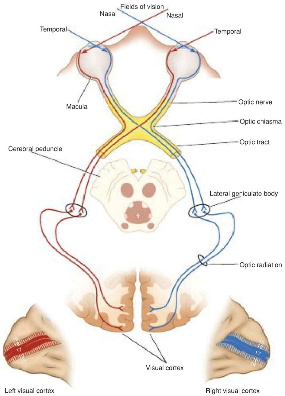
Figure 11-2 Optic pathway.
شکل ۱۱-۲ مسیر بینایی.
Visual Reflexes
Several unique neuronal pathways exert involuntary control over our vision for optimal visual function, protection, and cognitive processing.
رفلکسهای بینایی
چندین مسیر عصبی منحصر به فرد، کنترل غیرارادی بر بینایی ما اعمال میکنند تا عملکرد بینایی، محافظت و پردازش شناختی بهینه حاصل شود.
Direct and Consensual Light Reflexes
If a light is shone into one eye, the pupils of both eyes normally constrict. The constriction of the pupil on which the light is shone is called the direct light reflex; the constriction of the opposite pupil, even though no light fell on that eye, is called the consensual light reflex (see Fig. 11-3).
رفلکسهای نوری مستقیم و توافقی
اگر نوری به یک چشم تابیده شود، مردمکهای هر دو چشم به طور معمول منقبض میشوند. انقباض مردمکی که نور به آن تابیده میشود، رفلکس نوری مستقیم نامیده میشود؛ انقباض مردمک مقابل، حتی اگر هیچ نوری به آن چشم نخورده باشد، رفلکس نوری توافقی نامیده میشود (شکل 11-3 را ببینید).

Figure 11-3 Optic pathway and the visual reflexes.
شکل ۱۱-۳ مسیر بینایی و رفلکسهای بینایی.
The afferent impulses travel through the optic nerve, optic chiasma, and optic tract. Here, a small number of fibers leave the optic tract and synapse on nerve cells in the pretectal nucleus, which lies close to the superior colliculus. The impulses are passed by axons of the pretectal nerve cells to the parasympathetic nuclei (Edinger-Westphal nuclei) of the third cranial nerve on both sides. Here, the fibers synapse and the parasympathetic nerves travel through the third cranial nerve to the ciliary ganglion in the orbit. Finally, postganglionic parasympathetic fibers pass through the short ciliary nerves to the eyeball and the constrictor pupillae muscle of the iris. Both pupils constrict in the consensual light reflex because the pretectal nucleus sends fibers to the parasympathetic nuclei on both sides of the midbrain. The fibers that cross the median plane do so close to the cerebral aqueduct in the posterior commissure.
تکانههای آوران از طریق عصب بینایی، کیاسمای بینایی و دستگاه بینایی حرکت میکنند. در اینجا، تعداد کمی از فیبرها دستگاه بینایی را ترک میکنند و روی سلولهای عصبی در هسته پرهتکتال، که نزدیک به کولیکولوس فوقانی قرار دارد، سیناپس میکنند. تکانهها توسط آکسونهای سلولهای عصبی پرهتکتال به هستههای پاراسمپاتیک (هستههای ادینگر-وستفال) عصب جمجمهای سوم در هر دو طرف منتقل میشوند. در اینجا، فیبرها سیناپس میکنند و اعصاب پاراسمپاتیک از طریق عصب جمجمهای سوم به گانگلیون مژگانی در حدقه میروند. در نهایت، فیبرهای پاراسمپاتیک پسگانگلیونی از طریق اعصاب مژگانی کوتاه به کره چشم و عضله تنگکننده مردمک عنبیه عبور میکنند. هر دو مردمک در رفلکس نوری توافقی منقبض میشوند زیرا هسته پرهتکتال فیبرهایی را به هستههای پاراسمپاتیک در دو طرف مغز میانی میفرستد. فیبرهایی که از صفحه میانی عبور میکنند، این کار را نزدیک به قنات مغزی در رابط خلفی انجام میدهند.
Accommodation Reflex
When the eyes are directed from a distant to a near object, contraction of the medial recti brings about convergence of the ocular axes; the lens thickens to increase its refractive power by contraction of the ciliary muscle; and the pupils constrict to restrict the light waves to the thickest central part of the lens. The afferent impulses travel through the optic nerve, the optic chiasma, the optic tract, the lateral geniculate body, and the optic radiation to the visual cortex. The visual cortex is connected to the eye field of the frontal cortex. From here, cortical fibers descend through the internal capsule to the oculomotor nuclei in the midbrain. The oculomotor nerve travels to the medial recti muscles. Some of the descending cortical fibers synapse with the parasympathetic nuclei (Edinger-Westphal nuclei) of the third cranial nerve on both sides. Here, the fibers synapse, and the parasympathetic nerves travel through the third cranial nerve to the ciliary ganglion in the orbit. Finally, postganglionic parasympathetic fibers pass through the short ciliary nerves to the ciliary muscle and the constrictor pupillae muscle of the iris.
رفلکس تطابق
هنگامی که چشمها از یک شیء دور به یک شیء نزدیک هدایت میشوند، انقباض عضله راست داخلی باعث همگرایی محورهای چشمی میشود؛ عدسی ضخیم میشود تا قدرت انکساری خود را با انقباض عضله مژگانی افزایش دهد؛ و مردمکها منقبض میشوند تا امواج نور را به ضخیمترین قسمت مرکزی عدسی محدود کنند. تکانههای آوران از طریق عصب بینایی، کیاسمای بینایی، راه بینایی، جسم زانویی جانبی و تابش نوری به قشر بینایی منتقل میشوند. قشر بینایی به میدان بینایی قشر پیشانی متصل است. از اینجا، الیاف قشری از طریق کپسول داخلی به هستههای حرکتی چشم در مغز میانی میرسند. عصب حرکتی چشم به عضلات مستقیم داخلی میرود. برخی از الیاف قشری نزولی با هستههای پاراسمپاتیک (هستههای ادینگر-وستفال) عصب جمجمهای سوم در هر دو طرف سیناپس میکنند. در اینجا، فیبرها سیناپس میکنند و اعصاب پاراسمپاتیک از طریق عصب جمجمهای سوم به گانگلیون مژگانی در حدقه میروند. در نهایت، فیبرهای پاراسمپاتیک پسگانگلیونی از طریق اعصاب مژگانی کوتاه به عضله مژگانی و عضله تنگکننده مردمک عنبیه میروند.
Corneal Reflex
Light touching of the cornea or conjunctiva results in blinking of the eyelids. Afferent impulses from the cornea or conjunctiva travel through the ophthalmic division of the trigeminal nerve to the sensory nucleus of the trigeminal nerve (Fig. 11-4A). Internuncial neurons connect with the motor nucleus of the facial nerve on both sides through the medial longitudinal fasciculus. The facial nerve and its branches supply the orbicularis oculi muscle, which causes closure of the eyelids.
رفلکس قرنیه
لمس سبک قرنیه یا ملتحمه منجر به پلک زدن پلکها میشود. تکانههای آوران از قرنیه یا ملتحمه از طریق شاخه چشمی عصب سه قلو به هسته حسی عصب سه قلو منتقل میشوند (شکل 11-4A). نورونهای بینابینی از طریق فاسیکولوس طولی داخلی به هسته حرکتی عصب صورت در دو طرف متصل میشوند. عصب صورت و شاخههای آن عضله حلقوی چشم را تغذیه میکنند که باعث بسته شدن پلکها میشود.
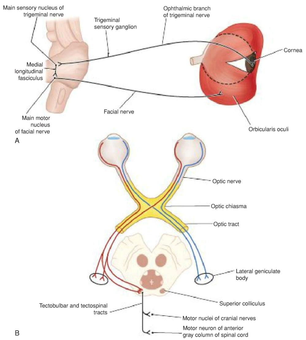
Figur 11-4 A: Corneal reflex. B: Visual body reflex.
شکل ۱۱-۴ الف: رفلکس قرنیه. ب: رفلکس بینایی بدن.
Visual Body Reflexes
The automatic scanning movements of the eyes and head that are made when reading, the automatic move- ment of the eyes, head, and neck toward the source of the visual stimulus, and the protective closing of the eyes and even the raising of the arm for protection are reflex actions that involve the following reflex arcs (see Fig. 11-4B). The visual impulses follow the optic nerves, optic chiasma, and optic tracts to the superior colliculi. Here, the impulses are relayed to the tectospinal and tectobulbar (tectonuclear) tracts and to the neurons of the anterior gray columns of the spinal cord and cranial motor nuclei.
رفلکسهای بینایی بدن
حرکات خودکار اسکن چشمها و سر که هنگام خواندن انجام میشوند، حرکت خودکار چشمها، سر و گردن به سمت منبع محرک بینایی، و بستن محافظ چشمها و حتی بالا بردن بازو برای محافظت، اعمال رفلکسی هستند که شامل قوسهای رفلکسی زیر میشوند (شکل 11-4B را ببینید). تکانههای بینایی از اعصاب بینایی، کیاسمای بینایی و مسیرهای بینایی به کولیکولهای فوقانی میروند. در اینجا، تکانهها به مسیرهای تکتوسپینال و تکتوبولبار (تکتونوکلئار) و به نورونهای ستونهای خاکستری قدامی نخاع و هستههای حرکتی جمجمه منتقل میشوند.
Pupillary Skin Reflex
The pupil will dilate if the skin is painfully stimulated by pinching. The afferent sensory fibers are believed to have connections with the efferent preganglionic sympathetic neurons in the lateral gray columns of the first and second thoracic segments of the spinal cord. The white rami communicantes of these segments pass to the sympathetic trunk, and the preganglionic fibers ascend to the superior cervical sympathetic ganglion. The postganglionic fibers pass through the internal carotid plexus, long ciliary nerves, and short ciliary nerves to the dilator pupillae muscle of the iris.
رفلکس پوستی مردمک
اگر پوست با نیشگون گرفتن به شدت تحریک شود، مردمک گشاد میشود. اعتقاد بر این است که فیبرهای حسی آوران با نورونهای سمپاتیک پیش عقدهای وابران در ستونهای خاکستری جانبی بخشهای اول و دوم سینهای نخاع ارتباط دارند. شاخههای سفید ارتباطی این بخشها به تنه سمپاتیک میروند و فیبرهای پیش عقدهای به گانگلیون سمپاتیک گردنی فوقانی صعود میکنند. فیبرهای پس عقدهای از طریق شبکه کاروتید داخلی، اعصاب مژگانی بلند و اعصاب مژگانی کوتاه به عضله گشادکننده مردمک عنبیه میروند.
«فصل یازدهم کتاب نورواناتومی بالینی اسنل»
🚀 با ما همراه شوید!
تازهترین مطالب و آموزشهای مغز و اعصاب را از دست ندهید. با فالو کردن کانال تلگرام آیندهنگاران مغز، از ما حمایت کنید!