پرسشهای مروری و مسائل بالینی اعصاب مغزی


دعای مطالعه [ نمایش ]
بِسْمِ الله الرَّحْمنِ الرَّحیمِ
اَللّهُمَّ اَخْرِجْنى مِنْ ظُلُماتِ الْوَهْمِ
خدايا مرا بيرون آور از تاريكىهاى وهم،
وَ اَكْرِمْنى بِنُورِ الْفَهْمِ
و به نور فهم گرامى ام بدار،
اَللّهُمَّ افْتَحْ عَلَيْنا اَبْوابَ رَحْمَتِكَ
خدايا درهاى رحمتت را به روى ما بگشا،
وَانْشُرْ عَلَيْنا خَزائِنَ عُلُومِكَ بِرَحْمَتِكَ يا اَرْحَمَ الرّاحِمينَ
و خزانههاى علومت را بر ما باز كن به امید رحمتت اى مهربانترين مهربانان.
آیندهنگاران مغز: «ما مغز را میشناسیم، تا آینده را بسازیم.»
کتاب «نوروآناتومی بالینی اسنل» اثر دکتر ریچارد اس. اسنل، با ویرایش جدید دکتر رایان اسپلیتگربر، همچنان بهعنوان مرجع طلایی این حوزه شناخته میشود. این اثر با تلفیق تازهترین پژوهشها و کاربردهای بالینی، پلی استوار میان دانش پایه و عمل پزشکی بنا میکند.
تلاشهای علمی و سالها تدریس ارزشمند استاد گرانقدر دکتر محمدتقی جغتایی الهامبخش ترجمه و گسترش این دانش در ایران بوده است.
ترجمه دقیق این کتاب توسط برند علمی آیندهنگاران مغز به مدیریت داریوش طاهری، امکان دسترسی فارسیزبانان به مرزهای نوین علوم اعصاب را فراهم کرده و رسالتی علمی در جهت شناخت عمیقتر مغز و ترسیم آیندهای روشنتر بر عهده گرفته است.
»» فصل ۱۱: هستههای اعصاب جمجمهای؛ بخش مسائل بالینی و پرسشهای مروری
»» Chapter 11: Cranial Nerve Nuclei
? Clinical Problem Solving
1. A 60-year-old woman is seen as an outpatient because she has suddenly developed double vision. She is watching her favorite television program when it suddenly occurs. She has no other symptoms. A complete physical examination shows that her right eye, when at rest, is turned medially, and she is unable to turn it laterally. A moderate amount of glucose is found in her urine, and she has an abnor- mally elevated blood glucose level. When closely questioned, she admits that recently she has noticed having to pass water more frequently, especially at night. She also says she often feels thirsty. She has lost 28 lb during the past 2 years. Using your knowl- edge of neuroanatomy, explain the problem in her right eye. Do you think a connection exists between her glucosuria, high blood glucose, polyuria, polydipsia, and weight loss and her eye condition?
؟ حل مسئله بالینی
۱. زنی 60 ساله به دلیل دوبینی ناگهانی به صورت سرپایی ویزیت میشود. او در حال تماشای برنامه تلویزیونی مورد علاقهاش است که ناگهان این اتفاق میافتد. او هیچ علامت دیگری ندارد. معاینه فیزیکی کامل نشان میدهد که چشم راست او، در حالت استراحت، به سمت داخل میچرخد و قادر به چرخاندن آن به سمت خارج نیست. مقدار متوسطی گلوکز در ادرار او یافت میشود و سطح گلوکز خون او به طور غیرطبیعی بالا است. وقتی از نزدیک مورد سوال قرار گرفت، اذعان کرد که اخیراً متوجه شده است که به خصوص در شب، نیاز به دفع مکرر آب دارد. او همچنین میگوید که اغلب احساس تشنگی میکند. او در طول 2 سال گذشته 28 پوند وزن کم کرده است. با استفاده از دانش خود در مورد نوروآناتومی، مشکل چشم راست او را توضیح دهید. آیا فکر میکنید ارتباطی بین گلوکوزوری، گلوکز خون بالا، پرادراری، پرنوشی و کاهش وزن و وضعیت چشم او وجود دارد؟
2. An 18-year-old man is admitted to the hospital unconscious after a serious motorcycle accident. A complete physical examination is performed and lateral and anteroposterior radiographs of the skull taken, which show that the patient has a fracture involving the anterior cranial fossa. It also is noted that he has a slight but continuous blood-stained watery discharge from his left nostril. Three days later, he regains consciousness, and a further physical examination reveals that he can no longer smell. This is tested by asking him to recognize the smell of coffee, oil of clove, and oil of peppermint. Using your knowledge of neuroanatomy, diagnose what is wrong with this patient. Is it possible for normal individuals with an acute sense of smell to be unable to recognize common scents? Could a tumor that had destroyed the olfactory cortex of one cere- bral hemisphere be responsible for the anosmia in this patient?
۲. مردی ۱۸ ساله پس از یک تصادف شدید موتورسیکلت، در حالت بیهوشی در بیمارستان بستری میشود. معاینه فیزیکی کامل انجام میشود و رادیوگرافیهای جانبی و قدامی-خلفی از جمجمه گرفته میشود که نشان میدهد بیمار دچار شکستگی در حفره جمجمه قدامی است. همچنین مشاهده میشود که او ترشحات آبکی خونآلود خفیف اما مداوم از سوراخ بینی چپ خود دارد. سه روز بعد، او به هوش میآید و معاینه فیزیکی بیشتر نشان میدهد که دیگر نمیتواند بو بکشد. این موضوع با درخواست از او برای تشخیص بوی قهوه، روغن میخک و روغن نعناع فلفلی آزمایش میشود. با استفاده از دانش خود در مورد نوروآناتومی، تشخیص دهید که مشکل این بیمار چیست. آیا افراد عادی با حس بویایی قوی میتوانند از تشخیص بوهای رایج ناتوان باشند؟ آیا توموری که قشر بویایی یک نیمکره مغزی را تخریب کرده است، میتواند مسئول آنوسمی در این بیمار باشد؟
3. A 72-year-old man with a known history of cerebrovascular problems visits his physician because 3 days previously, he had begun to have trouble reading the paper. He complains that the print started to tilt and that he is beginning to see double. He also says that he finds it difficult to see the steps when he descends the staircase to the physician’s office. On physical examination, the patient has weakness of movement of the right eye both downward and laterally. Using your knowledge of neuroanatomy, explain this patient’s signs and symptoms. If we assume that a cranial nerve nucleus is the site of the lesion, is it the right one or the left one that is involved?
۳. مردی ۷۲ ساله با سابقه شناخته شده مشکلات عروقی مغز به پزشک مراجعه میکند زیرا ۳ روز قبل در خواندن کاغذ مشکل داشته است. او شکایت دارد که نوشته شروع به کج شدن کرده و شروع به دوبینی کرده است. او همچنین میگوید که هنگام پایین آمدن از پلهها به سمت مطب پزشک، دیدن پلهها برایش دشوار است. در معاینه فیزیکی، بیمار دچار ضعف حرکتی چشم راست به سمت پایین و طرفین است. با استفاده از دانش خود در مورد نوروآناتومی، علائم و نشانههای این بیمار را توضیح دهید. اگر فرض کنیم که هسته عصب جمجمهای محل ضایعه است، آیا هسته عصب راست درگیر است یا چپ؟
4. A 73-year-old man consults his physician because he is becoming deaf. His only other complaints are that he did not think he was as tall as he used to be, and he is annoyed to find that each year he has to buy a size larger hat. The physician diagnoses osteitis deformans (Paget disease) and explains to the medical students that this is a disease of bones involving bone absorption and new bone formation. These bony changes lead to enlargement of the skull, deformities of the vertebral column, and bowing of the long bones of the legs. The physician asks the students whether a connection exists between the bone disease and the patient’s deafness and which other cranial nerve they would be particularly interested in testing. How would you have answered these questions?
۴. مردی ۷۳ ساله به دلیل ناشنوا شدن به پزشک مراجعه میکند. تنها شکایت دیگر او این است که فکر نمیکرد به اندازهی قبل قدبلند باشد و از اینکه هر سال مجبور است کلاهی با سایز بزرگتر بخرد، ناراحت است. پزشک، بیماری اوستئیت دفورمانس (بیماری پاژه) را تشخیص میدهد و برای دانشجویان پزشکی توضیح میدهد که این یک بیماری استخوانی است که شامل جذب استخوان و تشکیل استخوان جدید میشود. این تغییرات استخوانی منجر به بزرگ شدن جمجمه، بدشکلی ستون فقرات و خمیدگی استخوانهای بلند پاها میشود. پزشک از دانشجویان میپرسد که آیا ارتباطی بین بیماری استخوانی و ناشنوایی بیمار وجود دارد و آنها به طور خاص به آزمایش کدام عصب جمجمهای دیگر علاقهمند هستند. چگونه به این سؤالات پاسخ میدادید؟
5. A neurologist is visited by a 25-year-old man who complains of a feeling of heaviness in both legs and giddiness on walking. On examination, the patient has widely disseminated lesions involving the corticospinal tracts, the posterior white column, and the optic nerves. A diagnosis of multiple sclerosis is made. This disease of unknown origin primarily involves the white matter of the brain and spinal cord. Do you think this patient’s symptoms of verti- go could be accounted for by this disease?
۵. مردی ۲۵ ساله به متخصص مغز و اعصاب مراجعه میکند که از احساس سنگینی در هر دو پا و سرگیجه هنگام راه رفتن شکایت دارد. در معاینه، بیمار ضایعات منتشر گستردهای دارد که راههای قشری-نخاعی، ستون سفید خلفی و اعصاب بینایی را درگیر میکند. تشخیص بیماری اماس داده میشود. این بیماری با منشأ ناشناخته، عمدتاً ماده سفید مغز و نخاع را درگیر میکند. آیا فکر میکنید علائم سرگیجه این بیمار میتواند ناشی از این بیماری باشد؟
6. A 54-year-old woman with left-sided hemiplegia is examined by a fourth-year medical student. He very carefully tests each cranial nerve and notes any defects. During the examination, he stands behind the patient and gently grasps the trapezius muscles between his fingers and thumbs and asks the patient to shrug her shoulders against resistance. He is surprised to find no evidence of weakness in either trapezius muscle and no muscle wasting. Would you expect to find evidence of weakness or wasting in the trapezius muscles of a patient with hemiplegia?
۶. یک دانشجوی پزشکی سال چهارم، زنی ۵۴ ساله مبتلا به فلج نیمه بدن چپ را معاینه میکند. او با دقت هر عصب جمجمهای را آزمایش میکند و هرگونه نقصی را یادداشت میکند. در طول معاینه، او پشت بیمار میایستد و به آرامی عضلات ذوزنقهای را بین انگشتان و شست خود میگیرد و از بیمار میخواهد که شانههای خود را در برابر مقاومت بالا بیندازد. او با کمال تعجب هیچ نشانهای از ضعف در عضله ذوزنقهای و هیچ تحلیل عضلانی پیدا نمیکند. آیا انتظار دارید شواهدی از ضعف یا تحلیل در عضلات ذوزنقهای یک بیمار مبتلا به فلج نیمه بدن پیدا کنید؟
7. A 35-year-old man is admitted to the hospital with a complaint of severe pain of the right side of the forehead and the right eye. The pain started 3 weeks previously and has progressively increased since then. One week ago, he started to see double, and this morning, his wife notices that his right eye is turning out laterally. The physician in charge makes a careful neurologic workup on this patient and finds a lateral deviation of the right eye, dilatation of the right pupil with loss of direct and consensual light reflexes, paralysis of accommodation on the right, and paralysis of all right-sided ocular movement, except laterally. He initially advises the patient to have computed tomography and magnetic resonance imaging scans of the skull and later orders a right-sided carotid arteriogram. The film shows an aneurysm of the internal carotid artery on the right side. Explain the signs and symptoms of this patient. Relate the signs and symptoms to the aneurysm.
۷. مردی ۳۵ ساله با شکایت از درد شدید در سمت راست پیشانی و چشم راست در بیمارستان بستری میشود. این درد از ۳ هفته پیش شروع شده و از آن زمان به تدریج افزایش یافته است. یک هفته پیش، او شروع به دوبینی کرد و امروز صبح، همسرش متوجه شد که چشم راست او به سمت خارج میچرخد. پزشک مسئول، معاینه عصبی دقیقی روی این بیمار انجام میدهد و انحراف جانبی چشم راست، گشاد شدن مردمک راست با از دست دادن رفلکسهای نوری مستقیم و توافقی، فلج تطابق در سمت راست و فلج تمام حرکات چشم سمت راست، به جز سمت چپ، را تشخیص میدهد. او در ابتدا به بیمار توصیه میکند که سیتیاسکن و اسکن تصویربرداری رزونانس مغناطیسی از جمجمه انجام دهد و بعداً آرتریوگرام کاروتید سمت راست را تجویز میکند. فیلم، آنوریسم شریان کاروتید داخلی در سمت راست را نشان میدهد. علائم و نشانههای این بیمار را توضیح دهید. علائم و نشانهها را به آنوریسم ربط دهید.
8. During ward rounds, a neurologist demonstrates the signs and symptoms of neurosyphilis to a group of students. The patient is a 62-year-old man. The physician asks the students to note that both the patient’s pupils are small and fixed and are not altered by shining a light in the eyes or shading the eyes. However, the pupils narrow when the patient is asked to look from a distant object to the tip of his nose. Moreover, the pupils dilate again when he is looking into the distance. “This is a good example of the Argyll Robertson pupil,” said the physician. Using your knowledge of neuroanatomy, explain this curious pupillary reaction.
۸. در طول بازدیدهای بخش، یک متخصص مغز و اعصاب علائم و نشانههای نوروسیفلیس را به گروهی از دانشجویان نشان میدهد. بیمار مردی ۶۲ ساله است. پزشک از دانشجویان میخواهد توجه داشته باشند که هر دو مردمک چشم بیمار کوچک و ثابت هستند و با تاباندن نور به چشم یا سایه زدن تغییر نمیکنند. با این حال، وقتی از بیمار خواسته میشود از یک شیء دور به نوک بینی خود نگاه کند، مردمکها تنگ میشوند. علاوه بر این، وقتی به دوردست نگاه میکند، مردمکها دوباره گشاد میشوند. پزشک گفت: «این مثال خوبی از مردمک آرگیل رابرتسون است.» با استفاده از دانش خود در مورد نوروآناتومی، این واکنش عجیب مردمک را توضیح دهید.
9. Describe the effects of a lesion at the following points along the visual pathway of the right eye: (a) Section of the right optic nerve (b) Midline section of the optic chiasma (c) Section of the right optic tract (d) Section of the right optic radiation (e) Destruction of the cortex of the right occipital pole
۹. اثرات ضایعه در نقاط زیر در امتداد مسیر بینایی چشم راست را شرح دهید: (الف) بخشی از عصب بینایی راست (ب) بخشی از خط وسط کیاسمای بینایی (ج) بخشی از دستگاه بینایی راست (د) بخشی از تابش بینایی راست (ه) تخریب قشر قطب پس سری راست
10. A 58-year-old woman is diagnosed as having an advanced carcinoma of the nasopharynx with neoplastic infiltration of the posterior cranial fossa. How do you test for the integrity of the 9th, 10th, and 11th cranial nerves?
۱۰. برای یک زن ۵۸ ساله، کارسینوم پیشرفته نازوفارنکس با نفوذ نئوپلاستیک در حفره جمجمهای خلفی تشخیص داده شده است. چگونه سلامت اعصاب جمجمهای نهم، دهم و یازدهم را بررسی میکنید؟
11. A 32-year-old woman with syringomyelia is found on physical examination to have impairment of appreciation of pain and temperature of the face but preservation of light touch. Using your knowledge of neuroanatomy, explain this dissociated sensory loss in the face.
۱۱. در معاینه فیزیکی، مشخص میشود که یک زن ۳۲ ساله مبتلا به سیرنگومیلی، در درک درد و دمای صورت اختلال دارد، اما لمس سبک را حفظ میکند. با استفاده از دانش خود در مورد نوروآناتومی، این فقدان حسی تفکیکشده در صورت را توضیح دهید.
12. A 51-year-old man complaining of an agonizing, stabbing pain over the middle part of the right side of his face is seen in the emergency department. The stabs last a few seconds and are repeated several times. “The pain is the worst I have ever experienced,” he tells the physician. A draft of cold air on the right side of his face or the touching of a few hairs in the right temporal region of his scalp can trigger the pain. The patient has no other complaints and says he feels otherwise very fit. A complete physical examination of the cranial nerves reveals nothing abnormal. In particular, no evidence of sensory or motor loss of the right trigeminal nerve is seen. The patient indicates the area on the right side of his face in which he is experiencing the pain; it is seen to be in the distribution of the maxillary division of the trigeminal nerve. Using your knowledge of neuroanatomy, make the diagnosis.
۱۲. مردی ۵۱ ساله که از درد شدید و تیرکشنده در قسمت میانی سمت راست صورتش شکایت دارد، در بخش اورژانس ویزیت میشود. این درد چند ثانیه طول میکشد و چندین بار تکرار میشود. او به پزشک میگوید: «این درد بدترین دردی است که تا به حال تجربه کردهام.» وزش باد سرد به سمت راست صورتش یا لمس چند تار مو در ناحیه گیجگاهی سمت راست پوست سرش میتواند باعث درد شود. بیمار هیچ شکایت دیگری ندارد و میگوید که در غیر این صورت احساس خوبی دارد. معاینه فیزیکی کامل اعصاب جمجمهای هیچ چیز غیرطبیعی را نشان نمیدهد. به طور خاص، هیچ مدرکی دال بر از دست دادن حس یا حرکت عصب سه قلوی راست دیده نمیشود. بیمار ناحیهای را در سمت راست صورتش که در آن درد را تجربه میکند، نشان میدهد. به نظر میرسد که این ناحیه در محل توزیع شاخه فکی عصب سه قلو قرار دارد. با استفاده از دانش خود در مورد نوروآناتومی، تشخیص را انجام دهید.
13. A physician turned to a group of students and says, “I think this patient has an advanced neoplasm in the posterior cranial fossa with involvement of the medulla oblongata and in particular the nuclei of the vagus nerve.” What are the nuclei of the vagus nerve? Is it possible to have abnormal movements of the vocal cords in a patient with hemiplegia? Is it possible to have a solitary lesion of the vagal nuclei without involvement of other cranial nerve nuclei?
۱۳. پزشکی رو به گروهی از دانشجویان کرد و گفت: «فکر میکنم این بیمار یک نئوپلاسم پیشرفته در حفره جمجمهای خلفی با درگیری بصل النخاع و به ویژه هستههای عصب واگ دارد.» هستههای عصب واگ کدامند؟ آیا حرکات غیرطبیعی تارهای صوتی در بیمار مبتلا به همی پلژی امکانپذیر است؟ آیا ممکن است ضایعهای منفرد در هستههای واگ بدون درگیری سایر هستههای اعصاب جمجمهای وجود داشته باشد؟
√ Answers and Explanations to Clinical Problem Solving
1. The medial strabismus of her right eye, the diplopia, and the inability to turn the right eye laterally were due to paralysis of the right lateral rectus muscle caused by a lesion of the abducens nerve. Yes, a connection exists between the eye condition and the other symptoms. The glucosuria, high blood glucose, polyuria, polydipsia, and weight loss are the classic signs and symptoms of diabetes mellitus. The lesion of the abducens nerve was an example of diabetic neuropathy, a complication of untreated or poorly treated diabetes. Once the patient’s diabetes was carefully controlled, the right lateral rectus palsy disappeared after 3 months.
√ پاسخها و توضیحات مربوط به حل مسئله بالینی
۱. انحراف چشم راست، دوبینی و عدم توانایی در چرخاندن چشم راست به طرفین، ناشی از فلج عضله راست جانبی راست ناشی از ضایعه عصب ابدوسنس بود. بله، ارتباطی بین وضعیت چشم و سایر علائم وجود دارد. گلوکوزوری، قند خون بالا، پرادراری، پرنوشی و کاهش وزن از علائم و نشانههای کلاسیک دیابت شیرین هستند. ضایعه عصب ابدوسنس نمونهای از نوروپاتی دیابتی، عارضهای از دیابت درمان نشده یا درمان نشده، بود. پس از کنترل دقیق دیابت بیمار، فلج عضله راست جانبی راست پس از ۳ ماه از بین رفت.
2. This man suffered from anosmia secondary to a lesion involving both olfactory tracts. The watery discharge from the nose was due to a leak of cerebrospinal fluid through the fractured cribriform plate of the ethmoid bone. It was the fracture and the associated hemor- rhage that had damaged both olfactory tracts. Yes, many normal persons with an acute sense of smell cannot name common scents. No, a lesion of one olfactory cortex cannot produce complete anosmia because both olfactory tracts communicate with each other through the anterior commissure.
۲. این مرد از آنوسمی ثانویه به ضایعهای که هر دو مجرای بویایی را درگیر میکرد، رنج میبرد. ترشح آبکی از بینی به دلیل نشت مایع مغزی نخاعی از طریق صفحه مشبک شکسته استخوان اتموئید بود. این شکستگی و خونریزی مرتبط با آن بود که به هر دو مجرای بویایی آسیب رسانده بود. بله، بسیاری از افراد عادی با حس بویایی قوی نمیتوانند بوهای رایج را نام ببرند. خیر، ضایعه یک قشر بویایی نمیتواند آنوسمی کامل ایجاد کند زیرا هر دو مجرای بویایی از طریق رابط قدامی با یکدیگر ارتباط برقرار میکنند.
3. This patient has a paralysis of the right superior oblique muscle resulting from a lesion of the trochlear nerve. Since the trochlear nerves decussate on emergence from the midbrain, the left trochlear nucleus is the site of the lesion. This patient had a thrombosis of a small artery supplying the left trochlear nucleus. The difficulty in reading, the diplopia, and the difficulty in walking down stairs were due to the paralysis of the right superior oblique muscle.
۳. این بیمار دچار فلج عضله مایل فوقانی راست است که در اثر ضایعه عصب قرقرهای ایجاد شده است. از آنجایی که اعصاب قرقرهای هنگام خروج از مغز میانی به صورت متقاطع قرار میگیرند، هسته قرقرهای چپ محل ضایعه است. این بیمار دچار ترومبوز شریان کوچکی بود که به هسته قرقرهای چپ خونرسانی میکرد. مشکل در خواندن، دوبینی و مشکل در پایین آمدن از پلهها به دلیل فلج عضله مایل فوقانی راست بود.
4. As the result of the great increase in the thickness of the bones due to new bone formation in osteitis deformans, mental deterioration may occur owing to compression of the cerebral hemispheres. Those cranial nerves that pass through relatively small foramina in the skull are likely to be compressed by the new bone growth. The nerves commonly involved are the vestibulocochlear and facial nerves, following narrowing of the internal acoustic meatus. The olfactory and optic nerves also may be compressed as they pass through the cribriform plate and the optic canal, respectively.
۴. در نتیجه افزایش زیاد ضخامت استخوانها به دلیل تشکیل استخوان جدید در استئیت دفورمانس، ممکن است به دلیل فشرده شدن نیمکرههای مغزی، زوال عقل رخ دهد. آن دسته از اعصاب جمجمهای که از سوراخهای نسبتاً کوچک جمجمه عبور میکنند، احتمالاً توسط رشد استخوان جدید فشرده میشوند. اعصابی که معمولاً درگیر میشوند، اعصاب دهلیزی-حلزونی و صورت هستند که پس از باریک شدن مجرای شنوایی داخلی ایجاد میشوند. اعصاب بویایی و بینایی نیز ممکن است به ترتیب هنگام عبور از صفحه مشبک و کانال بینایی فشرده شوند.
5. Yes. Multiple sclerosis may affect white matter in widely disseminated areas of the central nervous system. Although remissions may occur, it is inevitably progressive. Thirty years later, when this patient died, numerous areas of sclerosis were found throughout the brainstem and white matter of the spinal cord. It was noted that the region of the vestibular nuclei beneath the floor of the fourth ventricle was involved in the disease process.
۵. بله. بیماری اماس ممکن است ماده سفید را در نواحی گسترده سیستم عصبی مرکزی تحت تأثیر قرار دهد. اگرچه ممکن است بهبودی رخ دهد، اما این بیماری ناگزیر پیشرونده است. سی سال بعد، هنگامی که این بیمار درگذشت، نواحی متعددی از اسکلروز در سراسر ساقه مغز و ماده سفید نخاع یافت شد. مشاهده شد که ناحیه هستههای دهلیزی زیر کف بطن چهارم در روند بیماری دخیل بوده است.
6. No. The trapezius muscle is supplied by the spinal part of the accessory nerve. The spinal nucleus of this nerve in the upper five cervical segments of the spinal cord receives cortical fibers from both cerebral hemispheres. This would account for the absence of muscular weakness in this patient with a left-sided hemiplegia. For a muscle to atrophy (except for disuse atrophy), the integrity of the monosynaptic reflex arc must be destroyed. This was not the case in this patient.
۶. خیر. عضله ذوزنقهای توسط قسمت نخاعی عصب فرعی تغذیه میشود. هسته نخاعی این عصب در پنج قطعه گردنی فوقانی نخاع، فیبرهای قشری را از هر دو نیمکره مغزی دریافت میکند. این امر میتواند دلیل عدم وجود ضعف عضلانی در این بیمار مبتلا به همی پلژی سمت چپ باشد. برای آتروفی یک عضله (به جز آتروفی ناشی از عدم استفاده)، باید یکپارچگی قوس رفلکس تک سیناپسی از بین رفته باشد. در این بیمار این مورد صدق نمیکرد.
7. The severe pain over the forehead and the right eye was due to irritation of the ophthalmic division of the trigeminal nerve by the slowly expanding aneurysm of the internal carotid artery as it was lying in the cavern- ous sinus. The double vision (diplopia) and the lateral deviation of the right eye were due to the unopposed action of the lateral rectus muscle (supplied by the abducens nerve). The dilatation of the right pupil with loss of direct and consensual light reflexes, paralysis of accommodation, and paralysis of all right-sided ocular movement except laterally were due to pressure on the right oculomotor nerve by the aneurysm. The nerve at this point is situated in the lateral wall of the cavernous sinus. Note that the lateral movement of the eyeball was accomplished by contracting the lateral rectus muscle (abducens nerve) and that the inferolateral movement was due to the contraction of the superior oblique muscle (trochlear nerve).
۷. درد شدید در پیشانی و چشم راست به دلیل تحریک شاخه چشمی عصب سه قلو توسط آنوریسم به آرامی در حال گسترش شریان کاروتید داخلی بود، زیرا در سینوس غاری قرار داشت. دوبینی (دیپلوپی) و انحراف جانبی چشم راست به دلیل عملکرد بدون مانع عضله رکتوس جانبی (که توسط عصب ابدوسنس تغذیه میشود) بود. گشاد شدن مردمک چشم راست با از دست دادن رفلکسهای نوری مستقیم و توافقی، فلج تطابق و فلج تمام حرکات چشم سمت راست به جز جانبی، به دلیل فشار آنوریسم بر عصب اکولوموتور راست بود. عصب در این نقطه در دیواره جانبی سینوس غاری قرار دارد. توجه داشته باشید که حرکت جانبی کره چشم با انقباض عضله رکتوس جانبی (عصب ابدوسنس) انجام میشد و حرکت تحتانی-جانبی به دلیل انقباض عضله مایل فوقانی (عصب تروکلئار) بود.
8. The Argyll Robertson pupil is a common finding in neurosyphilis, although it may occur in other diseases. The lesion is believed to be located where the pretectal fibers pass to the parasympathetic oculomotor nuclei on both sides of the midbrain. This lesion effectively destroys the direct and consensual light reflexes of both eyes but leaves the pathway for the accommodation reflex intact. (For details of pathway, see p. 329.)
۸. مردمک آرگیل رابرتسون یک یافته رایج در نوروسیفلیس است، اگرچه ممکن است در سایر بیماریها نیز رخ دهد. اعتقاد بر این است که این ضایعه در جایی قرار دارد که الیاف پرهتکتال به هستههای پاراسمپاتیک اکولوموتور در دو طرف مغز میانی منتقل میشوند. این ضایعه به طور موثری رفلکسهای نوری مستقیم و توافقی هر دو چشم را از بین میبرد، اما مسیر رفلکس تطابق را دست نخورده باقی میگذارد. (برای جزئیات مسیر، به صفحه ۳۲۹ مراجعه کنید.)
9. A lesion will have the following effects along the visual pathway of the right eye: (a) Complete blindness of the right eye (b) Bitemporal hemianopia (c) Left homonymous hemianopia (d) Left homonymous hemianopia (e) Left homonymous hemianopia, usually with some macular sparing owing to the very large area of the cortex allotted to the macula
۹. یک ضایعه اثرات زیر را در امتداد مسیر بینایی چشم راست خواهد داشت: (الف) نابینایی کامل چشم راست (ب) همیآنوپی دو گیجگاهی (ج) همیآنوپی همنام چپ (د) همیآنوپی همنام چپ (ه) همیآنوپی همنام چپ، معمولاً با مقداری از ماکولا به دلیل ناحیه بسیار بزرگی از قشر که به ماکولا اختصاص داده شده است.
10. The glossopharyngeal nerve supplies the posterior third of the tongue with fibers that subserve common sensations and taste. This may be tested easily. The vagus nerve, by means of its pharyngeal branch, supplies many muscles of the soft palate, and these may be tested by asking the patient to say “ah” and observing that normally the uvula is elevated in the midline. A lesion of the vagus nerve would result in the uvula being elevated to the opposite side. Additional tests may be carried out by observing the movements of the vocal cords through a laryngoscope.
۱۰. عصب زبانی-حلقی، یک سوم خلفی زبان را با فیبرهایی که به احساسات مشترک و چشایی کمک میکنند، تغذیه میکند. این را میتوان به راحتی آزمایش کرد. عصب واگ، به وسیله شاخه حلقی خود، بسیاری از عضلات کام نرم را تغذیه میکند و این عضلات را میتوان با درخواست از بیمار برای گفتن «آه» و مشاهده اینکه معمولاً زبان کوچک در خط وسط بالا قرار دارد، آزمایش کرد. ضایعه عصب واگ منجر به بالا رفتن زبان کوچک به سمت مقابل میشود. آزمایشهای اضافی را میتوان با مشاهده حرکات تارهای صوتی از طریق لارنگوسکوپ انجام داد.
The spinal part of the accessory nerve may be tested by asking the patient to shrug her shoulders by using the trapezius muscles or to rotate her head so that she looks upward to the opposite side by contracting the sternocleidomastoid muscles. Both muscles are innervated by the spinal part of the accessory nerve.
بخش نخاعی عصب فرعی را میتوان با درخواست از بیمار برای بالا انداختن شانههایش با استفاده از عضلات ذوزنقهای یا چرخاندن سر به طوری که با انقباض عضلات جناغی-چنبری-چانهای به سمت مقابل نگاه کند، آزمایش کرد. هر دو عضله توسط بخش نخاعی عصب فرعی عصبدهی میشوند.
11. The afferent fibers entering the central nervous system through the trigeminal nerve pass either to the main sensory nucleus in the pons or to the spinal nucleus situated in the medulla oblongata and the first two cervical segments of the spinal cord. The sensations of touch and pressure are served by the main sensory nucleus, while those of pain and temperature are served by the more inferiorly placed spinal nucleus. In this patient, the lesion of syringomyelia was situated in the medulla oblongata and the cervical part of the spinal cord, and the main sensory nucleus in the pons was intact.
۱۱. رشتههای آوران که از طریق عصب سهقلو وارد سیستم عصبی مرکزی میشوند، یا به هسته حسی اصلی در پل مغزی یا به هسته نخاعی واقع در بصل النخاع و دو بخش اول گردنی نخاع میروند. حس لمس و فشار توسط هسته حسی اصلی و حس درد و دما توسط هسته نخاعی که در قسمت پایینتر قرار دارد، منتقل میشود. در این بیمار، ضایعه سیرنگومیلی در بصل النخاع و قسمت گردنی نخاع قرار داشت و هسته حسی اصلی در پل مغزی سالم بود.
12. This patient exhibited the classic history of right-sided trigeminal neuralgia involving the maxillary division of that cranial nerve. The temporal region of the scalp, supplied by the auriculotemporal branch of the mandibular division of that nerve, was the trigger area for the initiation of the intense pain. Clearly, knowledge of the distribution of the branches of the trigeminal nerve and the diseases that can affect this nerve is essential for a physician to be able to make the diagnosis.
۱۲. این بیمار سابقه کلاسیک نورالژی تریژمینال سمت راست را نشان داد که شامل شاخه فکی آن عصب جمجمهای بود. ناحیه گیجگاهی پوست سر، که توسط شاخه گوشی-گیجگاهی شاخه فکی آن عصب تغذیه میشود، ناحیه محرک شروع درد شدید بود. واضح است که آگاهی از توزیع شاخههای عصب سهقلو و بیماریهایی که میتوانند این عصب را تحت تأثیر قرار دهند، برای پزشک ضروری است تا بتواند تشخیص را انجام دهد.
13. The vagal nuclei are the (a) main motor nucleus, (b) parasympathetic nucleus, and (c) sensory nucleus. The main motor and parasympathetic nuclei are controlled by both cerebral hemispheres; thus, hemiplegia will have no effect on the movement of the vocal cords. The vagal nuclei are practically continuous with the nuclei of the glossopharyngeal and accessory nerves, and these usually are involved together in lesions of the medulla oblongata.
۱۳. هستههای واگ (الف) هسته اصلی حرکتی، (ب) هسته پاراسمپاتیک و (ج) هسته حسی هستند. هستههای اصلی حرکتی و پاراسمپاتیک توسط هر دو نیمکره مغزی کنترل میشوند؛ بنابراین، فلج نیمه بدن هیچ تاثیری بر حرکت تارهای صوتی نخواهد داشت. هستههای واگ عملاً در امتداد هستههای اعصاب زبانی-حلقی و فرعی قرار دارند و این اعصاب معمولاً در ضایعات بصل النخاع با هم درگیر میشوند.
? Review Questions
Directions: Each of the numbered items in this section is followed by answers. Select the ONE lettered answer that is CORRECT.
؟ سوالات مروری
دستورالعمل: هر یک از موارد شمارهگذاری شده در این بخش با پاسخهایی دنبال میشوند. تنها پاسخ صحیح با یک حرف را انتخاب کنید.
1. The cranial nerve nuclei listed below have the following descending tracts terminating on them:
(a) The inferior salivatory nucleus of the glossopharyngeal nerve receives descending tracts from the thalamus.
(b) The nucleus of the abducens nerve receives only crossed corticobulbar tracts.
(c) The nucleus of the facial nerve supplying the muscles of the lower part of the face receives only crossed corticobulbar tracts.
(d) The trigeminal motor nucleus receives only uncrossed corticobulbar tracts.
(e) The nucleus of the trochlear nerve receives only crossed corticobulbar tracts.
۱. هستههای عصب جمجمهای که در زیر فهرست شدهاند، مسیرهای نزولی زیر را دارند که به آنها ختم میشوند:
(الف) هسته بزاقی تحتانی عصب زبانی-حلقی، مسیرهای نزولی را از تالاموس دریافت میکند.
(ب) هسته عصب ابدوسنس فقط مسیرهای قشری-بولبار متقاطع را دریافت میکند.
(ج) هسته عصب صورتی که عضلات قسمت پایینی صورت را تغذیه میکند، فقط مسیرهای قشری-بولبار متقاطع را دریافت میکند.
(د) هسته حرکتی سه قلو فقط مسیرهای قشری-بولبار متقاطع را دریافت میکند.
(ه) هسته عصب تروکلئار فقط مسیرهای قشری-بولبار متقاطع را دریافت میکند.
2. The nuclei associated with the facial nerve include the following:
(a) Spinal nucleus
(b) Inferior salivatory nucleus
(c) Nucleus ambiguus
(d) Main sensory nucleus
(e) Lacrimal nucleus
۲. هستههای مرتبط با عصب صورت شامل موارد زیر هستند:
(الف) هسته نخاعی
(ب) هسته بزاقی تحتانی
(ج) هسته مبهم
(د) هسته حسی اصلی
(ه) هسته اشکی
3. A patient with unilateral upper motor neuron paralysis of the facial muscles can smile with both sides of his face in response to a joke but not voluntarily. This can be explained by the following facts:
(a) The main corticobulbar fibers controlling voluntary movement of the facial muscles have been preserved.
(b) Reticular fibers, possibly originating in the hypothalamus and descending to the motor nuclei of the facial nerves, are damaged.
(c) The facial nerves are damaged.
(d) The muscles producing mimetic movements of the face are innervated by corticobulbar fibers that have a course separate from the main corticobulbar fibers.
(e) A lesion involving the lower motor neurons is present.
۳. بیماری که دچار فلج یک طرفه نورون حرکتی فوقانی عضلات صورت است، میتواند در پاسخ به یک شوخی با هر دو طرف صورت خود لبخند بزند، اما نه به صورت ارادی. این موضوع را میتوان با حقایق زیر توضیح داد:
(الف) فیبرهای اصلی قشری-بولبار که حرکت ارادی عضلات صورت را کنترل میکنند، حفظ شدهاند.
(ب) فیبرهای رتیکولار، که احتمالاً از هیپوتالاموس منشأ میگیرند و به هستههای حرکتی اعصاب صورت میرسند، آسیب دیدهاند.
(ج) اعصاب صورت آسیب دیدهاند.
(د) عضلاتی که حرکات تقلیدی صورت را ایجاد میکنند، توسط فیبرهای قشری-بولبار که مسیری جدا از فیبرهای اصلی قشری-بولبار دارند، عصبدهی میشوند.
(ه) ضایعهای در نورونهای حرکتی تحتانی وجود دارد.
Directions: Each of the numbered items or incomplete statements in this section is followed by answers or by completions of the statement. Select the ONE lettered answer or completion that is BEST in each case.
دستورالعمل: پس از هر یک از موارد شمارهگذاری شده یا عبارات ناقص در این بخش، پاسخها یا تکمیلهای عبارت آمده است. در هر مورد، پاسخ یا تکمیل تکحرفی را که بهترین است، انتخاب کنید.
4. Which of the following structures participates in the reception of sound?
(a) Trapezoid body
(b) Medial lemniscus
(c) Nucleus of the trigeminal lemniscus
(d) Inferior temporal gyrus
(e) Lateral geniculate body
۴. کدام یک از ساختارهای زیر در دریافت صدا نقش دارند؟
(الف) جسم ذوزنقهای
(ب) لمنیسکوس داخلی
(ج) هسته لمنیسکوس سه قلو
(د) شکنج گیجگاهی تحتانی
(ه) جسم زانویی خارجی
5. The cerebral cortex is necessary for which of the following visual reflexes?
(a) Corneal reflex
(b) Accommodation reflex
(c) Consensual light reflex
(d) Pupillary light reflex
(e) Visual body reflex
۵. قشر مغز برای کدام یک از رفلکسهای بینایی زیر ضروری است؟
(الف) رفلکس قرنیه
(ب) رفلکس تطابق
(ج) رفلکس نوری توافقی
(د) رفلکس نوری مردمک
(ه) رفلکس بینایی بدن
6. The nasal field of the right eye is projected to the:
(a) left lateral geniculate body.
(b) both banks of the left calcarine fissure.
(c) left optic tract.
(d) temporal retina of the right eye.
(e) left optic radiation.
۶. میدان دید بینی چشم راست به موارد زیر تصویر شده است:
(الف) جسم زانویی جانبی چپ.
(ب) هر دو کناره شیار کالکارین چپ.
(ج) مجرای بینایی چپ.
(د) شبکیه گیجگاهی چشم راست.
(ه) تابش بینایی چپ.
7. Right pupillary constriction associated with light directed at the left eye requires the:
(a) right optic radiation.
(b) left optic nerve.
(c) left Edinger-Westphal nucleus.
(d) left oculomotor nerve.
(e) right optic nerve.
۷. انقباض مردمک چشم راست که با نور تابیده شده به چشم چپ مرتبط است، نیازمند موارد زیر است:
(الف) تابش نور راست.
(ب) عصب بینایی چپ.
(ج) هسته ادینگر-وستفال چپ.
(د) عصب حرکتی چشم چپ.
(ه) عصب بینایی راست.
(b) The hypoglossal nerve conducts taste impulses from the posterior third of the tongue.
(c) The hypoglossal nerve emerges from the brainstem between the olive and the inferior cerebellar peduncle.
(d) The hypoglossal nerve carries with it fibers from the third and fourth cervical nerves.
(e) The fibers of the accessory nerve wind around the motor nucleus of the hypoglossal nerve beneath the floor of the fourth ventricle.
۸. جملهی صحیح مربوط به عصب زیرزبانی را انتخاب کنید:
(الف) ضایعهای که عصب زیرزبانی را درگیر میکند، هنگام بیرون آوردن زبان، منجر به انحراف زبان به همان سمت ضایعه میشود.
(ب) عصب زیرزبانی، تکانههای چشایی را از یک سوم خلفی زبان هدایت میکند.
(ج) عصب زیرزبانی از ساقه مغز بین زیتون و پایه مخچهای تحتانی خارج میشود.
(د) عصب زیرزبانی، رشتههایی از اعصاب گردنی سوم و چهارم را با خود حمل میکند.
(ه) رشتههای عصب فرعی به دور هسته حرکتی عصب زیرزبانی در زیر کف بطن چهارم میپیچند.
9. Select the lettered statement concerning the trigeminal nuclei that is correct:
(a) The main sensory nucleus lies within the medulla oblongata.
(b) The spinal nucleus extends inferiorly as far as the fifth cervical segment.
(c) Proprioceptive impulses from the muscles of mastication reach the mesencephalic nucleus along fibers that are part of the unipolar neurons of the nucleus.
(d) The sensations of pain and temperature terminate in the main sensory nucleus.
(e) The trigeminal lemniscus contains only efferent fibers from the ipsilateral sensory nuclei of the trigeminal nerve.
۹. جملهی صحیح مربوط به هستههای سهقلو را انتخاب کنید:
(الف) هستهی حسی اصلی در بصل النخاع قرار دارد.
(ب) هستهی نخاعی تا پنجمین قطعهی گردنی به سمت پایین امتداد دارد.
(ج) تکانههای حس عمقی از عضلات جونده در امتداد فیبرهایی که بخشی از نورونهای تکقطبی هسته هستند به هستهی مزانسفال میرسند.
(د) حس درد و دما در هستهی حسی اصلی خاتمه مییابند.
(ه) لمنیسکوس سهقلو فقط حاوی فیبرهای وابران از هستههای حسی همان طرف عصب سهقلو است.
Directions: Each of the numbered items in this section is followed by answers. Select the ONE lettered answer that is CORRECT.
دستورالعمل: هر یک از موارد شمارهگذاری شده در این بخش با پاسخهایی دنبال میشوند. پاسخی را که یک حرف دارد و صحیح است، انتخاب کنید.
10. The cranial nerves listed below are associated with the following functions:
(a) The spinal part of the accessory nerve shrugs the shoulder.
(b) The oculomotor nerve closes the eye.
(c) The trigeminal nerve is responsible for swallowing.
(d) The facial nerve receives the sensation of taste from the posterior two-thirds of the tongue.
(e) The glossopharyngeal nerve receives the sensation of touch from the anterior third of the tongue.
۱۰. اعصاب جمجمهای ذکر شده در زیر با عملکردهای زیر مرتبط هستند:
(الف) قسمت نخاعی عصب فرعی شانه را بالا میاندازد.
(ب) عصب حرکتی چشم چشم را میبندد.
(ج) عصب سه قلو مسئول بلع است.
(د) عصب صورت حس چشایی را از دو سوم خلفی زبان دریافت میکند.
(ه) عصب زبانی-حلقی حس لامسه را از یک سوم قدامی زبان دریافت میکند.
11. The following statements concern the cranial nerves involved in the process of vision:
(a) The nerve fibers of the optic nerve are surrounded by Schwann cells.
(b) The optic nerve is surrounded by an extension of the subarachnoid space.
(c) Internal ophthalmoplegia is a condition in which the oculomotor nerve supply to the dilator pupillae is lost, but the innervation of the extraocular muscles is spared.
(d) External ophthalmoplegia is a condition in which the oculomotor nerve supply to the extraocular muscles is spared, but the innervation of the sphincter pupillae and the ciliary muscle is lost.
(e) The optic nerve leaves the orbital cavity through the optic canal in the greater wing of the sphenoid bone.
۱۱. عبارات زیر مربوط به اعصاب جمجمهای دخیل در فرآیند بینایی است:
(الف) رشتههای عصبی عصب بینایی توسط سلولهای شوان احاطه شدهاند.
(ب) عصب بینایی توسط امتدادی از فضای زیر عنکبوتیه احاطه شده است.
(ج) فلج چشمی داخلی وضعیتی است که در آن عصب حرکتی چشم به مردمک گشادکننده از بین میرود، اما عصبدهی عضلات خارج چشمی حفظ میشود.
(د) فلج چشمی خارجی وضعیتی است که در آن عصب حرکتی چشم به عضلات خارج چشمی حفظ میشود، اما عصبدهی مردمک اسفنکتر و عضله مژگانی از بین میرود.
(ه) عصب بینایی از طریق کانال بینایی در بال بزرگ استخوان اسفنوئید، حفره کاسه چشم را ترک میکند.
12. The following statements concern the cranial nerves listed below:
(a) The main sensory nucleus of the trigeminal nerve lies in the brainstem medial to the motor nucleus.
(b) Proprioceptive impulses from the facial muscles end in the mesencephalic nucleus of the facial nerve.
(c) The facial nerve leaves the posterior cranial fossa with the vestibulocochlear nerve by passing through the stylomastoid foramen.
(d) The superior salivatory nucleus of the facial nerve innervates the parotid salivary gland.
(e) The olfactory receptor cells are located in the mucous membrane of the nasal cavity above the level of the superior concha.
۱۲. عبارات زیر مربوط به اعصاب جمجمهای ذکر شده در زیر است:
(الف) هسته حسی اصلی عصب سه قلو در ساقه مغز، در قسمت داخلی هسته حرکتی قرار دارد.
(ب) تکانههای حس عمقی از عضلات صورت در هسته مزانسفال عصب صورت به پایان میرسند.
(ج) عصب صورت با عبور از سوراخ استیلوماستوئید، حفره جمجمهای خلفی را به همراه عصب دهلیزی-حلزونی ترک میکند.
(د) هسته بزاقی فوقانی عصب صورت، غده بزاقی پاروتید را عصبدهی میکند.
(ه) سلولهای گیرنده بویایی در غشای مخاطی حفره بینی، بالاتر از سطح کونکای فوقانی قرار دارند.
Directions: Each case history is followed by questions. Read the case history, then select the ONE BEST lettered answer.
دستورالعمل: پس از هر شرح حال، سوالاتی مطرح شده است. شرح حال را بخوانید، سپس بهترین پاسخ با حروف الفبا را انتخاب کنید.
A 64-year-old man visited his physician because he had noticed a swelling on the right side of his neck. He men- tioned that he had suffered from a chronic cough for 6 months and was rapidly losing weight.
مردی ۶۴ ساله به دلیل مشاهده تورم در سمت راست گردنش به پزشک مراجعه کرد. او اظهار داشت که به مدت ۶ ماه از سرفه مزمن رنج میبرده و به سرعت در حال کاهش وزن بوده است.
(b) When he was asked to protrude his tongue, it turned to the right.
(c) The swelling on the right side of his neck was high up deep to the right sternocleidomastoid muscle and was hard and fixed.
(d) A chest radiograph revealed an advanced bronchogenic carcinoma of the right lung.
(e) The patient had no taste sensation on the anterior two-thirds of the tongue on the right side.
۱۳. در معاینه فیزیکی، علائم احتمالی زیر مشاهده شد به جز:
(الف) نیمه راست زبانش چروکیده و تحلیل رفته بود.
(ب) وقتی از او خواسته شد زبانش را بیرون بیاورد، به سمت راست برگشت.
(ج) تورم در سمت راست گردنش تا عمق عضله جناغی-چنبری-ماستوئید راست بالا آمده و سفت و ثابت بود.
(د) رادیوگرافی قفسه سینه، کارسینوم برونکوژنیک پیشرفته ریه راست را نشان داد.
(ه) بیمار در دو سوم قدامی زبان در سمت راست، حس چشایی نداشت.
14. The physician made the following correct conclusions except:
(a) The patient had numerous lung metastases in the deep cervical lymph nodes on the right side.
(b) A lesion of the right hypoglossal nerve existed at some point between the nucleus in the medulla oblongata and the tongue muscles supplied.
(c) One of the metastases had invaded the right hypoglossal nerve in the neck.
(d) The loss of weight could be explained by the presence of the advanced carcinoma in the lung.
(e) The tongue was wrinkled because the mucous membrane was atrophied.
۱۴. پزشک به جز موارد زیر، نتیجهگیریهای صحیح زیر را ارائه داد:
(الف) بیمار متاستازهای ریوی متعددی در غدد لنفاوی عمقی گردن در سمت راست داشت.
(ب) ضایعهای در عصب زیرزبانی راست در نقطهای بین هسته در بصل النخاع و عضلات زبان وجود داشت.
(ج) یکی از متاستازها به عصب زیرزبانی راست در گردن حمله کرده بود.
(د) کاهش وزن را میتوان با وجود کارسینوم پیشرفته در ریه توضیح داد.
(ه) زبان چروکیده بود زیرا غشای مخاطی آتروفی شده بود.
Directions: Each of the numbered incomplete statements in this section is followed by completions of the statement. Select the ONE lettered completion that is BEST in each case. For questions 15 through 23, study Figure 11-26, showing the inferior view of the brain.
دستورالعمل: پس از هر یک از جملات ناقص شمارهگذاری شده در این بخش، جملات تکمیلکنندهی آن جمله آمده است. در هر مورد، بهترین جملهی تکمیلشده با یک حرف را انتخاب کنید. برای سوالات ۱۵ تا ۲۳، شکل ۱۱-۲۶ را که نمای تحتانی مغز را نشان میدهد، مطالعه کنید.
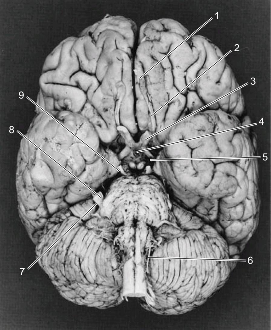
a) olfactory tract.
(b) olfactory nerve.
(c) anterior cerebral artery.
(d) olfactory bulb.
(e) inferior frontal gyrus.
۱۵. ساختار شماره ۱ عبارت است از:
الف) مجرای بویایی.
(ب) عصب بویایی.
(ج) شریان مغزی قدامی.
(د) پیاز بویایی.
(ه) شکنج پیشانی تحتانی.
16. Structure number 2 is the:
(a) inferior frontal gyrus.
(b) lateral olfactory stria.
(c) olfactory nerve.
(d) olfactory bulb.
(e) olfactory tract.
۱۶. ساختار شماره ۲ عبارت است از:
(الف) شکنج پیشانی تحتانی.
(ب) نوارهای بویایی جانبی.
(ج) عصب بویایی.
(د) پیاز بویایی.
(ه) مجرای بویایی.
(b) optic chiasma.
(c) anterior perforating substance.
(d) optic tract.
(e) oculomotor nerve.
۱۷. ساختار شماره ۳ عبارت است از:
(الف) عصب بینایی.
(ب) کیاسمای بینایی.
(ج) ماده سوراخ کننده قدامی.
(د) مجرای بینایی.
(ه) عصب حرکتی چشم.
(b) optic tract.
(c) optic chiasma.
(d) hypophysis cerebri.
(e) interpeduncular fossa.
۱۸. ساختار شماره ۴ عبارت است از:
(الف) عصب بینایی.
(ب) مجرای بینایی.
(ج) کیاسمای بینایی.
(د) هیپوفیز مغزی.
(ه) حفره بین پایهای.
(c) maxillary nerve.
(d) middle cerebral artery.
(e) optic tract.
۱۹. ساختار شماره ۵ عبارت است از:
(الف) ماده سوراخدار قدامی.
(ب) عصب حرکتی چشم.
(ج) عصب فکی.
(د) شریان مغزی میانی.
(ه) مجرای بینایی.
20. Structure number 6 is the:
(a) vertebral artery.
(b) spinal part of the accessory nerve.
(c) hypoglossal nerve.
(d) glossopharyngeal nerve.
(e) first cervical nerve.
۲۰. ساختار شماره ۶ عبارت است از:
(الف) شریان مهرهای.
(ب) بخش نخاعی عصب فرعی.
(ج) عصب زیرزبانی.
(د) عصب زبانی-حلقی.
(ه) عصب اول گردنی.
21. Structure number 7 is the:
(a) ophthalmic nerve.
(b) motor root of the trigeminal nerve.
(c) sensory root of the trigeminal nerve.
(d) flocculus of the cerebellum.
(e) vestibulocochlear nerve.
۲۱. ساختار شماره ۷ عبارت است از:
(الف) عصب چشمی.
(ب) ریشه حرکتی عصب سه قلو.
(ج) ریشه حسی عصب سه قلو.
(د) لخته مخچه.
(ه) عصب دهلیزی-حلزونی.
(b) sensory root of the trigeminal nerve.
(c) vestibular part of the eighth cranial nerve.
(d) maxillary nerve.
(e) abducens nerve.
۲۲. ساختار شماره ۸ عبارت است از:
(الف) ریشه حرکتی عصب سه قلو.
(ب) ریشه حسی عصب سه قلو.
(ج) بخش دهلیزی عصب هشتم جمجمهای.
(د) عصب فکی.
(ه) عصب ابدوسنس.
(b) abducens nerve.
(c) facial nerve.
(d) oculomotor nerve.
(e) vestibulocochlear nerve.
۲۳. ساختار شماره ۹ عبارت است از:
(الف) عصب قرقرهای.
(ب) عصب دورکننده.
(ج) عصب صورت.
(د) عصب حرکتی چشم.
(ه) عصب دهلیزی-حلزونی.
For questions 24 through 29, study Figure 11-27, showing a medial view of the right side of the brain following a median sagittal section.
برای سوالات ۲۴ تا ۲۹، شکل ۱۱-۲۷ را مطالعه کنید که نمای داخلی سمت راست مغز را پس از برش سهمی میانی نشان میدهد.

Figure 11-27 Medial view of the right side of the brain following a median sagittal section.
شکل ۱۱-۲۷ نمای داخلی سمت راست مغز پس از برش ساژیتال میانی.
24. Structure number 1 is the location of the nucleus of the:
(a) abducens nerve.
(b) trochlear nerve.
(c) trigeminal nerve.
(d) facial nerve.
(e) oculomotor nerve.
۲۴. ساختار شماره ۱ محل هستهی موارد زیر است:
(الف) عصب ابدوسنس.
(ب) عصب تروکلئار.
(ج) عصب سهقلو.
(د) عصب صورت.
(ه) عصب اوکولوموتور.
25. Structure number 2 is the location of the nucleus of the:
(a) trigeminal nerve.
(b) trochlear nerve.
(c) abducens nerve.
(d) oculomotor nerve.
(e) vestibulocochlear nerve.
۲۵. ساختار شماره ۲ محل هستهی موارد زیر است:
(الف) عصب سهقلو.
(ب) عصب قرقرهای.
(ج) عصب دورکننده.
(د) عصب حرکتی چشم.
(ه) عصب دهلیزی-حلقوی.
26. Structure number 3 is the:
(a) oculomotor nerve.
(b) trochlear nerve.
(c) trigeminal nerve.
(d) abducens nerve.
(e) facial nerve.
۲۶. ساختار شماره ۳ عبارت است از:
(الف) عصب حرکتی چشم.
(ب) عصب قرقرهای.
(ج) عصب سهقلو.
(د) عصب ابدوسنس.
(ه) عصب صورت.
(b) oculomotor nerve.
(c) trigeminal nerve.
(d) facial nerve.
(e) abducens nerve.
۲۷. ساختار شماره ۴ عبارت است از:
(الف) عصب قرقرهای.
(ب) عصب حرکتی چشم.
(ج) عصب سهقلو.
(د) عصب صورت.
(ه) عصب ابدوسنس.
(b) oculomotor nerve.
(c) trochlear nerve.
(d) abducens nerve.
(e) optic chiasma.
۲۸. ساختار شماره 5 عبارت است از:
(الف) لایه انتهایی.
(ب) عصب چشمی.
ج) عصب تروکلر.
د) عصب را می رباید.
(ه) کیاسما نوری.
(b) crista galli.
(c) olfactory stria.
(d) anterior cerebral artery.
(e) inferior frontal gyrus.
۲۹. ساختار شماره ۶ عبارت است از:
(الف) پیاز بویایی.
(ب) کریستا گالی.
(ج) نوارهای بویایی.
(د) شریان مغزی قدامی.
(ه) شکنج پیشانی تحتانی.
√ پاسخها و توضیحات مربوط به سوالات مروری
۱. ج صحیح است. هسته عصب صورتی که عضلات قسمت پایینی صورت را تغذیه میکند، فقط مسیرهای قشری-بولبار متقاطع را دریافت میکند (شکل ۱۱-۲۵ را ببینید). الف. هسته بزاقی تحتانی عصب زبانی-حلقی، مسیرهای نزولی را از هیپوتالاموس دریافت میکند. ب. هسته عصب ابدوسنس، مسیرهای قشری-بولبار متقاطع و غیر متقاطع را دریافت میکند. د. هسته حرکتی سه قلو، مسیرهای قشری-بولبار متقاطع و غیر متقاطع را دریافت میکند. ه. هسته عصب تروکلئار، مسیرهای قشری-بولبار متقاطع و غیر متقاطع را دریافت میکند.
۳. گزینه د صحیح است. در این بیمار، عضلاتی که حرکات تقلیدی صورت را ایجاد میکنند، توسط فیبرهای قشری-بولبار عصبدهی میشوند که مسیری جدا از فیبرهای اصلی قشری-بولبار دارند. الف) فیبرهای اصلی قشری-بولبار که حرکات عضلات ارادی صورت را در این بیمار کنترل میکنند، از بین رفتهاند. ب) فیبرهای مشبک، که احتمالاً از هیپوتالاموس منشأ میگیرند و به هستههای حرکتی اعصاب صورت میرسند، سالم هستند. ج) اعصاب صورت سالم هستند زیرا این بیمار قادر به حرکت عضلات صورت است. ه) نورونهای حرکتی تحتانی عصب صورت که عضلات صورت را تغذیه میکنند، سالم هستند.
4. A is correct. The trapezoid body participates in the reception of sound.
۴. الف صحیح است. جسم ذوزنقهای در دریافت صدا نقش دارد.
5. B is correct. The cerebral cortex is necessary for the accommodation reflex.
۵. ب صحیح است. قشر مغز برای رفلکس تطابق ضروری است.
6. D is correct. The nasal field of the right eye is projected to the temporal retina of the right eye. A. The nasal field of the right eye is projected to the right lateral geniculate body (see Fig. 11-2). B. The nasal field of the right eye is projected to both banks of the right calcarine fissure (see Fig. 11-2). C. The nasal field of the right eye is projected through the right optic tract (see Fig. 11-2). E. The nasal field of the right eye is projected through the right optic radiation (see Fig. 11-2).
۶. د صحیح است. میدان دید بینی چشم راست به شبکیه گیجگاهی چشم راست تصویر شده است. الف. میدان دید بینی چشم راست به جسم زانویی جانبی راست تصویر شده است (شکل ۱۱-۲ را ببینید). ب. میدان دید بینی چشم راست به هر دو کناره شکاف کالکارین راست تصویر شده است (شکل ۱۱-۲ را ببینید). ج. میدان دید بینی چشم راست از طریق مجرای بینایی راست تصویر شده است (شکل ۱۱-۲ را ببینید). ه. میدان دید بینی چشم راست از طریق تابش بینایی راست تصویر شده است (شکل ۱۱-۲ را ببینید).
7. B is correct. The right pupillary constriction associated with light directed at the left eye requires the left optic nerve (see Fig. 11-3). A. The right optic radiation is not required (see Fig. 11-3). C. The left Edinger-Westphal nucleus is not required (see Fig. 11-3). D. The right oculomotor nerve is required (see Fig. 11-3). E. The right optic nerve is not required (see Fig. 11-3).
۷. ب صحیح است. انقباض مردمک چشم راست که با نور تابیده شده به چشم چپ مرتبط است، به عصب بینایی چپ نیاز دارد (شکل ۱۱-۳ را ببینید). الف. تابش بینایی راست لازم نیست (شکل ۱۱-۳ را ببینید). ج. هسته ادینگر-وستفال چپ لازم نیست (شکل ۱۱-۳ را ببینید). د. عصب اوکولوموتور راست لازم است (شکل ۱۱-۳ را ببینید). ه. عصب بینایی راست لازم نیست (شکل ۱۱-۳ را ببینید).
8. A is correct.
۸. الف صحیح است.
9. C is correct.
۹. ج صحیح است.
10. A is correct. The spinal part of the accessory nerve supplies the trapezius muscle, which shrugs the shoulder. B. The facial nerve supplies the orbicularis oculi muscle, which closes the eye. C. The trigeminal nerve supplies the muscles of mastication responsible for chewing. D. The facial nerve receives the sensation of taste from the anterior two-thirds of the tongue. E. The glossopharyngeal nerve receives the sensation of touch from the pos- terior third of the tongue.
۱۰. الف صحیح است. بخش نخاعی عصب فرعی، عضله ذوزنقهای را که شانه را بالا میاندازد، عصب میدهد. ب. عصب صورت، عضله حلقوی چشم را که چشم را میبندد، عصب میدهد. ج. عصب سهقلو، عضلات جونده مسئول جویدن را عصب میدهد. د. عصب صورت، حس چشایی را از دو سوم قدامی زبان دریافت میکند. ه. عصب زبانی-حلقی، حس لامسه را از یک سوم خلفی زبان دریافت میکند.
11. B is correct. The optic nerve is surrounded by an extension of the subarachnoid space. A. The nerve fibers of the optic nerve are surrounded by oligodendrocytes. C. Internal ophthalmoplegia is a condition in which the oculomotor nerve supply to the sphincter pupillae and the ciliary muscle is lost, but the innervation of the extraocular muscles is spared. D. External ophthalmoplegia is a condition in which the oculomotor nerve supply to the extraocular muscles is lost, but the innervation of the sphincter pupillae and the ciliary muscle is spared. E. The optic nerve leaves the orbital cavity through the optic canal in the lesser wing of the sphenoid bone.
۱۱. ب صحیح است. عصب بینایی توسط امتدادی از فضای زیر عنکبوتیه احاطه شده است. الف. رشتههای عصبی عصب بینایی توسط الیگودندروسیتها احاطه شدهاند. ج. افتالموپلژی داخلی وضعیتی است که در آن عصبدهی عصب چشمی به مردمک اسفنکتر و عضله مژگانی از بین میرود، اما عصبدهی عضلات خارج چشمی حفظ میشود. د. افتالموپلژی خارجی وضعیتی است که در آن عصبدهی عصب چشمی به عضلات خارج چشمی از بین میرود، اما عصبدهی مردمک اسفنکتر و عضله مژگانی حفظ میشود. ه. عصب بینایی از طریق کانال بینایی در بال کوچک استخوان اسفنوئید، حفره کاسه چشم را ترک میکند.
12. E is correct. The olfactory receptor cells are located in the mucous membrane of the nasal cavity above the level of the superior concha. A. The main sensory nucleus of the trigeminal nerve lies in the brain- stem lateral to the motor nucleus (see Fig. 11-7). B. Proprioceptive impulses from the facial muscles end in the mesencephalic nucleus of the trigemi- nal nerve. C. The facial nerve leaves the posterior cranial fossa with the vestibulocochlear nerve and enters the internal acoustic meatus. D. The superior salivatory nucleus of the facial nerve innervates the submandibular and sublingual salivary glands.
۱۲. ه صحیح است. سلولهای گیرنده بویایی در غشای مخاطی حفره بینی، بالاتر از سطح شاخک فوقانی قرار دارند. الف. هسته حسی اصلی عصب سه قلو در ساقه مغز و در کنار هسته حرکتی قرار دارد (شکل ۱۱-۷ را ببینید). ب. تکانههای حس عمقی از عضلات صورت در هسته مزانسفال عصب سه قلو ختم میشوند. ج. عصب صورت به همراه عصب دهلیزی-حلزونی از حفره جمجمهای خلفی خارج شده و وارد مجرای شنوایی داخلی میشود. د. هسته بزاقی فوقانی عصب صورت، غدد بزاقی زیر فکی و زیر زبانی را عصبدهی میکند.
13. E is correct. The taste sensation from the mucous membrane covering the anterior two-thirds of the tongue is conducted in the facial nerves and the chorda tympani nerves, which are a considerable distance from the metastases in the deep cervical lymph nodes in the neck.
۱۳. گزینه E صحیح است. حس چشایی از غشای مخاطی که دو سوم قدامی زبان را پوشانده است، به اعصاب صورت و اعصاب طناب صماخ منتقل میشود که فاصله قابل توجهی از متاستازهای غدد لنفاوی عمقی گردن دارند.
14. E is correct. The wasted right half of the tongue and the pointing of the protruded tongue to the right side indicated a lesion of the right hypoglossal nerve. The tongue muscles on the right side had atrophied and diminished in size, resulting in the wrinkling of the overlying normal mucous membrane.
۱۴. گزینه E صحیح است. تحلیل رفتن نیمه راست زبان و انحراف زبان بیرونزده به سمت راست، نشاندهنده ضایعه عصب زیرزبانی راست بود. عضلات زبان در سمت راست تحلیل رفته و اندازه آنها کاهش یافته بود که منجر به چروکیدگی غشای مخاطی طبیعی روی آن شده بود.
The answers to questions 15 through 23 pertain to Figure 11-26, which shows the inferior view of the brain.
پاسخ سوالات ۱۵ تا ۲۳ مربوط به شکل ۱۱-۲۶ است که نمای پایینتر مغز را نشان میدهد.
15. D is correct. Structure number 1 is the olfactory bulb.
۱۵. د صحیح است. ساختار شماره ۱ پیاز بویایی است.
۱۶. ه صحیح است. ساختار شماره ۲ مجرای بویایی است.
۱۷. الف صحیح است. ساختار شماره ۳ عصب بینایی است.
۱۸. د صحیح است. ساختار شماره ۴ هیپوفیز مغزی است.
۱۹. ه صحیح است. ساختار شماره ۵ مجرای بینایی است.
۲۰. ب صحیح است. ساختار شماره ۶ قسمت نخاعی عصب فرعی است.
۲۱. ج صحیح است. ساختار شماره ۷ ریشه حسی عصب سه قلو است.
۲۲. الف صحیح است. ساختار شماره ۸ ریشه حرکتی عصب سه قلو است.
۲۳. د صحیح است. ساختار شماره ۹ عصب حرکتی چشم است.
The answers to questions 24 through 29 pertain to Figure 11-27, showing a medial view of the right side of the brain following a median sagittal section.
پاسخ سوالات ۲۴ تا ۲۹ مربوط به شکل ۱۱-۲۷ است که نمای داخلی سمت راست مغز را پس از برش سهمی میانی نشان میدهد.
24. E is correct. Structure number 1 is the nucleus of the oculomotor nerve in the tegmentum of the midbrain at the level of the superior colliculus.
۲۴. گزینه E صحیح است. ساختار شماره ۱، هسته عصب حرکتی چشم در تگمنتوم مغز میانی در سطح کولیکولوس فوقانی است.
25. B is correct. Structure number 2 is the nucleus of the trochlear nerve in the tegmentum of the midbrain at the level of the inferior colliculus.
۲۵. ب صحیح است. ساختار شماره ۲ هسته عصب تروکلئار در تگمنتوم مغز میانی در سطح کولیکولوس تحتانی است.
26. C is correct. Structure number 3 is the trigeminal nerve emerging on the anterior surface of the pons.
۲۶. ج صحیح است. ساختار شماره ۳ عصب سه قلو است که از سطح قدامی پل مغزی خارج میشود.
27. B is correct. Structure number 4 is the oculomotor nerve emerging from the anterior surface of the midbrain in the interpeduncular fossa.
۲۷. ب صحیح است. ساختار شماره ۴ عصب حرکتی چشم است که از سطح قدامی مغز میانی در حفره بین پایهای خارج میشود.
28. E is correct. Structure number 5 is the optic chiasma.
۲۸. ه صحیح است. ساختار شماره ۵ کیاسمای بینایی است.
29. A is correct. Structure number 6 is the olfactory bulb.
۲۹. الف صحیح است. ساختار شماره ۶ پیاز بویایی است.
«فصل یازدهم کتاب نورواناتومی بالینی اسنل»
🚀 با ما همراه شوید!
تازهترین مطالب و آموزشهای مغز و اعصاب را از دست ندهید. با فالو کردن کانال تلگرام آیندهنگاران مغز، از ما حمایت کنید!