هستههای قاعدهای: عقدههای قاعدهای؛ فصل دهم نوروآناتومی بالینی اسنل


دعای مطالعه [ نمایش ]
بِسْمِ الله الرَّحْمنِ الرَّحیمِ
اَللّهُمَّ اَخْرِجْنى مِنْ ظُلُماتِ الْوَهْمِ
خدايا مرا بيرون آور از تاريكىهاى وهم،
وَ اَكْرِمْنى بِنُورِ الْفَهْمِ
و به نور فهم گرامى ام بدار،
اَللّهُمَّ افْتَحْ عَلَيْنا اَبْوابَ رَحْمَتِكَ
خدايا درهاى رحمتت را به روى ما بگشا،
وَانْشُرْ عَلَيْنا خَزائِنَ عُلُومِكَ بِرَحْمَتِكَ يا اَرْحَمَ الرّاحِمينَ
و خزانههاى علومت را بر ما باز كن به امید رحمتت اى مهربانترين مهربانان.
آیندهنگاران مغز: «ما مغز را میشناسیم، تا آینده را بسازیم.»
کتاب «نوروآناتومی بالینی اسنل» اثر دکتر ریچارد اس. اسنل، با ویرایش جدید دکتر رایان اسپلیتگربر، همچنان بهعنوان مرجع طلایی این حوزه شناخته میشود. این اثر با تلفیق تازهترین پژوهشها و کاربردهای بالینی، پلی استوار میان دانش پایه و عمل پزشکی بنا میکند.
تلاشهای علمی و سالها تدریس ارزشمند استاد گرانقدر آقای دکتر محمدتقی جغتایی الهامبخش ترجمه و گسترش این دانش در ایران بوده است.
ترجمه دقیق این کتاب توسط برند علمی آیندهنگاران مغز به مدیریت داریوش طاهری، امکان دسترسی فارسیزبانان به مرزهای نوین علوم اعصاب را فراهم کرده و رسالتی علمی در جهت شناخت عمیقتر مغز و ترسیم آیندهای روشنتر بر عهده گرفته است.
»» فصل ۱۰: هستههای قاعدهای (عقدههای قاعدهای)؛ بخش مبانی و اصول
»» Chapter 10: Basal Nuclei (Basal Ganglia)
CHAPTER OBJECTIVE
• To describe the basal nuclei and their connections
• To relate basal nuclei functions to diseases commonly affecting this area of the nervous system
اهداف فصل
• توصیف هستههای قاعدهای و ارتباطات آنها
• ارتباط دادن عملکردهای هستههای قاعدهای با بیماریهایی که معمولاً این ناحیه از سیستم عصبی را تحت تأثیر قرار میدهند
A 58-year-old man goes to a neurologist because he has noticed the development of a slight tremor of his left hand. The tremors involve all of the fingers and the thumb and are present at rest but cease during voluntary movement.
مردی ۵۸ ساله به دلیل لرزش خفیف دست چپش به متخصص مغز و اعصاب مراجعه میکند. این لرزش تمام انگشتان و شست را درگیر میکند و در حالت استراحت وجود دارد اما در حین حرکت ارادی متوقف میشود.
On examination, the patient tends to perform all his movements slowly, and his face has very little expression and is almost masklike. On passively moving the patient’s arms, the neurologist finds that the muscles show increased tone, with a slight jerky resistance to the movements. When asked to stand up straight, the patient does so but with a stooped posture, and, when he walks, he does so by shuffling across the examining room.
در معاینه، بیمار تمایل دارد تمام حرکات خود را به آرامی انجام دهد و صورتش حالت بسیار کمی دارد و تقریباً ماسک مانند است. متخصص مغز و اعصاب با حرکت دادن غیرفعال بازوهای بیمار، متوجه میشود که عضلات افزایش تون نشان میدهند و در برابر حرکات، مقاومت کمی دارند. وقتی از بیمار خواسته میشود صاف بایستد، این کار را با حالت خمیده انجام میدهد و وقتی راه میرود، این کار را با راه رفتن روی زمین در اتاق معاینه انجام میدهد.
The neurologist makes the diagnosis of Parkinson disease, based on her knowledge of the structure and function of the basal ganglia and their connections to the substantia nigra of the midbrain. She is able to prescribe appropriate drug therapy, which results in a great improvement in the hand tremors.
متخصص مغز و اعصاب، بر اساس دانش خود از ساختار و عملکرد عقدههای قاعدهای و ارتباط آنها با جسم سیاه مغز میانی، بیماری پارکینسون را تشخیص میدهد. او قادر به تجویز دارو درمانی مناسب است که منجر به بهبود قابل توجه لرزش دست میشود.
The basal nuclei play an important role in the control of posture and voluntary movement. Unlike many other parts of the nervous system concerned with motor control, the basal nuclei have no direct input or output connections with the spinal cord.
هستههای قاعدهای نقش مهمی در کنترل وضعیت بدن و حرکات ارادی دارند. برخلاف بسیاری از بخشهای دیگر سیستم عصبی که با کنترل حرکتی مرتبط هستند، هستههای قاعدهای هیچ ارتباط ورودی یا خروجی مستقیمی با نخاع ندارند.

TERMINOLOGY
The term basal nuclei is applied to a collection of masses of gray matter situated within each cerebral hemisphere. They are the corpus striatum, the amygdaloid nucleus, and the claustrum.
اصطلاحات
اصطلاح هستههای قاعدهای به مجموعهای از تودههای ماده خاکستری که در هر نیمکره مغزی قرار دارند، اطلاق میشود. این تودهها عبارتند از جسم مخطط، هسته آمیگدالوئید و کلاستروم.
Clinicians and neuroscientists use a variety of different terminologies to describe the basal nuclei. A summary of the terminologies commonly used is shown in Table 10-1. The subthalamic nuclei, the substantia nigra, and the red nucleus are functionally closely related to the basal nuclei, but they should not be included with them.
پزشکان و دانشمندان علوم اعصاب از اصطلاحات مختلفی برای توصیف هستههای قاعدهای استفاده میکنند. خلاصهای از اصطلاحات رایج در جدول 10-1 نشان داده شده است. هستههای زیرتالاموسی، جسم سیاه و هسته قرمز از نظر عملکردی ارتباط نزدیکی با هستههای قاعدهای دارند، اما نباید آنها را با هستههای قاعدهای در نظر گرفت.
The interconnections of the basal nuclei are complex, but in this account, only the more important pathways are considered. The basal nuclei play an important role in the control of posture and voluntary movement.
ارتباطات هستههای قاعدهای پیچیده است، اما در این بررسی، تنها مسیرهای مهمتر در نظر گرفته شدهاند. هستههای قاعدهای نقش مهمی در کنترل وضعیت بدن و حرکات ارادی دارند.
Table 10-1 Terminology Commonly Used to Describe the Basal Nuclei
جدول ۱۰-۱ اصطلاحات رایج برای توصیف هستههای قاعدهای

a The term basal has been used in the past to denote the position of the nuclei at the base of the forebrain.
الف اصطلاح پایه در گذشته برای اشاره به موقعیت هستهها در پایه مغز پیشین به کار میرفته است.
CORPUS STRIATUM
The corpus striatum (Fig. 10-1; see also Atlas Plate 5) is situated lateral to the thalamus and is almost completely divided by a band of nerve fibers, the internal capsule, into the caudate nucleus and the lentiform nucleus. The term striatum is used here because of the striated appearance produced by the strands of gray matter passing through the internal capsule and connecting the caudate nucleus to the putamen of the lentiform nucleus (see below).
جسم مخطط
جسم مخطط (شکل 10-1؛ همچنین به صفحه اطلس 5 مراجعه کنید) در کنار تالاموس قرار دارد و تقریباً به طور کامل توسط نواری از الیاف عصبی، کپسول داخلی، به هسته دمدار و هسته عدسی شکل تقسیم میشود. اصطلاح جسم مخطط در اینجا به دلیل ظاهر مخطط ایجاد شده توسط رشتههای ماده خاکستری که از کپسول داخلی عبور میکنند و هسته دمدار را به پوتامن هسته عدسی شکل متصل میکنند، استفاده میشود (به پایین مراجعه کنید).

Figure 10-1 Lateral view of the right cerebral hemisphere dissected to show the position of the different basal nuclei.
شکل ۱۰-۱نمای جانبی نیمکره راست مغز که برای نشان دادن موقعیت هستههای قاعدهای مختلف تشریح شده است.
Caudate Nucleus
The caudate nucleus is a large C-shaped mass of gray matter that is closely related to the lateral ventricle and lies lateral to the thalamus. The lateral surface of the nucleus is related to the internal capsule, which separates it from the lentiform nucleus (Fig. 10-2). For purposes of description, it can be divided into a head, a body, and a tail.
هسته دمی
هسته دمی یک توده بزرگ C شکل از ماده خاکستری است که ارتباط نزدیکی با بطن جانبی دارد و در سمت خارجی تالاموس قرار گرفته است. سطح جانبی هسته به کپسول داخلی متصل است که آن را از هسته عدسی شکل جدا میکند (شکل 10-2). برای توصیف، میتوان آن را به یک سر، یک بدنه و یک دم تقسیم کرد.
The head of the caudate nucleus is large and rounded and forms the lateral wall of the anterior horn of the lateral ventricle (see also Atlas Plate 5). The head is continuous inferiorly with the putamen of the lentiform nucleus (the caudate nucleus and the putamen are sometimes referred to as the neostriatum or striatum). Just superior to this point of union, strands of gray matter pass through the internal capsule, giving the region a striated appearance, hence the term corpus striatum.
سر هسته دمدار بزرگ و گرد است و دیواره جانبی شاخ قدامی بطن جانبی را تشکیل میدهد (همچنین به صفحه ۵ اطلس مراجعه کنید). سر در امتداد پایین پوتامن هسته عدسی شکل قرار دارد (هسته دمدار و پوتامن گاهی اوقات نئواستریاتوم یا جسم مخطط نامیده میشوند). درست در بالای این نقطه اتصال، رشتههایی از ماده خاکستری از کپسول داخلی عبور میکنند و به این ناحیه ظاهری مخطط میدهند، از این رو به آن جسم مخطط میگویند.
The body of the caudate nucleus is long and narrow and is continuous with the head in the region of the interventricular foramen. The body of the caudate nucleus forms part of the floor of the body of the lateral ventricle.
بدنه هسته دمدار بلند و باریک است و در ناحیه سوراخ بین بطنی با سر آن در یک امتداد قرار دارد. بدنه هسته دمدار بخشی از کف بدنه بطن جانبی را تشکیل میدهد.
The tail of the caudate nucleus is long and slender and is continuous with the body in the region of the posterior end of the thalamus. It follows the contour of the lateral ventricle and continues forward in the roof of the inferior horn of the lateral ventricle. It terminates anteriorly in the amygdaloid nucleus (see Fig. 10-1).
دم هسته دمدار بلند و باریک است و در ناحیه انتهای خلفی تالاموس با بدنه آن در یک امتداد قرار دارد. این دم از امتداد بطن جانبی پیروی میکند و در سقف شاخ تحتانی بطن جانبی به سمت جلو ادامه مییابد. در قسمت قدامی در هسته آمیگدالوئید خاتمه مییابد (شکل 10-1 را ببینید).
Lentiform Nucleus
The lentiform nucleus is a wedge-shaped mass of gray matter whose broad convex base is directed laterally and whose blade is directed medially (see Fig. 10-2; see also Atlas Plate 5). It is buried deep in the white matter of the cerebral hemisphere and is related medially to the internal capsule, which separates it from the caudate nucleus and the thalamus. The lentiform nucleus is related laterally to a thin sheet of white matter, the external capsule, which separates it from a thin sheet of gray matter, called the claustrum. The claustrum, in turn, separates the external capsule from the subcortical white matter of the insula. A vertical plate of white matter divides the nucleus into a larger, darker lateral portion, the putamen, and an inner lighter portion, the globus pallidus. The paleness of the globus pallidus is due to the presence of a high concentration of myelinated nerve fibers. Inferiorly at its anterior end, the putamen is continuous with the head of the caudate nucleus (see Fig. 10-1).
هسته عدسی شکل
هسته عدسی شکل، تودهای گوهای شکل از ماده خاکستری است که قاعده محدب پهن آن به سمت طرفین و تیغه آن به سمت داخل است (شکل 10-2 را ببینید؛ همچنین به صفحه اطلس 5 مراجعه کنید). این هسته در اعماق ماده سفید نیمکره مغزی قرار دارد و از سمت داخل به کپسول داخلی متصل است که آن را از هسته دمدار و تالاموس جدا میکند. هسته عدسی شکل از سمت خارج به یک صفحه نازک از ماده سفید، کپسول خارجی، متصل است که آن را از یک صفحه نازک از ماده خاکستری به نام کلاستروم جدا میکند. کلاستروم نیز به نوبه خود، کپسول خارجی را از ماده سفید زیر قشری اینسولا جدا میکند. یک صفحه عمودی از ماده سفید، هسته را به یک بخش جانبی بزرگتر و تیرهتر، پوتامن، و یک بخش داخلی روشنتر، گلوبوس پالیدوس، تقسیم میکند. رنگپریدگی گلوبوس پالیدوس به دلیل وجود غلظت بالای فیبرهای عصبی میلیندار است. پوتامن در انتهای قدامی خود، در امتداد سر هسته دمدار قرار دارد (شکل 10-1 را ببینید).

Figure 10-2 Horizontal section of the cerebrum, as seen from above, showing the relationships of the different basal nuclei.
شکل ۱۰-۲ برش افقی مخ، از نمای بالا، که روابط هستههای قاعدهای مختلف را نشان میدهد.
AMYGDALOID NUCLEUS
The amygdaloid nucleus is situated in the temporal lobe close to the uncus (see Fig. 10-1). The amygdaloid nucleus is considered to be part of the limbic system and is described in Chapter 9. Through its connections, it can influence the body’s response to environmental changes. In the sense of fear, for example, it can change the heart rate, blood pressure, skin color, and rate of respiration.
هسته آمیگدالوئید
هسته آمیگدالوئید در لوب گیجگاهی نزدیک به آنکوس قرار دارد (شکل 10-1 را ببینید). هسته آمیگدالوئید بخشی از سیستم لیمبیک در نظر گرفته میشود و در فصل 9 توضیح داده شده است. از طریق ارتباطاتش، میتواند بر پاسخ بدن به تغییرات محیطی تأثیر بگذارد. به عنوان مثال، در احساس ترس، میتواند ضربان قلب، فشار خون، رنگ پوست و میزان تنفس را تغییر دهد.
SUBSTANTIA NIGRA AND SUBTHALAMIC NUCLEI
The substantia nigra of the midbrain and the subthalamic nuclei of the diencephalon are functionally closely related to the activities of the basal nuclei and are described elsewhere (see pp. 211 and 253). The neurons of the substantia nigra are dopaminergic and inhibitory and have many connections to the corpus striatum. The neurons of the subthalamic nuclei are glutaminergic and excitatory and have many connections to the globus pallidus and substantia nigra.
هستههای جسم سیاه و زیرتالاموس
جسم سیاه مغز میانی و هستههای زیرتالاموس دیانسفالون از نظر عملکردی ارتباط نزدیکی با فعالیتهای هستههای قاعدهای دارند و در جای دیگری توضیح داده شدهاند (به صفحات ۲۱۱ و ۲۵۳ مراجعه کنید). نورونهای جسم سیاه دوپامینرژیک و مهاری هستند و ارتباطات زیادی با جسم مخطط دارند. نورونهای هستههای زیرتالاموس گلوتامینرژیک و تحریکی هستند و ارتباطات زیادی با گلوبوس پالیدوس و جسم سیاه دارند.
CLAUSTRUM
The claustrum is a thin sheet of gray matter that is separated from the lateral surface of the lentiform nucleus by the external capsule (see Fig. 10-2). Lateral to the claustrum is the subcortical white matter of the insula. The function of the claustrum is unknown.
کلاستروم
کلاستروم یک صفحه نازک از ماده خاکستری است که توسط کپسول خارجی از سطح جانبی هسته عدسی شکل جدا شده است (شکل 10-2 را ببینید). در سمت جانبی کلاستروم، ماده سفید زیر قشری اینسولا قرار دارد. عملکرد کلاستروم ناشناخته است.
CONNECTIONS OF THE CORPUS STRIATUM AND GLOBUS PALLIDUS
The caudate nucleus and the putamen form the main sites for receiving input to the basal nuclei. The globus pallidus forms the major site from which the output leaves the basal nuclei.
اتصالات جسم مخطط و گلوبوس پالیدوس
هسته دمدار و پوتامن محلهای اصلی دریافت ورودی به هستههای قاعدهای را تشکیل میدهند. گلوبوس پالیدوس محل اصلی خروج خروجی از هستههای قاعدهای را تشکیل میدهد.
They receive no direct input from or output to the spinal cord.
آنها هیچ ورودی یا خروجی مستقیمی از نخاع دریافت نمیکنند.
Corpus Striatum Afferent Fibers
Projections to the corpus striatum include corticostriate, thalamostriate, nigrostriatal, and brainstem striatal fibers.
فیبرهای آوران جسم مخطط
برآمدگیهای جسم مخطط شامل فیبرهای کورتیکواستریات، تالاموستریاتال، نیگرواستریاتال و جسم مخطط ساقه مغز هستند.
Corticostriate Fibers
All parts of the cerebral cortex send axons to the caudate nucleus and the putamen (Fig. 10-3). Each part of the cerebral cortex projects to a specific part of the caudate-putamen complex. Most of the projections are from the cortex of the same side. The largest input is from the sensory motor cortex. Glutamate is the neurotransmitter of the corticostriate fibers (Fig. 10-4).
فیبرهای کورتیکواستریات
تمام بخشهای قشر مغز، آکسونها را به هسته دمدار و پوتامن میفرستند (شکل 10-3). هر بخش از قشر مغز به بخش خاصی از کمپلکس دمدار-پوتامن متصل میشود. بیشتر این اتصالات از قشر همان سمت هستند. بزرگترین ورودی از قشر حسی-حرکتی است. گلوتامات انتقالدهنده عصبی فیبرهای کورتیکواستریات است (شکل 10-4).

Figure 10-3 Some of the main connections between the cerebral cortex, the basal nuclei, the thalamic nuclei, the brainstem, and the spinal cord.
شکل ۱۰-۳ برخی از اتصالات اصلی بین قشر مغز، هستههای قاعدهای، هستههای تالاموس، ساقه مغز و نخاع.

Figure 10-4 Basal nuclei pathways showing the known neurotransmitters.
شکل ۱۰-۴ مسیرهای هستههای قاعدهای که انتقالدهندههای عصبی شناختهشده را نشان میدهند.
Thalamostriate Fibers
The intralaminar nuclei of the thalamus send large numbers of axons to the caudate nucleus and the putamen (see Fig. 10-3).
الیاف تالاموستریاتیک
هستههای اینترالمینی تالاموس تعداد زیادی آکسون را به هسته دمدار و پوتامن میفرستند (شکل 10-3 را ببینید).
Nigrostriatal Fibers
Neurons in the substantia nigra send axons to the caudate nucleus and the putamen (see Figs. 10-3 and 10-4) and liberate dopamine at their terminals as the neurotransmitter. These fibers are believed to be inhibitory in function.
فیبرهای نیگرواستریاتال
نورونهای جسم سیاه، آکسونها را به هسته دمدار و پوتامن میفرستند (شکلهای 10-3 و 10-4 را ببینید) و دوپامین را در پایانههای خود به عنوان انتقالدهنده عصبی آزاد میکنند. اعتقاد بر این است که این فیبرها عملکرد مهاری دارند.
Brainstem Striatal Fibers
Ascending fibers from the brainstem end in the caudate nucleus and putamen and liberate serotonin at their terminals as the neurotransmitter. These fibers are thought to be inhibitory in function.
فیبرهای مخطط ساقه مغز
فیبرهای صعودی از ساقه مغز به هسته دمدار و پوتامن ختم میشوند و در پایانههای خود سروتونین را به عنوان انتقالدهنده عصبی آزاد میکنند. تصور میشود که این فیبرها عملکرد مهاری دارند.
Corpus Striatum Efferent Fibers
Projections from the corpus striatum include striatopallidal and striatonigral fibers.
فیبرهای وابران جسم مخطط
برآمدگیهای جسم مخطط شامل فیبرهای استریاتوپالیدال و استریاتونیگرال هستند.
Striatopallidal Fibers
Striatopallidal fibers pass from the caudate nucleus and putamen to the globus pallidus (see Fig. 10-3). They have y-aminobutyric acid (GABA) as their neurotransmitter (see Fig. 10-4).
فیبرهای استریاتوپالیدی
فیبرهای استریاتوپالیدی از هسته دمدار و پوتامن به گلوبوس پالیدوس میروند (شکل 10-3 را ببینید). آنها دارای γ-آمینوبوتیریک اسید (GABA) به عنوان انتقالدهنده عصبی خود هستند (شکل 10-4 را ببینید).
Striatonigral Fibers
Striatonigral fibers pass from the caudate nucleus and putamen to the substantia nigra (see Fig. 10-3). Some of the fibers use GABA or acetylcholine as the neurotransmitter, while others use substance P (see Fig. 10-4).
فیبرهای استریاتونیگرال
فیبرهای استریاتونیگرال از هسته دمدار و پوتامن به جسم سیاه میروند (شکل 10-3 را ببینید). برخی از این فیبرها از گابا یا استیل کولین به عنوان انتقالدهنده عصبی استفاده میکنند، در حالی که برخی دیگر از ماده P استفاده میکنند (شکل 10-4 را ببینید).
Globus Pallidus Afferent Fibers
Striatopallidal fibers pass from the caudate nucleus and putamen to the globus pallidus. As noted previously, these fibers have GABA as their neurotransmitter (Fig. 10-4).
فیبرهای آوران گلوبوس پالیدوس
فیبرهای استریاتوپالیدال از هسته دمدار و پوتامن به گلوبوس پالیدوس میروند. همانطور که قبلاً اشاره شد، این فیبرها گابا را به عنوان انتقالدهنده عصبی خود دارند (شکل 10-4).
Globus Pallidus Efferent Fibers
Pallidofugal fibers are complicated and can be divided into groups: (1) the ansa lenticularis, which pass to the thalamic nuclei; (2) the fasciculus lenticularis, which pass to the subthalamus; (3) the pallidotegmental fibers, which terminate in the caudal tegmentum of the midbrain; and (4) the pallidosubthalamic fibers, which pass to the subthalamic nuclei.
فیبرهای وابران گلوبوس پالیدوس
فیبرهای پالیدوفوگال پیچیده هستند و میتوان آنها را به گروههایی تقسیم کرد: (1) آنسا لنتیکولاریس، که به هستههای تالاموس میروند؛ (2) فاسیکولوس لنتیکولاریس، که به ساب تالاموس میروند؛ (3) فیبرهای پالیدوتگمنتال، که در تگمنتوم دمی مغز میانی خاتمه مییابند؛ و (4) فیبرهای پالیدوساب تالاموس، که به هستههای ساب تالاموس میروند.
BASAL NUCLEI FUNCTIONS
The basal nuclei (Fig. 10-5) are joined together and connected with many different regions of the nervous system by a very complex number of neurons.
وظایف هستههای قاعدهای
هستههای قاعدهای (شکل 10-5) توسط تعداد بسیار پیچیدهای از نورونها به یکدیگر متصل شده و با مناطق مختلف سیستم عصبی مرتبط هستند.
Basically, the corpus striatum receives afferent information from most of the cerebral cortex, the thalamus, the subthalamus, and the brainstem, including the substantia nigra. The information is integrated within the corpus striatum, and the outflow passes back to the areas listed above. This circular pathway is believed to function as follows.
اساساً، جسم مخطط اطلاعات آوران را از بیشتر قشر مغز، تالاموس، زیرتالاموس و ساقه مغز، از جمله جسم سیاه، دریافت میکند. اطلاعات در جسم مخطط ادغام میشوند و جریان خروجی به نواحی ذکر شده در بالا باز میگردد. اعتقاد بر این است که این مسیر دایرهای به شرح زیر عمل میکند.
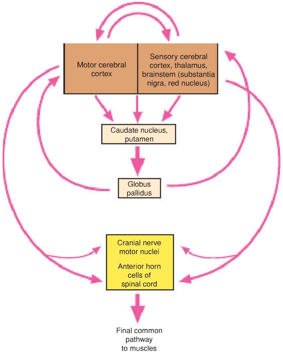
Figure 10-5 Diagram showing the main functional connections of the basal nuclei and how they can influence muscle activity.
شکل ۱۰-۵ نموداری که ارتباطات عملکردی اصلی هستههای قاعدهای و نحوهی تأثیر آنها بر فعالیت عضلات را نشان میدهد.
The activity of the basal nuclei is initiated by information received from the premotor and supplemental areas of the motor cortex, the primary sensory cortex, the thalamus, and the brainstem. The outflow from the basal nuclei is channeled through the globus pallidus, which then influences the activities of the motor areas of the cerebral cortex or other motor centers in the brainstem. Thus, the basal nuclei control muscular movements by influencing the cerebral cortex and have no direct control through descending pathways to the brainstem and spinal cord. In this way, the basal nuclei assist in the regulation of voluntary movement and the learning of motor skills.
فعالیت هستههای قاعدهای توسط اطلاعات دریافتی از نواحی پیشحرکتی و تکمیلی قشر حرکتی، قشر حسی اولیه، تالاموس و ساقه مغز آغاز میشود. جریان خروجی از هستههای قاعدهای از طریق گلوبوس پالیدوس هدایت میشود که سپس بر فعالیتهای نواحی حرکتی قشر مغز یا سایر مراکز حرکتی در ساقه مغز تأثیر میگذارد. بنابراین، هستههای قاعدهای با تأثیرگذاری بر قشر مغز، حرکات عضلانی را کنترل میکنند و هیچ کنترل مستقیمی از طریق مسیرهای نزولی به ساقه مغز و نخاع ندارند. به این ترتیب، هستههای قاعدهای در تنظیم حرکات ارادی و یادگیری مهارتهای حرکتی کمک میکنند.
Writing the letters of the alphabet, drawing a diagram, passing a football, using the vocal cords in talking and singing, and using the eye muscles when looking at an object are a few examples where the basal nuclei influence the skilled cortical motor activities.
نوشتن حروف الفبا، رسم نمودار، پاس دادن توپ فوتبال، استفاده از تارهای صوتی در صحبت کردن و آواز خواندن و استفاده از عضلات چشم هنگام نگاه کردن به یک شیء، چند نمونه از مواردی هستند که هستههای قاعدهای بر فعالیتهای حرکتی ماهرانه قشر مغز تأثیر میگذارند.
Destruction of the primary motor cerebral cortex prevents the individual from performing fine discrete movements of the hands and feet on the opposite side of the body (see p. 290). However, the individual is still capable of performing gross crude movements of the opposite limbs. If destruction of the corpus striatum then takes place, paralysis of the remaining movements of the opposite side of the body occurs.
تخریب قشر اولیه حرکتی مغز مانع از انجام حرکات ظریف و مجزای دستها و پاها در سمت مقابل بدن میشود (به صفحه ۲۹۰ مراجعه کنید). با این حال، فرد هنوز قادر به انجام حرکات کلی و خام اندامهای مخالف است. اگر تخریب جسم مخطط رخ دهد، فلج حرکات باقیمانده سمت مقابل بدن رخ میدهد.
The basal nuclei not only influence the execution of a particular movement of, say, the limbs but also help prepare for the movements. This may be achieved by controlling the axial and girdle movements of the body and the positioning of the proximal parts of the limbs. The activity in certain neurons of the globus pallidus increases before active movements take place in the distal limb muscles. This important preparatory function enables the trunk and limbs to be placed in appropriate positions before the primary motor part of the cerebral cortex activates discrete movements in the hands and feet.
هستههای قاعدهای نه تنها بر اجرای یک حرکت خاص مثلاً اندامها تأثیر میگذارند، بلکه به آمادهسازی برای حرکات نیز کمک میکنند. این امر ممکن است با کنترل حرکات محوری و کمربندی بدن و موقعیتیابی قسمتهای پروگزیمال اندامها حاصل شود. فعالیت در نورونهای خاصی از گلوبوس پالیدوس قبل از انجام حرکات فعال در عضلات اندام دیستال افزایش مییابد. این عملکرد آمادهسازی مهم، تنه و اندامها را قادر میسازد تا قبل از اینکه قسمت اولیه حرکتی قشر مغز حرکات مجزا را در دستها و پاها فعال کند، در موقعیتهای مناسب قرار گیرند.
«فصل یازدهم کتاب نورواناتومی بالینی اسنل»
🚀 با ما همراه شوید!
تازهترین مطالب و آموزشهای مغز و اعصاب را از دست ندهید. با فالو کردن کانال تلگرام آیندهنگاران مغز، از ما حمایت کنید!