
علوم اعصاب پروس؛ رابطه بین شار یونی و تغییرات پتانسیل پس سیناپسی؛ سیناپس سهگانه


دعای مطالعه [ نمایش ]
بِسْمِ الله الرَّحْمنِ الرَّحیمِ
اَللّهُمَّ اَخْرِجْنى مِنْ ظُلُماتِ الْوَهْمِ
خدايا مرا بيرون آور از تاريكىهاى وهم،
وَ اَكْرِمْنى بِنُورِ الْفَهْمِ
و به نور فهم گرامى ام بدار،
اَللّهُمَّ افْتَحْ عَلَيْنا اَبْوابَ رَحْمَتِكَ
خدايا درهاى رحمتت را به روى ما بگشا،
وَانْشُرْ عَلَيْنا خَزائِنَ عُلُومِكَ بِرَحْمَتِكَ يا اَرْحَمَ الرّاحِمينَ
و خزانههاى علومت را بر ما باز كن به امید رحمتت اى مهربانترين مهربانان.
آیندهنگاران مغز: «ما مغز را میشناسیم، تا آینده را بسازیم.»
کتاب «علوم اعصاب» اثر پروس و همکاران بهعنوان یکی از جامعترین و معتبرترین منابع در حوزه علوم اعصاب، همچنان مرجع کلیدی برای درک پیچیدگیهای مغز و سیستم عصبی است. این اثر با بهرهگیری از تازهترین پژوهشها و توضیحات دقیق درباره سازوکارهای عصبی، پلی میان دانش پایه علوم اعصاب و کاربردهای بالینی ایجاد میکند و نقشی بیبدیل در آموزش، پژوهش و ارتقای دانش مغز و اعصاب ایفا مینماید.
ترجمه دقیق و علمی این شاهکار توسط برند علمی «آیندهنگاران مغز» به مدیریت داریوش طاهری، دسترسی فارسیزبانان به مرزهای نوین دانش علوم اعصاب را ممکن ساخته و رسالتی علمی برای ارتقای آموزش، فهم عمیقتر عملکرد مغز و سیستم عصبی و توسعه روشهای نوین در حوزه سلامت عصبی فراهم آورده است.
» کتاب علوم اعصاب پروس
» » فصل ۵: انتقال سیناپسی؛ قسمت هفتم
» Neuroscience; Dale Purves, et al.
»» Chapter 5: Synaptic Transmission
در حال ویرایش
Relationship between Ion Fluxes and Postsynaptic Potential Changes
Defining the ion fluxes occurring during the EPC permits understanding of how these ion fluxes generate the EPP. If the membrane potential of the muscle fiber is kept at EK (approximately –100 mV), the EPC will arise entirely from an influx of Na+ because at this potential there is no driving force on K+. In the absence of a voltage clamp to prevent postsynaptic membrane potential changes, such an influx of Na+ would cause a large depolarization and yield a large depolarizing EPP (Figure 5.18A). At the usual resting membrane potential of –90 mV, there is a small driving force on K+, but a much greater one on Na+. This means that much more Na+ flows into the muscle cell than K+ flows out (Figure 5.18B, left); the net influx of cations causes an EPC somewhat smaller than that measured at –100 mV and yields a depolarizing EPP that is also somewhat smaller than the EPP measured at –100 mV (Figure 5.18B, right). Thus, at the resting membrane potential, the EPP is generated primarily by Na+ influx, along with a small efflux of K+. At the reversal potential of 0 mV, Na+ influx and K+ efflux are exactly balanced, so no net current flows during the opening of channels by ACh binding (Figure 5.18C). This yields neither an EPC nor an EPP. At potentials more positive than Erev the balance reverses; for example, at ENa there is no influx of Na+ and a large efflux of K+ because of the large driving force on K+ (Figure 5.18D). This produces an outward EPC and a hyperpolarizing EPP. In summary, the polarity and magnitude of the EPC (Figure 5.18E) depend on the electrochemical driving force on the permeant ions, which in turn determines the polarity and magnitude of the EPP (Figure 5.18F). EPPs will depolarize when the membrane potential is more negative than Erev , and hyperpo-larize when the membrane potential is more positive than Erev. The general rule, then, is that the action of a transmitter drives the postsynaptic membrane potential toward Erev for the particular ion channels being activated.
رابطه بین شار یونی و تغییرات پتانسیل پس سیناپسی
تعریف شار یونی که در طول EPC رخ میدهد، امکان درک چگونگی تولید EPP توسط این شارهای یونی را فراهم میکند. اگر پتانسیل غشای فیبر عضلانی در EK (تقریباً 100- میلیولت) نگه داشته شود، EPC کاملاً از هجوم +Na ناشی میشود زیرا در این پتانسیل هیچ نیروی محرکهای بر +K وجود ندارد. در غیاب یک گیره ولتاژ برای جلوگیری از تغییرات پتانسیل غشای پس سیناپسی، چنین هجومی از +Na باعث دپلاریزاسیون زیادی میشود و EPP دپلاریزه کننده بزرگی را به همراه دارد (شکل 5.18A). در پتانسیل استراحت معمول غشاء 90- میلیولت، نیروی محرکه کمی بر +K وجود دارد، اما نیروی محرکه بر +Na بسیار بیشتر است. این بدان معناست که +Na بسیار بیشتری نسبت به +K از سلول عضلانی خارج میشود (شکل 5.18B، سمت چپ). ورود خالص کاتیونها باعث ایجاد EPC تا حدودی کوچکتر از EPP اندازهگیری شده در 100- میلیولت میشود و EPP دپلاریزه کنندهای را ایجاد میکند که آن هم تا حدودی کوچکتر از EPP اندازهگیری شده در 100- میلیولت است (شکل 5.18B، سمت راست). بنابراین، در پتانسیل استراحت غشاء، EPP در درجه اول توسط ورود +Na، همراه با خروج کمی +K تولید میشود. در پتانسیل معکوس 0 میلیولت، ورود +Na و خروج +K دقیقاً متعادل هستند، بنابراین هیچ جریان خالصی در طول باز شدن کانالها توسط اتصال ACh جریان نمییابد (شکل 5.18C). این نه EPC و نه EPP ایجاد میکند. در پتانسیلهای مثبتتر از Erev، تعادل معکوس میشود. به عنوان مثال، در ENa به دلیل نیروی محرکه بزرگ روی +K، ورود +Na و خروج زیاد +K وجود ندارد (شکل 5.18D). این امر باعث ایجاد EPC به سمت بیرون و EPP فوق قطبی میشود. به طور خلاصه، قطبیت و بزرگی EPC (شکل 5.18E) به نیروی محرکه الکتروشیمیایی روی یونهای نفوذکننده بستگی دارد که به نوبه خود قطبیت و بزرگی EPP را تعیین میکند (شکل 5.18F). EPPها وقتی پتانسیل غشا منفیتر از Erev باشد، دپلاریزه میشوند و وقتی پتانسیل غشا مثبتتر از Erev باشد، هایپرپلاریزه میشوند. بنابراین، قاعده کلی این است که عمل یک فرستنده، پتانسیل غشای پس سیناپسی را به سمت Erev برای کانالهای یونی خاص فعال شده هدایت میکند.
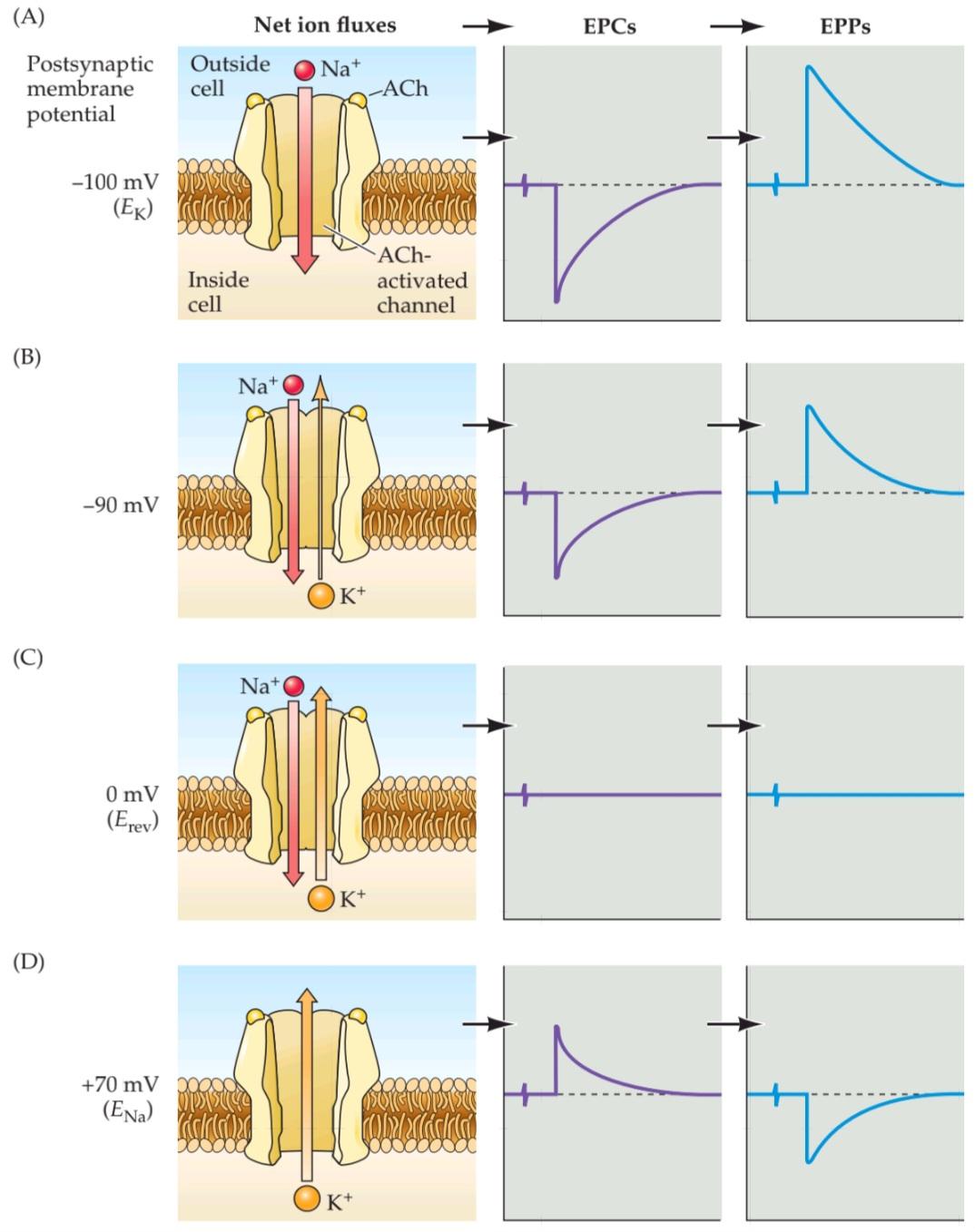

FIGURE 5.18 Na+ and K+ movements during EPCs and EPPs. (A–D) Each of the postsynaptic potentials indicated at the left results in different relative fluxes of Na+ and K+ (net ion fluxes). These ion flux-es determine the amplitude and polarity of the EPCs, which in turn determine the EPPs. Note that at about 0 mV the Na+ flux is exactly balanced by an opposite K+ flux, resulting in no net current flow, and hence no change in the membrane potential. (E) EPCs are inward at potentials more negative than Erev and outward at potentials more positive than Erev. (F) EPPs depolarize the postsynaptic cell at potentials more negative than Erev. At potentials more positive than Erev, EPPs hyperpolarize the cell.
شکل ۵.۱۸ حرکات +Na و +K در طول EPCها و EPPها. (A-D) هر یک از پتانسیلهای پسسیناپسی نشان داده شده در سمت چپ منجر به شارهای نسبی متفاوتی از +Na و +K (شارهای خالص یون) میشود. این شارهای یونی دامنه و قطبیت EPCها را تعیین میکنند که به نوبه خود EPPها را تعیین میکنند. توجه داشته باشید که در حدود 0 میلیولت، شار +Na دقیقاً با شار +K مخالف متعادل میشود و در نتیجه هیچ جریان خالصی جریان نمییابد و از این رو هیچ تغییری در پتانسیل غشا ایجاد نمیشود. (E) EPCها در پتانسیلهای منفیتر از Erev به سمت داخل و در پتانسیلهای مثبتتر از Erev به سمت خارج هستند. (F) EPPها در پتانسیلهای منفیتر از Erev سلول پسسیناپسی را دپلاریزه میکنند. در پتانسیلهای مثبتتر از Erev، EPPها سلول را هایپرپلاریزه میکنند.
Although this discussion has focused on the neuromuscular junction, similar mechanisms generate postsynaptic responses at all chemical synapses: Transmitter binding to postsynaptic receptors produces a postsynaptic conductance change as ion channels are opened (or sometimes closed). The postsynaptic conductance is increased if—as at the neuromuscular junction—channels are opened, and is decreased if channels are closed. This conductance change typically generates an electrical current, the postsynaptic current (PSC), which in turn changes the postsynaptic membrane potential to produce a postsynaptic potential (PSP). As in the specific case of the EPP at the neuromuscular junction, PSPs are depolarizing if their reversal potential is more positive than the resting membrane potential and hyperpolarizing if their reversal potential is more negative.
اگرچه این بحث بر روی اتصال عصبی-عضلانی متمرکز شده است، مکانیسمهای مشابهی پاسخهای پسسیناپسی را در تمام سیناپسهای شیمیایی ایجاد میکنند: اتصال فرستنده به گیرندههای پسسیناپسی با باز شدن (یا گاهی بسته شدن) کانالهای یونی، تغییر رسانایی پسسیناپسی ایجاد میکند. اگر کانالها باز باشند – مانند اتصال عصبی-عضلانی – رسانایی پسسیناپسی افزایش مییابد و اگر کانالها بسته باشند، کاهش مییابد. این تغییر رسانایی معمولاً یک جریان الکتریکی، جریان پسسیناپسی (PSC) ایجاد میکند که به نوبه خود پتانسیل غشای پسسیناپسی را برای تولید پتانسیل پسسیناپسی (PSP) تغییر میدهد. همانطور که در مورد خاص EPP در اتصال عصبی-عضلانی، PSPها اگر پتانسیل برگشت آنها مثبتتر از پتانسیل غشای استراحت باشد، دپلاریزه میشوند و اگر پتانسیل برگشت آنها منفیتر باشد، هایپرپلاریزه میشوند.
The conductance changes and the PSPs that typically accompany them are the ultimate outcome of most chemical synaptic transmission, concluding a sequence of electrical and chemical events that begins with the invasion of an action potential into the terminals of a presynaptic neuron. In many ways, the events that produce PSPs at synapses are similar to those that generate action potentials in axons; in both cases, conductance changes produced by ion channels lead to ion current flow that changes the membrane potential.
تغییرات رسانایی و PSPهایی که معمولاً با آنها همراه هستند، نتیجه نهایی اکثر انتقالهای سیناپسی شیمیایی هستند و به دنبالهای از رویدادهای الکتریکی و شیمیایی ختم میشوند که با هجوم یک پتانسیل عمل به پایانههای یک نورون پیشسیناپسی آغاز میشود. از بسیاری جهات، رویدادهایی که PSPها را در سیناپسها تولید میکنند، مشابه رویدادهایی هستند که پتانسیلهای عمل را در آکسونها ایجاد میکنند. در هر دو مورد، تغییرات رسانایی ایجاد شده توسط کانالهای یونی منجر به جریان جریان یونی میشود که پتانسیل غشا را تغییر میدهد.
Excitatory and Inhibitory Postsynaptic Potentials
PSPs ultimately alter the probability that an action potential will be produced in the postsynaptic cell. At the neuromuscular junction, synaptic action increases the probability that an action potential will occur in the postsynaptic muscle cell; indeed, the large amplitude of the EPP ensures that an action potential always is triggered. At many other synapses, PSPs similarly increase the probability of firing a postsynaptic action potential. However, still other synapses actually decrease the probability that the postsynaptic cell will generate an action potential. PSPs are called excitatory (or EPSPs) if they increase the likelihood of a postsynaptic action potential occurring, and inhibitory (or IPSPs) if they decrease this likelihood. Given that most neurons receive inputs from both excitatory and inhibitory synapses, it is important to understand more precisely the mechanisms that determine whether a particular synapse excites or inhibits its postsynaptic partner.
پتانسیلهای پسسیناپسی تحریکی و مهاری
PSPها در نهایت احتمال تولید یک پتانسیل عمل در سلول پسسیناپسی را تغییر میدهند. در محل اتصال عصبی-عضلانی، عمل سیناپسی احتمال وقوع یک پتانسیل عمل در سلول عضلانی پسسیناپسی را افزایش میدهد؛ در واقع، دامنه بزرگ EPP تضمین میکند که یک پتانسیل عمل همیشه فعال میشود. در بسیاری از سیناپسهای دیگر، PSPها به طور مشابه احتمال شلیک یک پتانسیل عمل پسسیناپسی را افزایش میدهند. با این حال، سیناپسهای دیگر در واقع احتمال تولید یک پتانسیل عمل توسط سلول پسسیناپسی را کاهش میدهند. PSPها اگر احتمال وقوع یک پتانسیل عمل پسسیناپسی را افزایش دهند، تحریکی (یا EPSP) و اگر این احتمال را کاهش دهند، مهاری (یا IPSP) نامیده میشوند. با توجه به اینکه اکثر نورونها ورودیها را از هر دو سیناپس تحریکی و مهاری دریافت میکنند، درک دقیقتر مکانیسمهایی که تعیین میکنند یک سیناپس خاص، شریک پسسیناپسی خود را تحریک یا مهار میکند، مهم است.
The principles of excitation just described for the neuromuscular junction are pertinent to all excitatory synapses. The principles of postsynaptic inhibition are much the same as for excitation, and they are also quite general. In both cases, neurotransmitters binding to receptors open or close ion channels in the postsynaptic cell. Whether a postsynaptic response is an EPSP or an IPSP depends on the type of channel that is coupled to the receptor, and on the concentration of permeant ions inside and outside the cell. In fact, the only distinction between postsynaptic excitation and inhibition is the reversal potential of the PSP in relation to the threshold voltage for generating action potentials in the postsynaptic cell.
اصول تحریک که برای اتصال عصبی-عضلانی شرح داده شد، برای همه سیناپسهای تحریکی نیز صادق است. اصول مهار پسسیناپسی تقریباً مشابه با تحریک است و همچنین کاملاً عمومی است. در هر دو مورد، انتقالدهندههای عصبی متصل به گیرندهها، کانالهای یونی را در سلول پسسیناپسی باز یا بسته میکنند. اینکه یک پاسخ پسسیناپسی EPSP باشد یا IPSP، به نوع کانالی که به گیرنده متصل است و به غلظت یونهای نفوذپذیر در داخل و خارج سلول بستگی دارد. در واقع، تنها تمایز بین تحریک پسسیناپسی و مهار، پتانسیل معکوس PSP در رابطه با ولتاژ آستانه برای تولید پتانسیلهای عمل در سلول پسسیناپسی است.
Consider, for example, a neuronal synapse that uses glutamate as the transmitter. Many such synapses have receptors that, like the ACh receptors at neuromuscular synapses, open ion channels that are nonselectively permeable to cations (see Chapter 6). When these glutamate receptors are activated, both Na+ and K+ flow across the postsynaptic membrane, yielding an Erev of approximately 0 mV for the resulting postsynaptic current. If the resting potential of the postsynaptic neuron is –60 mV, the resulting EPSP will depolarize by bringing the postsynaptic membrane potential toward 0 mV. For the hypothetical neuron shown in Figure 5.19A, the action potential threshold voltage is –40 mV. Thus, a glutamate-induced EPSP will increase the probability that this neuron produces an action potential, defining the synapse as excitatory.
به عنوان مثال، یک سیناپس عصبی را در نظر بگیرید که از گلوتامات به عنوان فرستنده استفاده میکند. بسیاری از این سیناپسها گیرندههایی دارند که مانند گیرندههای ACh در سیناپسهای عصبی-عضلانی، کانالهای یونی را باز میکنند که به طور غیرانتخابی نسبت به کاتیونها نفوذپذیر هستند (به فصل 6 مراجعه کنید). وقتی این گیرندههای گلوتامات فعال میشوند، هم +Na و هم +K از غشای پسسیناپسی عبور میکنند و Erev تقریباً 0 میلیولت برای جریان پسسیناپسی حاصل ایجاد میکنند. اگر پتانسیل استراحت نورون پسسیناپسی 60- میلیولت باشد، EPSP حاصل با رساندن پتانسیل غشای پسسیناپسی به سمت 0 میلیولت، دپلاریزه میشود. برای نورون فرضی نشان داده شده در شکل 5.19A، ولتاژ آستانه پتانسیل عمل 40- میلیولت است. بنابراین، EPSP القا شده توسط گلوتامات احتمال تولید پتانسیل عمل توسط این نورون را افزایش میدهد و سیناپس را به عنوان سیناپس تحریکی تعریف میکند.
As an example of inhibitory postsynaptic action, consider a neuronal synapse that uses GABA as its transmitter. At such synapses, the GABA receptors typically open channels that are selectively permeable to Cl–, and the action of GABA causes Cl– to flow across the postsynaptic membrane. Consider a case where ECl is –70 mV, as is the case for some neu-rons, so that the postsynaptic resting potential of –60 mV isless negative than ECl. The resulting positive electrochemical driving force (Vm – Erev) will cause negatively charged Cl– to flow into the cell and produce a hyperpolarizing IPSP (Figure 5.19B). This hyperpolarizing IPSP will take the postsynaptic membrane away from the action potential threshold of –40 mV, clearly inhibiting the postsynaptic cell.
به عنوان مثالی از عمل مهاری پس سیناپسی، یک سیناپس عصبی را در نظر بگیرید که از GABA به عنوان فرستنده خود استفاده میکند. در چنین سیناپسهایی، گیرندههای GABA معمولاً کانالهایی را باز میکنند که به طور انتخابی نسبت به Cl- نفوذپذیر هستند و عمل GABA باعث میشود Cl- از غشای پس سیناپسی جریان یابد. موردی را در نظر بگیرید که ECl برابر با 70- میلیولت باشد، همانطور که برای برخی از نورونها صادق است، به طوری که پتانسیل استراحت پس سیناپسی 60- میلیولت کمتر از ECl منفی است. نیروی محرک الکتروشیمیایی مثبت حاصل (Vm – Erev) باعث میشود Cl- با بار منفی به داخل سلول جریان یابد و یک IPSP فوق قطبی ایجاد کند (شکل 5.19B). این IPSP فوق قطبی، غشای پس سیناپسی را از آستانه پتانسیل عمل 40- میلیولت دور میکند و به وضوح سلول پس سیناپسی را مهار میکند.
Surprisingly, inhibitory synapses need not produce hyperpolarizing IPSPs. For instance, if ECl were –50 mV instead of –70 mV, then the negative electrochemical driving force would cause Cl– to flow out of the cell and produce a depolarizing IPSP (Figure 5.19C). However, the synapse would still be inhibitory: Given that the reversal potential of the IPSP still is more negative than the action potential threshold (–40 mV), the depolarizing IPSP would inhibit because the postsynaptic membrane potential would be kept more negative than the threshold for action potential initiation. Another way to think about this peculiar situation is that if another excitatory input onto this neuron brought the cell’s membrane potential to –41 mV—just below the threshold for firing an action potential—the IPSP would then hyperpolarize the membrane potential toward –50 mV, bringing the potential away from the action potential threshold. Thus, while EPSPs depolarize the postsynaptic cell, IPSPs can either hyperpolarize or depolarize; indeed, an inhibitory conductance change may produce no potential change at all and still exert an inhibitory effect by making it more difficult for an EPSP to evoke an action potential in the postsynaptic cell.
جالب اینجاست که سیناپسهای مهاری نیازی به تولید IPSP هایپرپلاریزه کننده ندارند. برای مثال، اگر ECl به جای 70- میلیولت، 50- میلیولت باشد، نیروی محرک الکتروشیمیایی منفی باعث میشود Cl- از سلول خارج شود و IPSP دپلاریزه کننده تولید کند (شکل 5.19C). با این حال، سیناپس همچنان مهاری خواهد بود: با توجه به اینکه پتانسیل معکوس IPSP هنوز منفیتر از آستانه پتانسیل عمل (40- میلیولت) است، IPSP دپلاریزه کننده مهار میکند زیرا پتانسیل غشای پس سیناپسی منفیتر از آستانه شروع پتانسیل عمل نگه داشته میشود. راه دیگری برای فکر کردن در مورد این وضعیت عجیب این است که اگر یک ورودی تحریکی دیگر به این نورون، پتانسیل غشای سلول را به 41- میلیولت – درست زیر آستانه شلیک پتانسیل عمل – برساند، IPSP پتانسیل غشا را به سمت 50- میلیولت هایپرپلاریزه میکند و پتانسیل را از آستانه پتانسیل عمل دور میکند. بنابراین، در حالی که EPSP ها سلول پس سیناپسی را دپلاریزه میکنند، IPSP ها میتوانند یا هایپرپلاریزه یا دپلاریزه شوند؛ در واقع، یک تغییر رسانایی مهاری ممکن است هیچ تغییر پتانسیلی ایجاد نکند و همچنان با دشوارتر کردن ایجاد پتانسیل عمل در سلول پس سیناپسی توسط EPSP، اثر مهاری خود را اعمال کند.
Although the particulars of postsynaptic action can be complex, a simple rule distinguishes postsynaptic excitation from inhibition: An EPSP has a reversal potential more positive than the action potential threshold, whereas an IPSP has a reversal potential more negative than threshold (Figure 5.19D). Intuitively, this rule can be understood by realizing that an EPSP will tend to depolarize the membrane potential so that it exceeds threshold, whereas an IPSP will always act to keep the membrane potential more negative than the threshold potential.
اگرچه جزئیات عمل پس سیناپسی میتواند پیچیده باشد، اما یک قانون ساده، تحریک پس سیناپسی را از مهار متمایز میکند: یک EPSP پتانسیل معکوسی مثبتتر از آستانه پتانسیل عمل دارد، در حالی که یک IPSP پتانسیل معکوسی منفیتر از آستانه دارد (شکل 5.19D). به طور شهودی، این قانون را میتوان با درک این نکته درک کرد که یک EPSP تمایل دارد پتانسیل غشا را دپلاریزه کند تا از آستانه فراتر رود، در حالی که یک IPSP همیشه طوری عمل میکند که پتانسیل غشا را منفیتر از پتانسیل آستانه نگه دارد.


FIGURE 5.19 Reversal potentials and threshold potentials determine postsynaptic excitation and inhibition. (A) If the reversal potential for a PSP (0 mV) is more positive than the action potential threshold (–40 mV), the effect of a transmitter is excitatory, and it generates EPSPs. (B) If the reversal potential for a PSP is more negative than the action potential threshold, the transmitter is inhibitory and generates IPSPs. (C) IPSPs can nonetheless depolarize the postsynaptic cell if their reversal potential is between the resting potential and the action potential threshold. (D) The general rule of postsynaptic action is: If the reversal potential is more positive than threshold, excitation results; inhibition occurs if the reversal potential is more negative than threshold.
شکل ۵.۱۹ پتانسیلهای معکوس و پتانسیلهای آستانه، تحریک و مهار پسسیناپسی را تعیین میکنند. (الف) اگر پتانسیل معکوس برای یک PSP (0 میلیولت) مثبتتر از آستانه پتانسیل عمل (40- میلیولت) باشد، اثر یک فرستنده تحریکی است و EPSP تولید میکند. (ب) اگر پتانسیل معکوس برای یک PSP منفیتر از آستانه پتانسیل عمل باشد، فرستنده مهاری است و IPSP تولید میکند. (ج) با این وجود، IPSPها میتوانند سلول پسسیناپسی را دپلاریزه کنند اگر پتانسیل معکوس آنها بین پتانسیل استراحت و آستانه پتانسیل عمل باشد. (د) قانون کلی عمل پسسیناپسی این است: اگر پتانسیل معکوس مثبتتر از آستانه باشد، تحریک حاصل میشود؛ اگر پتانسیل معکوس منفیتر از آستانه باشد، مهار رخ میدهد.
Summation of Synaptic Potentials
The PSPs produced at most synapses in the brain are much smaller than those at the neuromuscular junction; indeed, EPSPs produced by individual excitatory synapses may be only a fraction of a millivolt and are usually well below the threshold for generating postsynaptic action potentials. How, then, can such synapses transmit information if their PSPs are subthreshold? The answer is that neurons in the central nervous system are typically innervated by thousands of synapses, and the PSPs produced by each active synapse can sum together—in space and in time—to determine the behavior of the postsynaptic neuron.
جمع پتانسیلهای سیناپسی
PSP های تولید شده در اکثر سیناپسهای مغز بسیار کوچکتر از آنهایی هستند که در محل اتصال عصبی-عضلانی وجود دارند. در واقع، EPSP های تولید شده توسط سیناپسهای تحریکی منفرد ممکن است تنها کسری از میلیولت باشند و معمولاً بسیار پایینتر از آستانه تولید پتانسیلهای عمل پس سیناپسی هستند. پس چگونه چنین سیناپسهایی میتوانند اطلاعات را منتقل کنند اگر PSP های آنها زیرآستانه باشند؟ پاسخ این است که نورونها در سیستم عصبی مرکزی معمولاً توسط هزاران سیناپس عصبدهی میشوند و PSP های تولید شده توسط هر سیناپس فعال میتوانند – در فضا و در زمان – با هم جمع شوند تا رفتار نورون پس سیناپسی را تعیین کنند.
Consider the highly simplified case of a neuron that is innervated by two excitatory synapses, each generating a subthreshold EPSP, and an inhibitory synapse that produces an IPSP (Figure 5.20A). While activation of either one of the excitatory synapses alone (E1 or E2 in Figure 5.20B) produces a subthreshold EPSP, activation of both excitatory synapses at about the same time causes the two EPSPs to sum together. If the sum of the two EPSPs (E1 + E2) depolarizes the postsynaptic neuron sufficiently to reach the threshold potential, a postsynaptic action potential results. Summation thus allows subthreshold EPSPs to influence action potential production. Likewise, an IPSP generated by an inhibitory synapse (I) can sum (algebraically speaking) with a subthreshold EPSP to reduce its amplitude (E1 + I) or can sum with suprathreshold EPSPs to prevent the postsynaptic neuron from reaching threshold (E1 + I + E2).
مورد بسیار سادهشدهای از یک نورون را در نظر بگیرید که توسط دو سیناپس تحریکی عصبدهی میشود، که هر کدام یک EPSP زیرآستانهای تولید میکنند، و یک سیناپس مهاری که IPSP تولید میکند (شکل 5.20A). در حالی که فعال شدن هر یک از سیناپسهای تحریکی به تنهایی (E1 یا E2 در شکل 5.20B) یک EPSP زیرآستانهای تولید میکند، فعال شدن هر دو سیناپس تحریکی تقریباً همزمان باعث میشود که دو EPSP با هم جمع شوند. اگر مجموع دو EPSP (E1 + E2) نورون پسسیناپسی را به اندازه کافی دپلاریزه کند تا به پتانسیل آستانه برسد، یک پتانسیل عمل پسسیناپسی حاصل میشود. بنابراین، جمع شدن به EPSPهای زیرآستانهای اجازه میدهد تا بر تولید پتانسیل عمل تأثیر بگذارند. به همین ترتیب، یک IPSP تولید شده توسط یک سیناپس مهاری (I) میتواند (به صورت جبری) با یک EPSP زیرآستانهای جمع شود تا دامنه آن کاهش یابد (E1 + I) یا میتواند با EPSP های فوقآستانهای جمع شود تا از رسیدن نورون پسسیناپسی به آستانه (E1 + I + E2) جلوگیری شود.
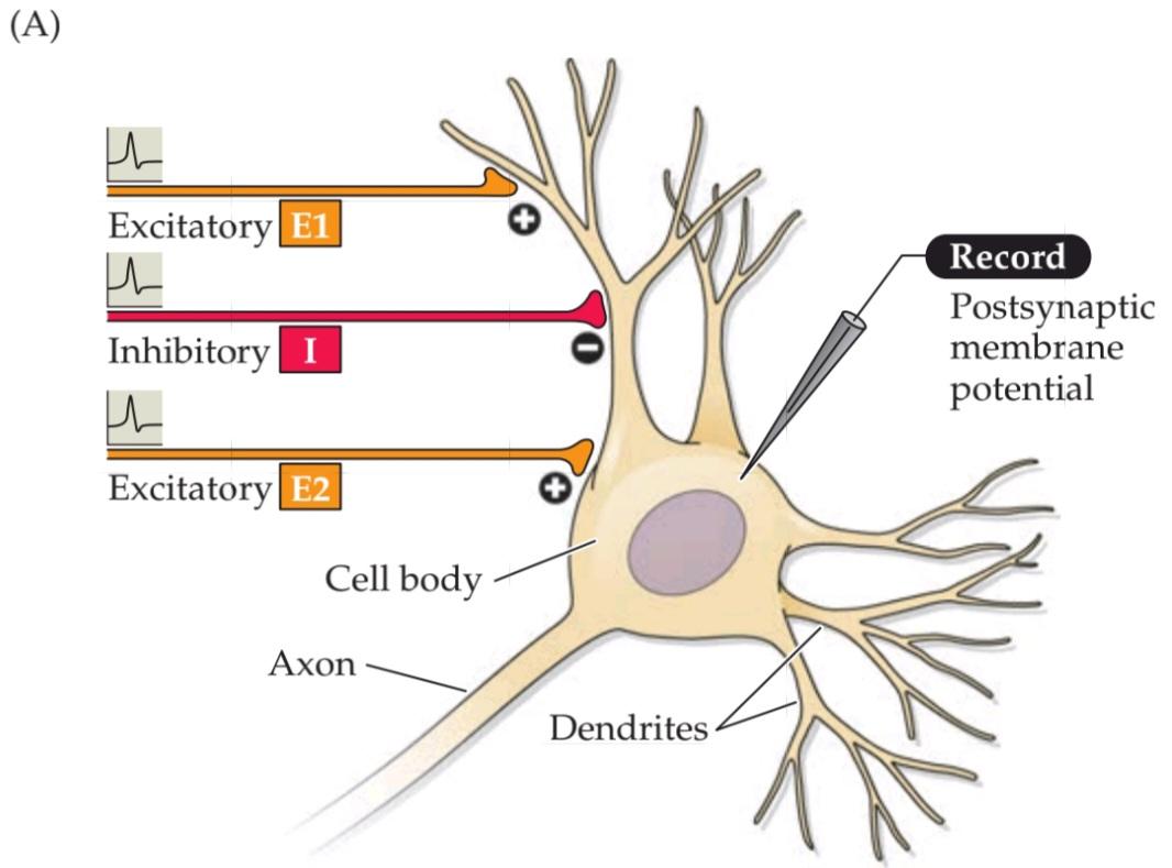

FIGURE 5.20 Summation of postsynaptic potentials.(A) A microelectrode records the postsynaptic potentials produced by the activity of two excitatory synapses (E1 and E2) and an inhibitory synapse (I). (B) Electrical responses to synaptic activation. Stimulating either excitatory synapse (E1 or E2) produces a subthreshold EPSP, whereas stimulating both synapses at the same time (E1 + E2) produces a suprathreshold EPSP that evokes a postsynaptic action potential (shown in blue). Activation of the inhibitory synapse alone (I) results in a hyperpolarizing IPSP. Summing this IPSP (dashed red line) with the EPSP (dashed yellow line) produced by one excitatory synapse (E1 + I) reduces the amplitude of the EPSP (solid orange line), while summing it with the suprathreshold EPSP produced by activating synapses E1 and E2 keeps the postsynaptic neuron below threshold, so that no action potential is evoked.
شکل۵.۲۰ جمعبندی پتانسیلهای پسسیناپسی. (الف) یک میکروالکترود پتانسیلهای پسسیناپسی تولید شده توسط فعالیت دو سیناپس تحریکی (E1 و E2) و یک سیناپس مهاری (I) را ثبت میکند. (ب) پاسخهای الکتریکی به فعالسازی سیناپسی. تحریک هر یک از سیناپسهای تحریکی (E1 یا E2) یک EPSP زیرآستانهای تولید میکند، در حالی که تحریک همزمان هر دو سیناپس (E1 + E2) یک EPSP فوقآستانهای تولید میکند که یک پتانسیل عمل پسسیناپسی را ایجاد میکند (با رنگ آبی نشان داده شده است). فعال شدن سیناپس مهاری به تنهایی (I) منجر به IPSP فوققطبی میشود. جمع این IPSP (خط قرمز خطچین) با EPSP (خط زرد خطچین) تولید شده توسط یک سیناپس تحریکی (E1 + I) دامنه EPSP (خط نارنجی پررنگ) را کاهش میدهد، در حالی که جمع آن با EPSP فوق آستانه تولید شده توسط سیناپسهای فعال E1 و E2، نورون پسسیناپسی را زیر آستانه نگه میدارد، به طوری که هیچ پتانسیل عملی برانگیخته نمیشود.
In short, the summation of EPSPs and IPSPs by a postsynaptic neuron permits a neuron to integrate the electrical information provided by all the inhibitory and excitatory synapses acting on it at any moment. Whether the sum of active synaptic inputs results in the production of an action potential depends on the balance between excitation and inhibition. If the sum of all EPSPs and IPSPs results in a depolarization of sufficient amplitude to the membrane potential above threshold, then the postsynaptic cell will produce an action potential. Conversely, if inhibition prevails, then the postsynaptic cell will remain silent. Typically, the balance between EPSPs and IPSPs changes continually over time, depending on the number of excitatory and inhibitory synapses active at a given moment and the magnitude of the current at each active synapse. Summation is therefore a tug-of-war between all excitatory and inhibitory postsynaptic currents; the outcome of the contest determines whether or not a postsynaptic neuron fires an action potential and thereby becomes an active element in the neural circuits to which it belongs.
به طور خلاصه، جمع EPSPها و IPSPها توسط یک نورون پس سیناپسی به یک نورون اجازه میدهد تا اطلاعات الکتریکی ارائه شده توسط تمام سیناپسهای مهاری و تحریکی که در هر لحظه بر روی آن عمل میکنند را ادغام کند. اینکه آیا مجموع ورودیهای سیناپسی فعال منجر به تولید پتانسیل عمل میشود یا خیر، به تعادل بین تحریک و مهار بستگی دارد. اگر مجموع تمام EPSPها و IPSPها منجر به دپلاریزاسیون با دامنه کافی برای پتانسیل غشاء بالاتر از آستانه شود، سلول پس سیناپسی یک پتانسیل عمل تولید خواهد کرد. برعکس، اگر مهار غالب باشد، سلول پس سیناپسی ساکت خواهد ماند. به طور معمول، تعادل بین EPSPها و IPSPها به مرور زمان به طور مداوم تغییر میکند، بسته به تعداد سیناپسهای تحریکی و مهاری فعال در یک لحظه معین و بزرگی جریان در هر سیناپس فعال. بنابراین جمعبندی، یک مسابقه طنابکشی بین تمام جریانهای پس سیناپسی تحریکی و مهاری است. نتیجهی این رقابت تعیین میکند که آیا یک نورون پسسیناپسی پتانسیل عمل ایجاد میکند یا خیر و در نتیجه به عنصری فعال در مدارهای عصبی که به آن تعلق دارد تبدیل میشود یا خیر.
In conclusion, at chemical synapses neurotransmitter release from presynaptic terminals initiates a series of postsynaptic events that culminate in a transient change in the probability of a postsynaptic action potential occurring (Figure 5.21). Such synaptic signaling allows neurons to form the intricate synaptic circuits that play fundamental roles in brain information processing. Recent work further suggests that glial cells also contribute to synaptic signaling, adding another dimension to information processing in the brain (Box 5A).
در نتیجه، در سیناپسهای شیمیایی، آزادسازی انتقالدهندههای عصبی از پایانههای پیشسیناپسی، مجموعهای از رویدادهای پسسیناپسی را آغاز میکند که منجر به تغییر گذرا در احتمال وقوع پتانسیل عمل پسسیناپسی میشود (شکل 5.21). چنین سیگنالدهی سیناپسی به نورونها اجازه میدهد تا مدارهای سیناپسی پیچیدهای را تشکیل دهند که نقشهای اساسی در پردازش اطلاعات مغز ایفا میکنند. تحقیقات اخیر نشان میدهد که سلولهای گلیال نیز در سیگنالدهی سیناپسی نقش دارند و بُعد دیگری به پردازش اطلاعات در مغز میافزایند (کادر 5A).
BOX 5A ◊ The Tripartite Synapse
Up to this point, the discussion has considered signaling between presynaptic neurons and their postsynaptic targets to be a private dialogue between these two cells. However, recent work suggests that this synaptic conver-sation may involve glial cells as well.
کادر ۵A ◊ سیناپس سهجانبه
تا این لحظه، بحث، سیگنالدهی بین نورونهای پیشسیناپسی و اهداف پسسیناپسی آنها را به عنوان یک گفتگوی خصوصی بین این دو سلول در نظر گرفته است. با این حال، تحقیقات اخیر نشان میدهد که این گفتگوی سیناپسی ممکن است سلولهای گلیال را نیز درگیر کند.
As mentioned in Chapter 1, glial cells support neurons in several ways. For example, it is well established that glia regulate the extracellular environment by removing K+ that accumulates during action potential generation and by removing neurotransmitters at the conclusion of synaptic transmission. Consistent with such roles, glia seem to occupy virtually all of the non-neuronal volume of the brain. This means that glia are found in very close association with synapses (see Figure A); a given synapse typically is no more than a few hundred nanometers away from a glial cell. Glia form exceedingly fine processes that completely envelop synapses (see Figure B), an intimate association that raises the possibility of a signaling role for glia at synapses.
همانطور که در فصل 1 ذکر شد، سلولهای گلیال از چندین طریق از نورونها پشتیبانی میکنند. به عنوان مثال، به خوبی ثابت شده است که گلیا با حذف +K که در طول تولید پتانسیل عمل تجمع مییابد و با حذف انتقالدهندههای عصبی در پایان انتقال سیناپسی، محیط خارج سلولی را تنظیم میکند. مطابق با چنین نقشهایی، به نظر میرسد گلیا تقریباً تمام حجم غیر نورونی مغز را اشغال میکند. این بدان معناست که گلیا در ارتباط بسیار نزدیکی با سیناپسها یافت میشوند (شکل A را ببینید)؛ یک سیناپس مشخص معمولاً بیش از چند صد نانومتر از یک سلول گلیال فاصله ندارد. گلیا زوائد بسیار ظریفی را تشکیل میدهند که سیناپسها را به طور کامل میپوشانند (شکل B را ببینید)، ارتباط نزدیکی که احتمال نقش سیگنالدهی گلیا در سیناپسها را افزایش میدهد.
(A) Electron microscopy image of a synapse between a presynaptic terminal (pre) and postsynaptic neuron (post), with a glial cell (astrocyte; astro) immediately adjacent to the synapse.
(الف) تصویر میکروسکوپ الکترونی از یک سیناپس بین یک نورون پیشسیناپسی (پیش) و یک نورون پسسیناپسی (پست)، با یک سلول گلیال (آستروسیت؛ آسترو) که بلافاصله در مجاورت سیناپس قرار دارد.
(B) Three-dimensional structure of the contact between a glial cell (blue) surrounding dendrites of four postsynaptic neurons (different colors).
(ب) ساختار سهبعدی تماس بین یک سلول گلیال (آبی) که دندریتهای چهار نورون پسسیناپسی (رنگهای مختلف) را احاطه کرده است.

The first support for such a role came from the discovery that glia respond to application of neurotransmitters. The list of neurotransmitters that elicit responses in glia now includes acetylcholine, glutamate, GABA, and many others. These responses are mediated by the same sorts of neurotransmitter receptors that are employed in synaptic signaling, most often the metabotropic receptors that are coupled to intracellular signaling cascades (see Figure 5.14B). In some cases, neurotransmitters produce changes in the membrane potential of glia. More often, these neurotransmitters cause transient changes in intracellular calcium concentration within the glial cell. These intracellular calcium signals often are observed to trigger calcium waves that spread both within a single glial cell and also propagate between glia (see Figure C).
اولین تایید برای چنین نقشی از کشف این موضوع ناشی شد که گلیا به کاربرد انتقالدهندههای عصبی پاسخ میدهد. فهرست انتقالدهندههای عصبی که در گلیا پاسخ ایجاد میکنند، اکنون شامل استیل کولین، گلوتامات، گابا و بسیاری دیگر است. این پاسخها توسط همان نوع گیرندههای انتقالدهنده عصبی که در سیگنالدهی سیناپسی به کار میروند، و اغلب گیرندههای متابوتروپیک که به آبشارهای سیگنالدهی درون سلولی متصل هستند، واسطهگری میشوند (شکل 5.14B را ببینید). در برخی موارد، انتقالدهندههای عصبی تغییراتی در پتانسیل غشای گلیا ایجاد میکنند. اغلب، این انتقالدهندههای عصبی باعث تغییرات گذرا در غلظت کلسیم درون سلولی در سلول گلیال میشوند. این سیگنالهای کلسیم درون سلولی اغلب مشاهده میشوند که امواج کلسیم را تحریک میکنند که هم در یک سلول گلیال واحد پخش میشوند و هم بین گلیاها منتشر میشوند (شکل C را ببینید).
(C) Application of glutamate (at arrow) increases intracellular Ca2+ (white) in cultured glia. Examination of these cells at different times indicates that the calciumsignals spread as a wave through neighboring glia.
(ج) استفاده از گلوتامات (در فلش) باعث افزایش +Ca2 درون سلولی (سفید) در گلیاهای کشت شده میشود. بررسی این سلولها در زمانهای مختلف نشان میدهد که سیگنالهای کلسیم به صورت موجی از طریق گلیاهای همسایه پخش میشوند.
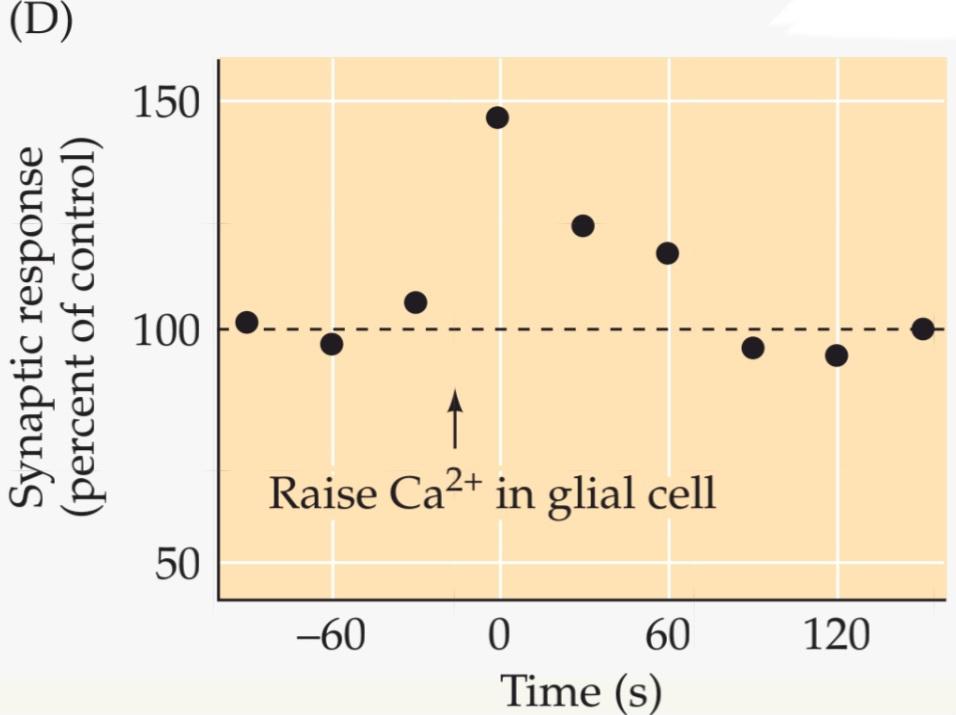
 (D) Transient elevation of Ca2+ whitin a single glial cell (arrow) enhances transmission at an excitatory synapse in the hippocampus. (A.B from witcher et al., 2007; C from Cornell-Bell et al. 1990; D after Perea and Araque, 2007.)
(د) افزایش گذرای کلسیم در یک سلول گلیال (فلش) انتقال را در یک سیناپس تحریکی در هیپوکامپ افزایش میدهد. (A.B از witcher و همکاران، ۲۰۰۷؛ C از Cornell-Bell و همکاران، ۱۹۹۰؛ D پس از Perea و Araque، ۲۰۰۷.)
These transient rises in intracellular calcium serve as second messenger signals (see Chapter 7) that trigger a number of physiological responses in glia. The most remarkable response is the release of several molecules—such as glutamate, GABA, and ATP—that are traditionally considered to be neurotransmitters. Release of such “gliotransmitters” occurs both via the calcium-triggered exocytotic mechanisms employed in presynaptic terminals of neurons (see Figure 5.4C), as well as via unconventional release mechanisms such as permeation through certain ion channels.
این افزایشهای گذرا در کلسیم درون سلولی به عنوان سیگنالهای پیامرسان ثانویه عمل میکنند (به فصل 7 مراجعه کنید) که تعدادی از پاسخهای فیزیولوژیکی را در گلیا آغاز میکنند. قابل توجهترین پاسخ، آزادسازی چندین مولکول – مانند گلوتامات، GABA و ATP – است که به طور سنتی به عنوان انتقالدهندههای عصبی در نظر گرفته میشوند. آزادسازی چنین “گلیوترنسمیترهایی” هم از طریق مکانیسمهای اگزوسیتوزی ناشی از کلسیم که در پایانههای پیشسیناپسی نورونها به کار میروند (به شکل 5.4C مراجعه کنید) و هم از طریق مکانیسمهای آزادسازی غیرمتعارف مانند نفوذ از طریق کانالهای یونی خاص رخ میدهد.
The ability both to respond to and to release neurotransmitters potentially makes glia participants in synaptic signaling. Indeed, release of neurotransmitters from a variety of presynaptic terminals has been found to elicit responses in glia. Furthermore, release of gliotransmitters has been found to regulate transmission at numerous synapses (see Figure D). In some cases, glia regulate release of transmitters from presynaptic terminals, while in other cases they alter postsynaptic responsiveness. Glia can also alter the ability of synapses to undergo activity-dependent, plastic changes in synaptic transmission (see Chapter 8).
توانایی پاسخ دادن و آزاد کردن انتقالدهندههای عصبی به طور بالقوه باعث میشود که گلیا در سیگنالدهی سیناپسی شرکت کند. در واقع، مشخص شده است که آزاد شدن انتقالدهندههای عصبی از انواع پایانههای پیشسیناپسی، پاسخهایی را در گلیا ایجاد میکند. علاوه بر این، مشخص شده است که آزاد شدن انتقالدهندههای گلیوترانسمیتر، انتقال را در سیناپسهای متعدد تنظیم میکند (شکل D را ببینید). در برخی موارد، گلیا آزاد شدن انتقالدهندهها را از پایانههای پیشسیناپسی تنظیم میکند، در حالی که در موارد دیگر، آنها پاسخدهی پسسیناپسی را تغییر میدهند. گلیا همچنین میتواند توانایی سیناپسها را برای انجام تغییرات انعطافپذیر وابسته به فعالیت در انتقال سیناپسی تغییر دهد (به فصل 8 مراجعه کنید).
This ability of glia to participate in synaptic signaling has led to the concept of the tripartite synapse, a three-way junction involving the presynaptic terminal, the postsynaptic process, and neighboring glia. While there is still debate about the physiological significance of such neuron–glia interactions, the ability of glia to release neurotransmitters, similar to presynaptic terminals, and to respond to neurotransmitters, similar to postsynaptic neurons, is dramatically changing our view of brain signaling mechanisms.
این توانایی گلیا برای شرکت در سیگنالینگ سیناپسی منجر به مفهوم سیناپس سهجانبه شده است، یک اتصال سهطرفه شامل ترمینال پیشسیناپسی، فرآیند پسسیناپسی و گلیاهای همسایه. در حالی که هنوز در مورد اهمیت فیزیولوژیکی چنین تعاملات نورون-گلیا بحث وجود دارد، توانایی گلیا در آزادسازی انتقالدهندههای عصبی، مشابه ترمینالهای پیشسیناپسی، و پاسخ به انتقالدهندههای عصبی، مشابه نورونهای پسسیناپسی، به طور چشمگیری دیدگاه ما را در مورد مکانیسمهای سیگنالینگ مغز تغییر میدهد.
Summary
Synapses communicate the information carried by action potentials from one neuron to the next in neural circuits. The mechanisms underlying postsynaptic potentials generated during synaptic transmission are closely related to the mechanisms that generate other types of neuronal electrical signals, namely, ionic flow through membrane channels. In the case of electrical synapses, these channels are connexons; direct but passive flow of current through connexons is the basis for transmission. In the case of chemical synapses, channels with smaller and more selective pores are activated by the binding of neurotransmitters to postsynaptic receptors following release of the neurotransmitters from the presynaptic terminal. The large number of neurotransmitters in the nervous system can be divided into two broad classes: small-molecule transmitters and neuropeptides. Neurotransmitters are synthesized from defined precursors by regulated enzymatic pathways, packaged into one of several types of synaptic vesicles, and released into the synaptic cleft in a Ca2+-dependent manner. Transmitter agents are released presynaptically in units, or quanta, reflecting their storage within synaptic vesicles. Vesicles discharge their contents into the synaptic cleft when the presynaptic depolarization generated by the invasion of an action potential opens voltage-gated calcium channels, allowing Ca2+ to enter the presynaptic terminal. Calcium triggers neurotransmitter release by binding to the Ca2+ sensor protein, synaptotagmin, working in concert with SNARE proteins found on both vesicle and plasma membranes. Postsynaptic receptors are a diverse group of proteins that transduce binding of neurotransmitters into electrical signals by opening or closing postsynaptic ion channels. Two broadly different families of neurotransmitter receptors have evolved to carry out the postsynaptic signaling actions of neurotransmitters. The postsynaptic currents produced by the synchronous opening or closing of ion channels change the conductance of the postsynaptic cell, thus increasing or decreasing its excitability. Conductance changes that increase the probability of firing an action potential are excitatory, whereas those that decrease the probability of generating an action potential are inhibitory. Because postsynaptic neurons are usually innervated by many different inputs, the integrated effect of the conductance changes underlying all EPSPs and IPSPs produced in a postsynaptic cell at any moment determines whether or not the cell fires an action potential. The postsynaptic effects of neurotransmitters are terminated by the degradation of the transmitter in the synaptic cleft, by transport of the transmitter back into cells, or by diffusion out of the synaptic cleft. The response elicited at a given synapse depends on the type of neurotransmitter released and the postsynaptic complement of receptors and associated channels.
خلاصه
سیناپسها اطلاعات حمل شده توسط پتانسیلهای عمل را از یک نورون به نورون بعدی در مدارهای عصبی منتقل میکنند. مکانیسمهای زیربنایی پتانسیلهای پسسیناپسی تولید شده در طول انتقال سیناپسی، ارتباط نزدیکی با مکانیسمهایی دارند که انواع دیگر سیگنالهای الکتریکی عصبی، یعنی جریان یونی از طریق کانالهای غشایی را تولید میکنند. در مورد سیناپسهای الکتریکی، این کانالها کانکسون هستند؛ جریان مستقیم اما غیرفعال جریان از طریق کانکسونها اساس انتقال است. در مورد سیناپسهای شیمیایی، کانالهایی با منافذ کوچکتر و انتخابیتر با اتصال انتقالدهندههای عصبی به گیرندههای پسسیناپسی پس از آزادسازی انتقالدهندههای عصبی از ترمینال پیشسیناپسی فعال میشوند. تعداد زیادی از انتقالدهندههای عصبی در سیستم عصبی را میتوان به دو دسته گسترده تقسیم کرد: انتقالدهندههای مولکولی کوچک و نوروپپتیدها. انتقالدهندههای عصبی از پیشسازهای تعریفشده توسط مسیرهای آنزیمی تنظیمشده سنتز میشوند، در یکی از چندین نوع وزیکول سیناپسی بستهبندی میشوند و به صورت وابسته به +Ca2 به شکاف سیناپسی آزاد میشوند. عوامل انتقالدهنده به صورت پیشسیناپسی در واحدها یا کوانتومها آزاد میشوند که نشاندهنده ذخیره آنها در وزیکولهای سیناپسی است. وزیکولها محتویات خود را به شکاف سیناپسی تخلیه میکنند، زمانی که دپلاریزاسیون پیشسیناپسی ناشی از تهاجم یک پتانسیل عمل، کانالهای کلسیم وابسته به ولتاژ را باز میکند و به +Ca2 اجازه ورود به ترمینال پیشسیناپسی را میدهد. کلسیم با اتصال به پروتئین حسگر +Ca2، سیناپتوتاگمین، که با پروتئینهای SNARE موجود در غشاهای وزیکول و پلاسما هماهنگ است، آزادسازی انتقالدهنده عصبی را آغاز میکند. گیرندههای پسسیناپسی گروه متنوعی از پروتئینها هستند که با باز یا بسته کردن کانالهای یونی پسسیناپسی، اتصال انتقالدهندههای عصبی را به سیگنالهای الکتریکی تبدیل میکنند. دو خانواده کاملاً متفاوت از گیرندههای انتقالدهنده عصبی برای انجام اعمال سیگنالدهی پسسیناپسی انتقالدهندههای عصبی تکامل یافتهاند. جریانهای پسسیناپسی تولید شده توسط باز یا بسته شدن همزمان کانالهای یونی، رسانایی سلول پسسیناپسی را تغییر میدهند و در نتیجه تحریکپذیری آن را افزایش یا کاهش میدهند. تغییرات رسانایی که احتمال شلیک یک پتانسیل عمل را افزایش میدهند، تحریکی هستند، در حالی که آنهایی که احتمال تولید یک پتانسیل عمل را کاهش میدهند، مهاری هستند. از آنجا که نورونهای پسسیناپسی معمولاً توسط ورودیهای مختلف زیادی عصبدهی میشوند، اثر یکپارچه تغییرات رسانایی که زیربنای همه EPSPها و IPSPهای تولید شده در یک سلول پسسیناپسی در هر لحظه است، تعیین میکند که آیا سلول پتانسیل عمل شلیک میکند یا خیر. اثرات پسسیناپسی انتقالدهندههای عصبی با تخریب انتقالدهنده در شکاف سیناپسی، با انتقال مجدد انتقالدهنده به داخل سلولها یا با انتشار به خارج از شکاف سیناپسی خاتمه مییابد. پاسخ ایجاد شده در یک سیناپس معین به نوع انتقالدهنده عصبی آزاد شده و مکمل پسسیناپسی گیرندهها و کانالهای مرتبط بستگی دارد.

FIGURE 5.21 Overview of postsynaptic signaling. Neurotransmitter released from presynaptic terminals binds to its cognate postsynaptic receptor, which causes the opening or closing of specific postsynaptic ion channels. The resulting conductance change causes current to flow, which may change the membrane potential. The postsynaptic cell sums (or integrates) all of the EPSPs and IPSPs, resulting in moment-to-moment control of action potential generation.
شکل ۵.۲۱ مروری بر سیگنالینگ پسسیناپسی. انتقالدهنده عصبی آزاد شده از پایانههای پیشسیناپسی به گیرنده پسسیناپسی همریشه خود متصل میشود که باعث باز یا بسته شدن کانالهای یونی پسسیناپسی خاص میشود. تغییر رسانایی حاصل باعث ایجاد جریان میشود که ممکن است پتانسیل غشا را تغییر دهد. سلول پسسیناپسی تمام EPSPها و IPSPها را جمع (یا ادغام) میکند و در نتیجه کنترل لحظه به لحظه تولید پتانسیل عمل را به دست میآورد.
ADDITIONAL READING
کلیک کنید «Reviews»
Ackermann, F., J. A. Gregory and L. Brodin (2012) Key events in synaptic vesicle endocytosis. Chapter 1 in Molecular Regulation of Endocytosis, B. Ceresa (ed.). Rijeka, Croatia: Intech.
Augustine, G. J. and H. Kasai (2006) Bernard Katz, quantal transmitter release, and the foundations of presynaptic physiology. J. Physiol. (Lond.) 578: 623–625.
Hestrin, S. and M. Galarreta (2005) Electrical synapses define networks of neocortical GABAergic neurons. Trends Neurosci. 28: 304–309.
Lefkowitz, R. J. (2007) Seven transmembrane receptors: something old, something new. Acta Physiol. (Oxf.) 190: 9–19.
Rizzoli, S. O. (2014) Synaptic vesicle recycling: steps and principles. EMBO J. 33: 788–822.
کلیک کنید «Important original papers»
Adler, E., M. Adler, G. J. Augustine, M. P. Charlton and S. N. Duffy (1991) Alien intracellular calcium chelators attenuate
neurotransmitter release at the squid giant synapse. J. Neurosci. 11: 1496–1507.
Augustine, G. J. and R. Eckert (1984) Divalent cations differentially support transmitter release at the squid giant synapse. J. Physiol. (Lond.) 346: 257–271.
Beierlein, M., J. R. Gibson and B. W. Connors (2000) A network of electrically coupled interneurons drives synchronized inhibition in neocortex. Nature Neurosci. 3: 904–910.
Boyd, I. A. and A. R. Martin (1955) The end-plate potential in mammalian muscle. J. Physiol. (Lond.) 132: 74–91.
Burette, A. C. and 6 others (2012) Electron tomographic analysis of synaptic ultrastructure. J. Comp. Neurol. 520: 2697–2711.
Curtis, D. R., J. W. Phillis and J. C. Watkins (1959) Chemical excitation of spinal neurons. Nature 183: 611–612.
Dale, H. H., W. Feldberg and M. Vogt (1936) Release of acetylcholine at voluntary motor nerve endings. J. Physiol. 86: 353–380.
del Castillo, J. and B. Katz (1954) Quantal components of the end plate potential. J. Physiol. (Lond.) 124: 560–573.
Fatt, P. and B. Katz (1951) An analysis of the end plate potential recorded with an intracellular electrode. J. Physiol. (Lond.) 115: 320–370.
Fatt, P. and B. Katz (1952) Spontaneous subthreshold activity at motor nerve endings. J. Physiol. (Lond.) 117: 109–128.
Fotin, A. and 6 others (2004) Molecular model for a complete clathrin lattice from electron cryomicroscopy. Nature 432: 573–579.
Furshpan, E. J. and D. D. Potter (1959) Transmission at the giant motor synapses of the crayfish. J. Physiol. (Lond.) 145: 289–325.
Harris, B. A., J. D. Robishaw, S. M. Mumby and A. G. Gilman (1985) Molecular cloning of complementary DNA for the alpha subunit of the G protein that stimulates adenylate cyclase. Science 229: 1274–1277.
Heuser, J. E. and T. S. Reese (1973) Evidence for recycling of synaptic vesicle membrane during transmitter release at the frog neuromuscular junction. J. Cell Biol. 57: 315–344.
Heuser, J. E. and 5 others (1979) Synaptic vesicle exocytosis captured by quick freezing and correlated with quantal transmitter release. J. Cell Biol. 81: 275–300.
Imig, C. and 8 others (2014) The morphological and molecular nature of synaptic vesicle priming at presynaptic active zones. Neuron 84: 416–431.
Loewi, O. (1921) Über humorale Übertragbarkeit der Herznerven-wirkung. Pflügers Arch. 189: 239–242.
Maeda, S. and 6 others (2009) Structure of the connexin 26 gap junction channel at 3.5 A˚ resolution. Nature 458: 597–602.
Miledi, R. (1973) Transmitter release induced by injection of calcium ions into nerve terminals. Proc. R. Soc. Lond. B 183: 421–425.
Neher, E. and B. Sakmann (1976) Single-channel currents recorded from membrane of denervated frog muscle fibres. Nature 260: 799–802.
Reubold, T. F. and 12 others (2015) Crystal structure of the dynamin tetramer. Nature 525: 404–408.
Smith, S. J., J. Buchanan, L. R. Osses, M. P. Charlton and G. J. Augustine (1993) The spatial distribution of calcium signals in squid presynaptic terminals. J. Physiol. (Lond.) 472: 573–593.
Sotelo, C., R. Llinas and R. Baker (1974) Structural study of inferior olivary nucleus of the cat: morphological correlates of electrotonic coupling. J. Neurophysiol. 37: 541–559.
Sutton, R. B., D. Fasshauer, R. Jahn and A. T. Brünger (1998) Crystal structure of a SNARE complex involved in synaptic exocytosis at 2.4 Å resolution. Nature 395: 347–353.
Takamori, S. and 21 others (2006) Molecular anatomy of a trafficking organelle. Cell 127: 831–846.
Takeuchi, A. and N. Takeuchi (1960) On the permeability of end-plate membrane during the action of transmitter. J. Physiol. (Lond.) 154: 52–67.
Wickman, K. and 7 others (1994) Recombinant Gbg activates the muscarinic-gated atrial potassium channel IKACh. Nature 368: 255–257.
Zhou, Q. and 19 others (2015) Architecture of the synaptotagmin-SNARE machinery for neuronal exocytosis. Nature 525: 62–67.
کلیک کنید «Books»
Katz, B. (1969) The Release of Neural Transmitter Substances. Liverpool: Liverpool University Press.
Nestler, E., S. Hyman, D. M. Holtzman and R. Malenka (2015) Molecular Neuropharmacology: A Foundation for Clinical Neuroscience, 3rd Edition. New York: McGraw Hill.
Peters, A., S. L. Palay and H. deF. Webster (1991) The Fine Structure of the Nervous System: Neurons and Their Supporting Cells, 3rd Edition. Oxford: Oxford University Press.
Go to the NEUROSCIENCE 6e Companion Website at oup-arc.com/access/purves-6e for Web Topics, Animations, Flashcards, and more. Go to DASHBOARD for additional resources and assessments.
🚀 با ما همراه شوید!
تازهترین مطالب و آموزشهای مغز و اعصاب را از دست ندهید. با فالو کردن کانال تلگرام آیندهنگاران مغز، از ما حمایت کنید!



