
علوم اعصاب پروس؛ گیرندههای انتقالدهنده عصبی: متابوتروپیک و یونوتروپیک


دعای مطالعه [ نمایش ]
بِسْمِ الله الرَّحْمنِ الرَّحیمِ
اَللّهُمَّ اَخْرِجْنى مِنْ ظُلُماتِ الْوَهْمِ
خدايا مرا بيرون آور از تاريكىهاى وهم،
وَ اَكْرِمْنى بِنُورِ الْفَهْمِ
و به نور فهم گرامى ام بدار،
اَللّهُمَّ افْتَحْ عَلَيْنا اَبْوابَ رَحْمَتِكَ
خدايا درهاى رحمتت را به روى ما بگشا،
وَانْشُرْ عَلَيْنا خَزائِنَ عُلُومِكَ بِرَحْمَتِكَ يا اَرْحَمَ الرّاحِمينَ
و خزانههاى علومت را بر ما باز كن به امید رحمتت اى مهربانترين مهربانان.
آیندهنگاران مغز: «ما مغز را میشناسیم، تا آینده را بسازیم.»
کتاب «علوم اعصاب» اثر پروس و همکاران بهعنوان یکی از جامعترین و معتبرترین منابع در حوزه علوم اعصاب، همچنان مرجع کلیدی برای درک پیچیدگیهای مغز و سیستم عصبی است. این اثر با بهرهگیری از تازهترین پژوهشها و توضیحات دقیق درباره سازوکارهای عصبی، پلی میان دانش پایه علوم اعصاب و کاربردهای بالینی ایجاد میکند و نقشی بیبدیل در آموزش، پژوهش و ارتقای دانش مغز و اعصاب ایفا مینماید.
ترجمه دقیق و علمی این شاهکار توسط برند علمی «آیندهنگاران مغز» به مدیریت داریوش طاهری، دسترسی فارسیزبانان به مرزهای نوین دانش علوم اعصاب را ممکن ساخته و رسالتی علمی برای ارتقای آموزش، فهم عمیقتر عملکرد مغز و سیستم عصبی و توسعه روشهای نوین در حوزه سلامت عصبی فراهم آورده است.
» کتاب علوم اعصاب پروس
» » فصل ۵: انتقال سیناپسی؛ قسمت ششم
» Neuroscience; Dale Purves, et al.
»» Chapter 5: Synaptic Transmission
در حال ویرایش
Neurotransmitter Receptors
The generation of postsynaptic electrical signals is also understood in considerable depth. Such studies began in 1907, when the British physiologist John N. Langley introduced the concept of receptor molecules to explain the specific and potent actions of certain chemicals on muscle and nerve cells. We now know that neurotransmitterreceptors are proteins that are embedded in the plasma membrane of postsynaptic cells and have an extracellular neurotransmitter binding site that detects the presence of neurotransmitters in the synaptic cleft.
گیرندههای انتقالدهنده عصبی
تولید سیگنالهای الکتریکی پسسیناپسی نیز به طور قابل توجهی درک شده است. چنین مطالعاتی در سال ۱۹۰۷ آغاز شد، زمانی که فیزیولوژیست بریتانیایی، جان ان. لانگلی، مفهوم مولکولهای گیرنده را برای توضیح اقدامات خاص و قوی برخی مواد شیمیایی بر روی سلولهای عضلانی و عصبی معرفی کرد. اکنون میدانیم که گیرندههای انتقالدهنده عصبی پروتئینهایی هستند که در غشای پلاسمایی سلولهای پسسیناپسی تعبیه شدهاند و دارای یک محل اتصال انتقالدهنده عصبی خارج سلولی هستند که وجود انتقالدهندههای عصبی را در شکاف سیناپسی تشخیص میدهد.
There are two broad families of receptor proteins that differ in their mechanism of transducing transmitter binding into postsynaptic responses. The receptors in one family contain a membrane-spanning domain that forms an ion channel (Figure 5.14A). These receptors combine transmitter-binding and channel functions into a single molecular entity and thus are called ionotropic receptors (Greek tropos, “to move in response to a stimulus”) or ligand-gated ion channels.
دو خانوادهی گسترده از پروتئینهای گیرنده وجود دارد که در مکانیسم انتقال اتصال فرستنده به پاسخهای پسسیناپسی متفاوت هستند. گیرندههای یک خانواده حاوی یک دامنهی غشایی هستند که یک کانال یونی را تشکیل میدهد (شکل 5.14A). این گیرندهها عملکردهای اتصال فرستنده و کانال را در یک موجودیت مولکولی واحد ترکیب میکنند و بنابراین گیرندههای یونوتروپیک (تروپوس یونانی، “حرکت در پاسخ به یک محرک”) یا کانالهای یونی دریچهدار لیگاندی نامیده میشوند.
The second family of neurotransmitter receptor is metabotropic receptors, so called because the eventual movement of ions through a channel depends on intervening metabolic steps. These receptors do not have ion channels as part of their structure; instead, they have an intracellular domain that indirectly affects channels though the activation of intermediate molecules called G-proteins (Figure 5.14B). Neurotransmitter binding to these receptors activates G-proteins, which then dissociate from the receptor and interact directly with ion channels or bind to other effector proteins, such as enzymes, that make intracellular messengers that open or close ion channels. Thus, G-proteins can be thought of as transducers that couple neurotransmitter binding to a receptor with regulation of postsynaptic ion channels. For this reason, metabotropic receptors are also called G-protein-coupled receptors. The postsynaptic signaling events initiated by metabotropic receptors are described in Chapter 7.
خانواده دوم گیرندههای انتقالدهنده عصبی، گیرندههای متابوتروپیک هستند، که به این دلیل به این نام خوانده میشوند که حرکت نهایی یونها از طریق یک کانال به مراحل متابولیکی مداخلهگر بستگی دارد. این گیرندهها کانالهای یونی را به عنوان بخشی از ساختار خود ندارند؛ در عوض، آنها یک دامنه درون سلولی دارند که به طور غیرمستقیم از طریق فعالسازی مولکولهای واسطهای به نام پروتئینهای G بر کانالها تأثیر میگذارد (شکل 5.14B). اتصال انتقالدهنده عصبی به این گیرندهها، پروتئینهای G را فعال میکند، که سپس از گیرنده جدا میشوند و مستقیماً با کانالهای یونی تعامل میکنند یا به سایر پروتئینهای مؤثر، مانند آنزیمها، متصل میشوند که پیامرسانهای درون سلولی را میسازند که کانالهای یونی را باز یا بسته میکنند. بنابراین، پروتئینهای G را میتوان به عنوان مبدلهایی در نظر گرفت که اتصال انتقالدهنده عصبی به یک گیرنده را با تنظیم کانالهای یونی پسسیناپسی جفت میکنند. به همین دلیل، گیرندههای متابوتروپیک، گیرندههای جفتشده با پروتئین G نیز نامیده میشوند. رویدادهای سیگنالینگ پسسیناپسی که توسط گیرندههای متابوتروپیک آغاز میشوند، در فصل 7 شرح داده شدهاند.


FIGURE 5.14 Two different types of neurotransmitter receptors. (A) Ligand-gated ion channels combine receptor and channel functions in a single protein complex. (B) Me tabotropic receptors usually activate G-proteins, which modulate ion channels directly or indirectly through intracellular effector enzymes and second messengers.
شکل ۵.۱۴ دو نوع مختلف از گیرندههای انتقالدهنده عصبی. (الف) کانالهای یونی دریچهدار لیگاندی، عملکردهای گیرنده و کانال را در یک کمپلکس پروتئینی واحد ترکیب میکنند. (ب) گیرندههای metabotropic معمولاً پروتئینهای G را فعال میکنند که کانالهای یونی را به طور مستقیم یا غیرمستقیم از طریق آنزیمهای مؤثر درون سلولی و پیامرسانهای ثانویه تنظیم میکنند.
These two families of postsynaptic receptors give rise to postsynaptic actions that range from less than a millisecond to minutes, hours, or even days. Ionotropic receptors generally mediate rapid postsynaptic effects. Examples are the EPP produced at neuromuscular synapses by ACh (see Figure 5.5B), as well as the postsynaptic responses produced at certain glutamatergic synapses and GABAergic synapses (see Figure 5.19B). In these cases, the postsynaptic potentials arise within a millisecond or two of an action potential invading the presynaptic terminal and last for only a few tens of milliseconds or less. In contrast, the activation of metabotropic receptors typically produces much slower responses, ranging from hundreds of milliseconds to minutes or even longer. The comparative slowness of metabotropic receptor actions reflects the fact that multiple proteins need to bind to each other sequentially in order to produce the final physiological response. Importantly, many transmitters can activate both ionotropic and metabotropic receptors to produce both fast and slow postsynaptic potentials (sometimes even at the same synapse).
این دو خانواده از گیرندههای پسسیناپسی باعث ایجاد اعمال پسسیناپسی میشوند که از کمتر از یک میلیثانیه تا چند دقیقه، چند ساعت یا حتی چند روز متغیر است. گیرندههای یونوتروپیک عموماً واسطه اثرات پسسیناپسی سریع هستند. به عنوان مثال میتوان به EPP تولید شده در سیناپسهای عصبی-عضلانی توسط ACh (شکل 5.5B را ببینید) و همچنین پاسخهای پسسیناپسی تولید شده در سیناپسهای گلوتاماترژیک خاص و سیناپسهای گاباارژیک اشاره کرد (شکل 5.19B را ببینید). در این موارد، پتانسیلهای پسسیناپسی در عرض یک یا دو میلیثانیه از یک پتانسیل عمل که به ترمینال پیشسیناپسی حمله میکند، ایجاد میشوند و تنها چند ده میلیثانیه یا کمتر دوام میآورند. در مقابل، فعال شدن گیرندههای متابوتروپیک معمولاً پاسخهای بسیار کندتری ایجاد میکند، از صدها میلیثانیه تا چند دقیقه یا حتی بیشتر. کندی نسبی اعمال گیرندههای متابوتروپیک نشان دهنده این واقعیت است که برای ایجاد پاسخ فیزیولوژیکی نهایی، پروتئینهای متعدد باید به طور متوالی به یکدیگر متصل شوند. نکته مهم این است که بسیاری از انتقالدهندهها میتوانند گیرندههای یونوتروپیک و متابوتروپیک را فعال کنند تا پتانسیلهای پسسیناپسی سریع و کند (گاهی حتی در همان سیناپس) تولید کنند.
Postsynaptic Membrane Permeability Changes during Synaptic Transmission
Just as studies of the neuromuscular synapse paved the way for understanding neurotransmitter release mechanisms, this peripheral synapse has been equally valuable for understanding the mechanisms that allow neurotransmitter receptors to generate postsynaptic signals. The binding of ACh to postsynaptic receptors opens ion channels in the muscle fiber membrane. This effect can be demonstrated directly by using the patch clamp method (see Box 4A) to measure the minute postsynaptic currents that flow when two molecules of individual ACh bind to receptors, as Erwin Neher and Bert Sakmann first did in 1976. Exposure of the extracellular surface of a patch of postsynaptic membrane to ACh causes single-channel currents to flow for a few milliseconds (Figure 5.15A). This shows that ACh binding to its receptors opens ligand-gated ion channels, much in the way that changes in membrane potential open voltage-gated ion channels (see Chapter 4).
تغییرات نفوذپذیری غشای پس سیناپسی در طول انتقال سیناپسی
همانطور که مطالعات سیناپس عصبی-عضلانی راه را برای درک مکانیسمهای آزادسازی انتقالدهنده عصبی هموار کرد، این سیناپس محیطی نیز به همان اندازه برای درک مکانیسمهایی که به گیرندههای انتقالدهنده عصبی اجازه میدهند سیگنالهای پس سیناپسی تولید کنند، ارزشمند بوده است. اتصال ACh به گیرندههای پس سیناپسی، کانالهای یونی را در غشای فیبر عضلانی باز میکند. این اثر را میتوان مستقیماً با استفاده از روش گیره وصله (به کادر 4A مراجعه کنید) برای اندازهگیری جریانهای پس سیناپسی جزئی که هنگام اتصال دو مولکول ACh منفرد به گیرندهها جریان مییابند، نشان داد، همانطور که اروین نهر و برت ساکمن برای اولین بار در سال 1976 انجام دادند. قرار گرفتن سطح خارج سلولی یک تکه از غشای پس سیناپسی در معرض ACh باعث میشود جریانهای تک کانالی برای چند میلیثانیه جریان یابند (شکل 5.15A). این نشان میدهد که اتصال ACh به گیرندههایش، کانالهای یونی دریچهدار لیگاندی را باز میکند، بسیار شبیه به روشی که پتانسیل غشا را تغییر میدهد و کانالهای یونی دریچهدار ولتاژی را باز میکند (به فصل 4 مراجعه کنید).
The electrical actions of ACh are greatly multiplied when an action potential in a presynaptic motor neuron causes the release of millions of molecules of ACh into the synaptic cleft. In this more physiological case, the transmitter molecules bind to many thousands of ACh receptors packed in a dense array on the postsynaptic membrane, transiently opening a very large number of postsynaptic ion channels. Although individual ACh receptors generate a microscopic current of only a few picoamperes (Figure 5.15B1), the opening of a large number of channels are opened synchronously when ACh is secreted from presynaptic terminals (Figure 5.15B2,3). The macroscopic current resulting from the summed opening of many ion channels is called the end plate current, or EPC. Because the EPC normally is inward, it causes the postsynaptic membrane potential to depolarize. This depolarizing change in potential is the EPP (Figure 5.15C), which typically triggers a postsynaptic action potential by opening voltage-gated Na+ and K+ channels (see Figure 5.5B).
وقتی یک پتانسیل عمل در یک نورون حرکتی پیشسیناپسی باعث آزاد شدن میلیونها مولکول ACh به داخل شکاف سیناپسی میشود، اعمال الکتریکی ACh به میزان زیادی افزایش مییابد. در این مورد فیزیولوژیکیتر، مولکولهای فرستنده به هزاران گیرنده ACh که در یک آرایه متراکم روی غشای پسسیناپسی قرار گرفتهاند، متصل میشوند و به طور موقت تعداد بسیار زیادی کانال یونی پسسیناپسی را باز میکنند. اگرچه گیرندههای ACh منفرد جریان میکروسکوپی تنها چند پیکوآمپر تولید میکنند (شکل 5.15B1)، باز شدن تعداد زیادی کانال به طور همزمان هنگام ترشح ACh از پایانههای پیشسیناپسی باز میشود (شکل 5.15B2,3). جریان ماکروسکوپی حاصل از مجموع باز شدن بسیاری از کانالهای یونی، جریان صفحه انتهایی یا EPC نامیده میشود. از آنجا که EPC معمولاً به سمت داخل است، باعث دپلاریزه شدن پتانسیل غشای پسسیناپسی میشود. این تغییر دپلاریزه کننده در پتانسیل، EPP است (شکل 5.15C)، که معمولاً با باز کردن کانالهای +Na و +K وابسته به ولتاژ، پتانسیل عمل پس سیناپسی را فعال میکند (شکل 5.5B را ببینید).
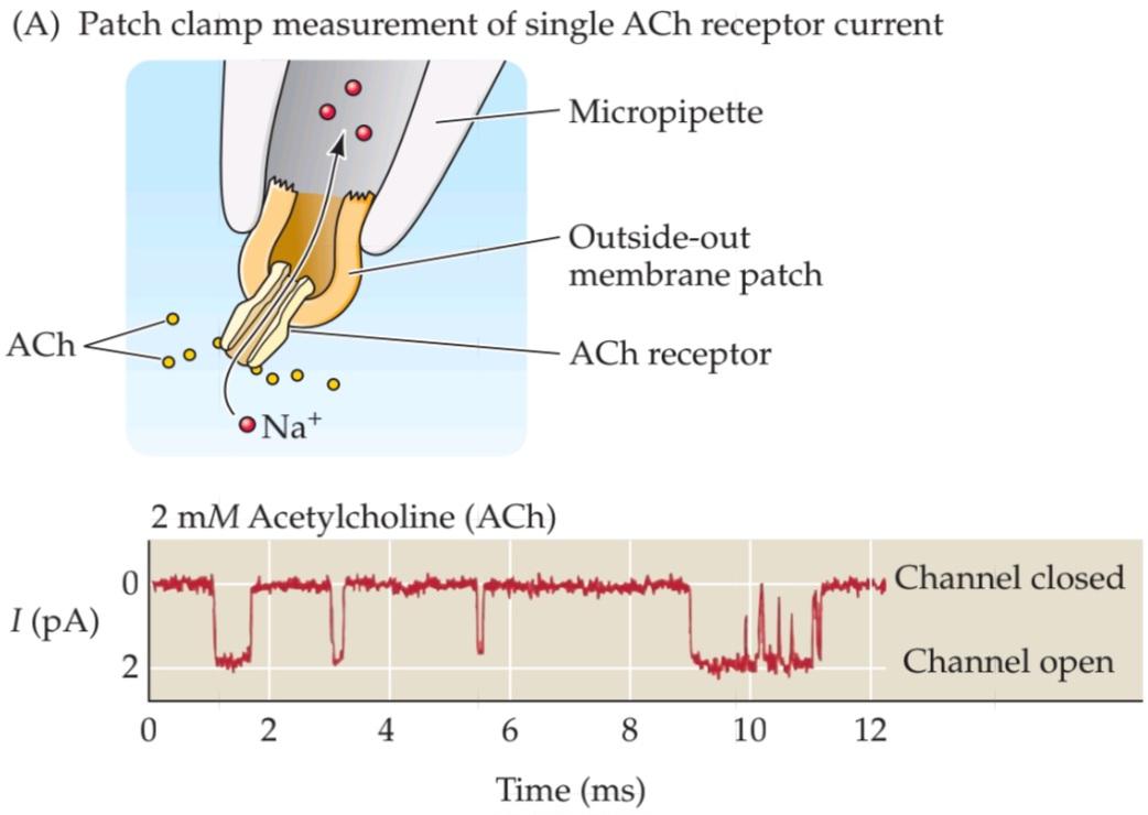


FIGURE 5.15 Activation of ACh receptors at neuromuscular synapses. (A) Patch clamp measurement of single ACh receptor currents from a patch of membrane removed from the postsynaptic muscle cell. When ACh is applied to the extracellular surface of the membrane, the repeated brief opening of a single channel can be observed as downward deflections corresponding to inward current (i.e., positive ions flowing into the cell). (B) Synchronized opening of many ACh-activated channels at a synapse being voltage clamped. (1) If a single channel is examined during the release of ACh from the presynaptic terminal, the channel can be seen to open transiently. (2) If several channels are examined together, ACh release opens the channels almost synchronously. (3) The opening of a very large number of postsynaptic channels produces a macroscopic EPC. (C) In a normal muscle cell (i.e., not being voltage clamped), the inward EPC depolarizes the postsynaptic muscle cell, giving rise to an EPP. Typically, this depolarization generates an action potential (not shown; see Figure 5.5B).
شکل ۵.۱۵ فعال شدن گیرندههای ACh در سیناپسهای عصبی-عضلانی. (الف) اندازهگیری جریانهای گیرنده ACh منفرد از یک تکه غشاء که از سلول عضلانی پس سیناپسی جدا شده است، با استفاده از گیره وصلهای. هنگامی که ACh به سطح خارج سلولی غشاء اعمال میشود، باز شدن کوتاه و مکرر یک کانال منفرد را میتوان به صورت انحرافهای رو به پایین مربوط به جریان داخلی (یعنی یونهای مثبت که به داخل سلول جریان مییابند) مشاهده کرد. (ب) باز شدن همزمان بسیاری از کانالهای فعال شده توسط ACh در سیناپسی که تحت فشار ولتاژ قرار دارد. (1) اگر یک کانال منفرد در حین آزادسازی ACh از پایانه پیش سیناپسی بررسی شود، میتوان مشاهده کرد که کانال به صورت گذرا باز میشود. (2) اگر چندین کانال با هم بررسی شوند، آزادسازی ACh کانالها را تقریباً همزمان باز میکند. (3) باز شدن تعداد بسیار زیادی از کانالهای پس سیناپسی یک EPC ماکروسکوپی ایجاد میکند. (ج) در یک سلول عضلانی طبیعی (یعنی بدون محدودیت ولتاژ)، EPC داخلی، سلول عضلانی پس سیناپسی را دپلاریزه میکند و منجر به EPP میشود. معمولاً این دپلاریزاسیون یک پتانسیل عمل ایجاد میکند (نشان داده نشده است؛ به شکل 5.5B مراجعه کنید).
The identity of the ions that flow during the EPC can be determined via the same approaches used to identify the roles of Na+ and K+ fluxes in the currents underlying action potentials (see Chapter 3). Key to such an analysis is identifying the membrane potential at which no current flows during transmitter action. When the potential of the postsynaptic muscle cell is controlled by the voltage clamp method (Figure 5.16A), the magnitude of the membrane potential clearly affects the amplitude and polarity of EPCs (Figure 5.16B). Thus, when the postsynaptic membrane potential is made more negative than the resting potential, the amplitude of the EPC becomes larger, whereas this current is reduced when the membrane potential is made more positive. At approximately 0 mV, no EPC is detected, and at even more positive potentials, the current reverses its polarity, becoming outward rather than inward (Figure 5.16C). The potential where the EPC reverses, about 0 mV in the case of the neuromuscular junction, is called the reversal potential.
هویت یونهایی که در طول EPC جریان مییابند را میتوان از طریق همان رویکردهایی که برای شناسایی نقش شارهای +Na و +K در جریانهای زمینهساز پتانسیلهای عمل استفاده میشوند، تعیین کرد (به فصل 3 مراجعه کنید). کلید چنین تحلیلی، شناسایی پتانسیل غشایی است که در آن هیچ جریانی در طول عمل فرستنده جریان نمییابد. هنگامی که پتانسیل سلول ماهیچه پس سیناپسی با روش گیره ولتاژ کنترل میشود (شکل 5.16A)، بزرگی پتانسیل غشا به وضوح بر دامنه و قطبیت EPCها تأثیر میگذارد (شکل 5.16B). بنابراین، هنگامی که پتانسیل غشای پس سیناپسی منفیتر از پتانسیل استراحت میشود، دامنه EPC بزرگتر میشود، در حالی که این جریان با مثبتتر شدن پتانسیل غشا کاهش مییابد. در تقریباً 0 میلیولت، هیچ EPC شناسایی نمیشود و در پتانسیلهای حتی مثبتتر، جریان قطبیت خود را معکوس میکند و به جای اینکه به سمت داخل باشد، به سمت خارج میرود (شکل 5.16C). پتانسیلی که در آن EPC معکوس میشود، حدود 0 میلیولت در مورد اتصال عصبی-عضلانی، پتانسیل معکوس نامیده میشود.
As was the case for currents flowing through voltage-gated ion channels (see Chapter 4), the magnitude of the EPC at any membrane potential is given by the product of the ion conductance activated by ACh (gACh) and the electrochemical driving force on the ions flowing through ligand-gated channels. Thus, the value of the EPC is given by the relationship
همانطور که در مورد جریانهای عبوری از کانالهای یونی دریچهدار ولتاژی صدق میکرد (به فصل ۴ مراجعه کنید)، مقدار EPC در هر پتانسیل غشایی توسط حاصلضرب رسانایی یونی فعال شده توسط ACh (gACh) و نیروی محرکه الکتروشیمیایی روی یونهای عبوری از کانالهای دریچهدار لیگاندی بدست میآید. بنابراین، مقدار EPC توسط رابطه زیر بدست میآید:

where Erev is the reversal potential for the EPC. This relationship predicts that the EPC will be an inward current at potentials more negative than Erev because the electrochemical driving force, Vm – Erev , is a negative number. Furthermore, the EPC will become smaller at potentials approaching Erev because the driving force is reduced. At potentials more positive than Erev , the EPC is outward because the driving force is reversed in direction (that is, positive). Because the channels opened by ACh are largely insensitive to membrane voltage, gACh will depend only on the number of channels opened by ACh, which depends in turn on the concentration of ACh in the synaptic cleft. Thus, the magnitude and polarity of the postsynaptic membrane potential determine the direction and amplitude of the EPC solely by altering the driving force on ions flowing through the receptor channels opened by ACh.
که در آن Erev پتانسیل معکوس برای EPC است. این رابطه پیشبینی میکند که EPC در پتانسیلهای منفیتر از Erev یک جریان رو به داخل خواهد بود زیرا نیروی محرکه الکتروشیمیایی، Vm – Erev، یک عدد منفی است. علاوه بر این، EPC در پتانسیلهای نزدیک به Erev کوچکتر میشود زیرا نیروی محرکه کاهش مییابد. در پتانسیلهای مثبتتر از Erev، EPC به سمت خارج است زیرا نیروی محرکه در جهت معکوس (یعنی مثبت) است. از آنجا که کانالهای باز شده توسط ACh تا حد زیادی به ولتاژ غشا حساس نیستند، gACh فقط به تعداد کانالهای باز شده توسط ACh بستگی دارد که به نوبه خود به غلظت ACh در شکاف سیناپسی بستگی دارد. بنابراین، بزرگی و قطبیت پتانسیل غشای پس سیناپسی، جهت و دامنه EPC را تنها با تغییر نیروی محرکه روی یونهای جاری از طریق کانالهای گیرنده باز شده توسط ACh تعیین میکند.
When Vm is at the reversal potential, Vm – Erev is equal to 0 and there is no net driving force on the ions that can permeate the receptor-activated channel. The identity of the ions that flow during the EPC can be deduced by observing how the reversal potential of the EPC compares with the equilibrium potential for various ion species (Figure 5.16D). For example, if ACh were to open an ion channel permeable only to K+, then the reversal potential of the EPC would be at the equilibrium potential for K+, which for a muscle cell is close to –100 mV. If the ACh-activated channels were permeable only to Na+, then the reversal potential of the current would be approximately +70 mV, the Na+ equilibrium potential of muscle cells; if these channels were permeable only to Cl–, then the reversal potential would be approximately –50 mV. By this reasoning, ACh-activated channels cannot be permeable to only one of these ions, because the reversal potential of the EPC is not near the equilibrium potential for any of them (see Figure 5.16C). However, if these channels were permeable to both Na+ and K+, then the reversal potential of the EPC would be between +70 mV and –100 mV.
وقتی Vm در پتانسیل معکوس باشد، Vm – Erev برابر با 0 است و هیچ نیروی محرکه خالصی بر روی یونهایی که میتوانند از کانال فعالشده توسط گیرنده عبور کنند، وجود ندارد. هویت یونهایی که در طول EPC جریان مییابند را میتوان با مشاهده چگونگی مقایسه پتانسیل معکوس EPC با پتانسیل تعادل برای گونههای مختلف یونی استنباط کرد (شکل 5.16D). به عنوان مثال، اگر ACh یک کانال یونی را که فقط به +K نفوذپذیر است باز کند، پتانسیل معکوس EPC در پتانسیل تعادل برای +K خواهد بود، که برای یک سلول عضلانی نزدیک به -100 میلیولت است. اگر کانالهای فعالشده توسط ACh فقط به +Na نفوذپذیر باشند، پتانسیل معکوس جریان تقریباً 70+ میلیولت، پتانسیل تعادل +Na سلولهای عضلانی، خواهد بود. اگر این کانالها فقط به Cl- نفوذپذیر باشند، پتانسیل معکوس تقریباً 50- میلیولت خواهد بود. با این استدلال، کانالهای فعالشده با ACh نمیتوانند فقط به یکی از این یونها نفوذپذیر باشند، زیرا پتانسیل برگشت EPC نزدیک به پتانسیل تعادل برای هیچکدام از آنها نیست (شکل 5.16C را ببینید). با این حال، اگر این کانالها به هر دو یون +Na و +K نفوذپذیر باشند، پتانسیل برگشت EPC بین 70+ میلیولت و 100- میلیولت خواهد بود.


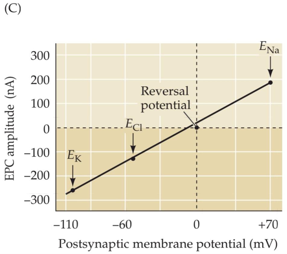

FIGURE 5.16 Influence of the postsynaptic membrane potential on end plate currents. (A) A postsynaptic muscle fiber is voltage clamped using two electrodes, while the presynaptic neuron is electrically stimulated to cause the release of ACh from presynaptic terminals. This experimental arrangement allows the recording of EPCs produced by ACh. (B) Amplitude and time course of EPCs generated by stimulating the presynaptic motor neuron while the postsynaptic cell is voltage clamped at four different membrane potentials. (C) The relationship between the peak amplitude of EPCs and postsynaptic membrane potential is nearly linear, with a reversal potential (the voltage at which the direction of the current changes from inward to outward) close to 0 mV. Also indicated on this graph are the equilibrium potentials of Na+, K+, and Cl– ions. (D) The identity of the ions permeating postsynaptic receptors is revealed by the reversal potential (Erev). Activation of postsynaptic channels permeable only to K+ (yellow) results in currents reversing at E , near –100 mV, while activation of terminalspostsynaptic Na+ channels results in currents reversing at ENa, near +70 mV (red). Cl–-selective currents reverse at ECl, near –50 mV (green). (A–C after Takeuchi and Takeuchi, 1960.)
شکل ۵.۱۶ تأثیر پتانسیل غشای پس سیناپسی بر جریانهای صفحه انتهایی. (الف) یک فیبر عضلانی پس سیناپسی با استفاده از دو الکترود تحت ولتاژ ثابت قرار میگیرد، در حالی که نورون پیشسیناپسی به صورت الکتریکی تحریک میشود تا باعث آزاد شدن استیل کولین از پایانههای پیشسیناپسی شود. این چیدمان آزمایشگاهی امکان ثبت EPCهای تولید شده توسط استیل کولین را فراهم میکند. (ب) دامنه و سیر زمانی EPCهای تولید شده با تحریک نورون حرکتی پیشسیناپسی در حالی که سلول پس سیناپسی در چهار پتانسیل غشایی مختلف تحت ولتاژ ثابت قرار میگیرد. (ج) رابطه بین دامنه اوج EPCها و پتانسیل غشای پس سیناپسی تقریباً خطی است، با پتانسیل معکوس (ولتاژی که در آن جهت جریان از داخل به خارج تغییر میکند) نزدیک به 0 میلیولت. همچنین در این نمودار پتانسیلهای تعادلی یونهای +Na+، K و Cl- نشان داده شده است. (د) هویت یونهای نفوذکننده به گیرندههای پس سیناپسی توسط پتانسیل معکوس (Erev) آشکار میشود. فعال شدن کانالهای پسسیناپسی که فقط به +K نفوذپذیر هستند (زرد) منجر به معکوس شدن جریانها در E، نزدیک به 100- میلیولت میشود، در حالی که فعال شدن کانالهای پسسیناپسی +Na ترمینال منجر به معکوس شدن جریانها در ENa، نزدیک به 70+ میلیولت (قرمز) میشود. جریانهای انتخابی Cl- در ECl، نزدیک به 50- میلیولت (سبز) معکوس میشوند. (A-C برگرفته از Takeuchi و Takeuchi، 1960.)
The fact that EPCs reverse at approximately 0 mV is therefore consistent with the idea that ACh-activated ion channels are almost equally permeable to both Na+ and K+. This hypothesis was tested in 1960, by the Japanese husband and wife team of Akira and Noriko Takeuchi, by altering the extracellular concentration of these two ions. As predicted, the magnitude and reversal potential of the EPC were changed by altering the concentration gradient of each ion. Lowering the external Na+ concentration, which makes ENa more negative, produces a negative shift in Erev (Figure 5.17A), whereas elevating external K+ concentration, which makes EK more positive, causes Erev to shift to a more positive potential (Figure 5.17B). Such experiments establish that the ACh-activated ion channels are in fact permeable to both Na+ and K+.
این واقعیت که EPCها تقریباً در 0 میلیولت معکوس میشوند، با این ایده سازگار است که کانالهای یونی فعالشده با ACh تقریباً به طور مساوی نسبت به +Na و +K نفوذپذیر هستند. این فرضیه در سال 1960 توسط تیم زن و شوهر ژاپنی آکیرا و نوریکو تاکوچی، با تغییر غلظت خارج سلولی این دو یون، مورد آزمایش قرار گرفت. همانطور که پیشبینی میشد، بزرگی و پتانسیل معکوس EPC با تغییر گرادیان غلظت هر یون تغییر کرد. کاهش غلظت +Na خارجی، که ENa را منفیتر میکند، باعث تغییر منفی در Erev میشود (شکل 5.17A)، در حالی که افزایش غلظت +K خارجی، که EK را مثبتتر میکند، باعث میشود Erev به پتانسیل مثبتتری تغییر کند (شکل 5.17B). چنین آزمایشهایی نشان میدهند که کانالهای یونی فعالشده با ACh در واقع نسبت به +Na و +K نفوذپذیر هستند.

FIGURE 5.17 Reversal potential of the end plate current changes when ion gradients change. (A) Lowering the external Na+ concentration causes EPCs to reverse at more negative potentials. (B) Raising the external K+ concentration makes the reversal potential more positive. (After Takeuchi and Takeuchi, 1960.)
شکل ۵.۱۷ پتانسیل معکوس جریان صفحه انتهایی با تغییر گرادیان یونی تغییر میکند. (الف) کاهش غلظت +Na خارجی باعث میشود EPCها در پتانسیلهای منفیتر معکوس شوند. (ب) افزایش غلظت +K خارجی پتانسیل معکوس را مثبتتر میکند. (برگرفته از Takeuchi و Takeuchi، ۱۹۶۰.)
Relationship between Ion Fluxes and Postsynaptic Potential Changes
🚀 با ما همراه شوید!
تازهترین مطالب و آموزشهای مغز و اعصاب را از دست ندهید. با فالو کردن کانال تلگرام آیندهنگاران مغز، از ما حمایت کنید!


 ورود/ ثبت نام با جیمیل
