
وریدهای مغزی، حلقه ویلیس و گردش خون مغزی


دعای مطالعه [ نمایش ]
بِسْمِ الله الرَّحْمنِ الرَّحیمِ
اَللّهُمَّ اَخْرِجْنى مِنْ ظُلُماتِ الْوَهْمِ
خدايا مرا بيرون آور از تاريكىهاى وهم،
وَ اَكْرِمْنى بِنُورِ الْفَهْمِ
و به نور فهم گرامى ام بدار،
اَللّهُمَّ افْتَحْ عَلَيْنا اَبْوابَ رَحْمَتِكَ
خدايا درهاى رحمتت را به روى ما بگشا،
وَانْشُرْ عَلَيْنا خَزائِنَ عُلُومِكَ بِرَحْمَتِكَ يا اَرْحَمَ الرّاحِمينَ
و خزانههاى علومت را بر ما باز كن به امید رحمتت اى مهربانترين مهربانان.
آیندهنگاران مغز: «ما مغز را میشناسیم، تا آینده را بسازیم.»
کتاب «نوروآناتومی بالینی اسنل» اثر دکتر ریچارد اس. اسنل، با ویرایش جدید دکتر رایان اسپلیتگربر، همچنان بهعنوان مرجع طلایی این حوزه شناخته میشود. این اثر با تلفیق تازهترین پژوهشها و کاربردهای بالینی، پلی استوار میان دانش پایه و عمل پزشکی بنا میکند.
تلاشهای علمی و سالها تدریس ارزشمند استاد گرانقدر آقای دکتر محمدتقی جغتایی الهامبخش ترجمه و گسترش این دانش در ایران بوده است.
ترجمه دقیق این کتاب توسط برند علمی آیندهنگاران مغز به مدیریت داریوش طاهری، امکان دسترسی فارسیزبانان به مرزهای نوین علوم اعصاب را فراهم کرده و رسالتی علمی در جهت شناخت عمیقتر مغز و ترسیم آیندهای روشنتر بر عهده گرفته است.
»» فصل ۱۷: خونرسانی به مغز و نخاع؛ بخش مبانی و اصول؛ قسمت دوم
»» Chapter 17: Blood Supply of the Braint and Spinal Cord
VEINS OF THE BRAIN
The veins of the brain have no muscular tissue in their very thin walls, and they possess no valves. They emerge from the brain and lie in the subarachnoid space. They pierce the arachnoid mater and the meningeal layer of the dura and drain into the cranial venous sinuses (Fig. 17-5).
وریدهای مغز
وریدهای مغز در دیوارههای بسیار نازک خود بافت عضلانی ندارند و دریچهای ندارند. آنها از مغز خارج میشوند و در فضای زیر عنکبوتیه قرار میگیرند. آنها ماده عنکبوتیه و لایه مننژی سختشامه را سوراخ میکنند و به سینوسهای وریدی جمجمهای تخلیه میشوند (شکل 17-5).
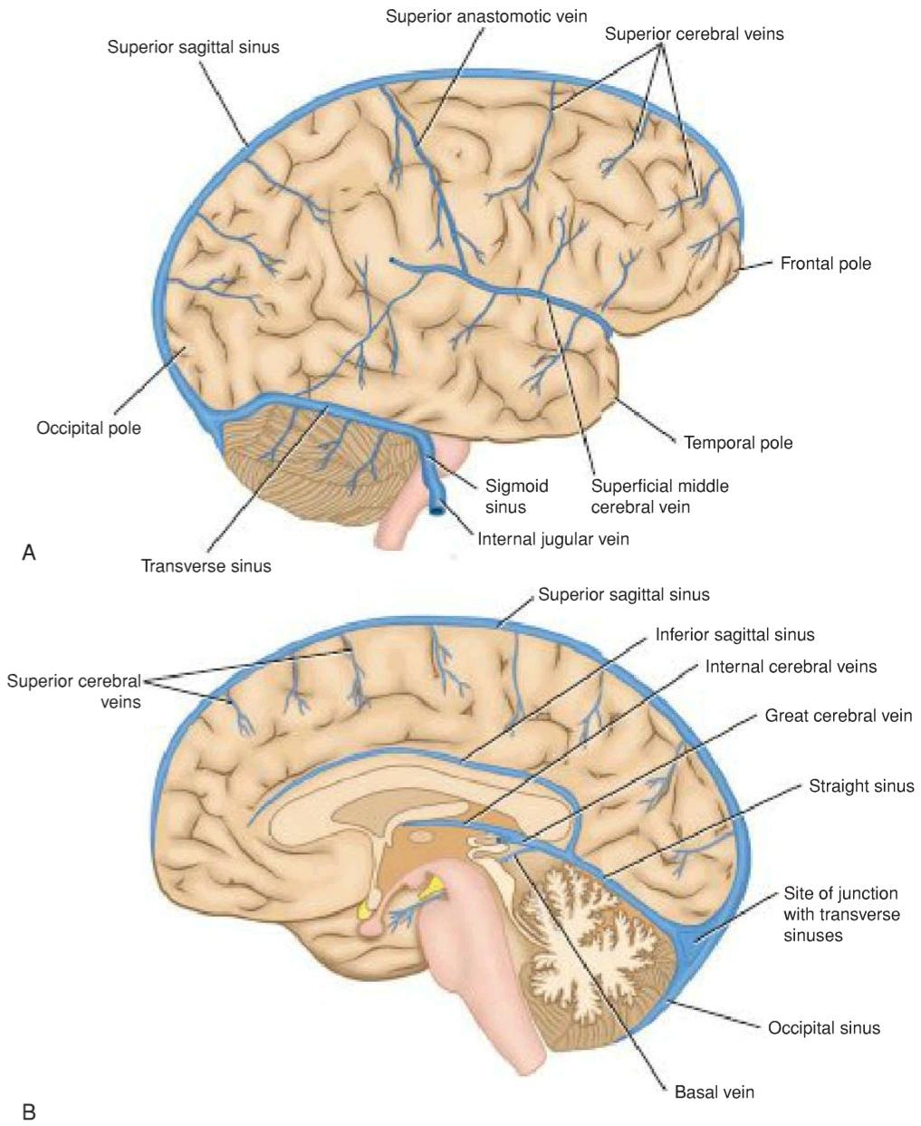
Figure 17-5 Venous drainage of the right cerebral hemisphere. A: Lateral surface. B: Medial surface.
شکل ۱۷-۵ تخلیه وریدی نیمکره راست مغز. الف: سطح جانبی. ب: سطح داخلی.
External Cerebral Veins
The superior cerebral veins pass upward over the lateral surface of the cerebral hemisphere and empty into the superior sagittal sinus.
وریدهای مغزی خارجی
وریدهای مغزی فوقانی از سطح جانبی نیمکره مغزی به سمت بالا عبور کرده و به سینوس ساژیتال فوقانی تخلیه میشوند.
The superficial middle cerebral vein drains the lateral surface of the cerebral hemisphere. It runs inferiorly in the lateral sulcus and empties into the cavernous sinus.
ورید مغزی میانی سطحی، سطح جانبی نیمکره مغزی را تخلیه میکند. این ورید در شیار جانبی به سمت پایین امتداد یافته و به سینوس غاری تخلیه میشود.
The deep middle cerebral vein drains the insula and is joined by the anterior cerebral and striate veins to form the basal vein. The basal vein ultimately joins the great cerebral vein, which in turn drains into the straight sinus.
ورید مغزی میانی عمقی، اینسولا را تخلیه میکند و توسط وریدهای مغزی قدامی و مخطط به هم متصل شده و ورید قاعدهای را تشکیل میدهد. ورید قاعدهای در نهایت به ورید بزرگ مغزی میپیوندد که به نوبه خود به سینوس مستقیم تخلیه میشود.
Internal Cerebral Veins
The two internal cerebral veins are formed by the union of the thalamostriate vein and the choroid vein at the interventricular foramen. The two veins run posteriorly in the tela choroidea of the third ventricle and unite beneath the splenium of the corpus callosum to form the great cerebral vein, which empties into the straight sinus.
وریدهای مغزی داخلی
دو ورید مغزی داخلی از به هم پیوستن ورید تالاموستریا و ورید مشیمیه در سوراخ بین بطنی تشکیل میشوند. این دو ورید در قسمت خلفی تلا مشیمیه بطن سوم امتداد یافته و در زیر اسپلنیوم جسم پینهای به هم میپیوندند و ورید مغزی بزرگ را تشکیل میدهند که به سینوس مستقیم تخلیه میشود.
Veins of Specific Brain Areas
The midbrain is drained by veins that open into the basal or great cerebral veins.
رگهای نواحی خاص مغز
مغز میانی توسط رگهایی که به رگهای قاعدهای یا بزرگ مغزی باز میشوند، تخلیه میشود.
The pons is drained by veins that open into the basal vein, cerebellar veins, or neighboring venous sinuses.
پل مغزی توسط رگهایی که به رگ قاعدهای، رگهای مخچهای یا سینوسهای وریدی مجاور باز میشوند، تخلیه میشود.
The medulla oblongata is drained by veins that open into the spinal veins and neighboring venous sinuses.
بصل النخاع توسط رگهایی که به رگهای نخاعی و سینوسهای وریدی مجاور باز میشوند، تخلیه میشود.
The cerebellum is drained by veins that empty into the great cerebral vein or adjacent venous sinuses.
مخچه توسط رگهایی که به رگ بزرگ مغزی یا سینوسهای وریدی مجاور تخلیه میشوند، تخلیه میشود.
BRAIN CAPILLARIES
The capillary blood supply to the brain is greater in the gray matter than in the white matter. This is to be expected, since the metabolic activity in the neuronal cell bodies in the gray matter is much greater than in the nerve processes in the white matter. The blood- brain barrier isolates the brain tissue from the rest of the body and is formed by the tight junctions that exist between the endothelial cells in the capillary beds (see pp. 452-453).
مویرگهای مغز
خونرسانی مویرگی به مغز در ماده خاکستری بیشتر از ماده سفید است. این قابل انتظار است، زیرا فعالیت متابولیک در اجسام سلولی نورونی در ماده خاکستری بسیار بیشتر از فرآیندهای عصبی در ماده سفید است. سد خونی-مغزی، بافت مغز را از بقیه بدن جدا میکند و توسط اتصالات محکمی که بین سلولهای اندوتلیال در بسترهای مویرگی وجود دارد، تشکیل میشود (به صفحات ۴۵۲-۴۵۳ مراجعه کنید).
CEREBRAL CIRCULATION
The blood flow to the brain must deliver oxygen, glucose, and other nutrients to the nervous tissue and remove carbon dioxide, lactic acid, and other metabolic by-products. The brain has been shown to be supplied with arterial blood from the two internal carotid arteries and the two vertebral arteries. The blood supply to half of the brain is provided by the internal carotid and vertebral arteries on that side, and their respective streams come together in the posterior communicating artery at a point where the pressure of the two is equal and they do not mix (Fig. 17-6). If, however, the internal carotid or vertebral artery is occluded, the blood passes forward or backward across that point to compensate for the reduction in blood flow. The arterial circle also permits the blood to flow across the midline, as shown when the internal carotid or vertebral artery on one side is occluded. It also has been shown that the two streams of blood from the vertebral arteries remain separate and on the same side of the lumen of the basilar artery and do not mix.
گردش خون مغزی
جریان خون به مغز باید اکسیژن، گلوکز و سایر مواد مغذی را به بافت عصبی برساند و دی اکسید کربن، اسید لاکتیک و سایر محصولات جانبی متابولیک را از آن خارج کند. نشان داده شده است که مغز از طریق دو شریان کاروتید داخلی و دو شریان مهرهای، خون شریانی دریافت میکند. خونرسانی به نیمی از مغز توسط شریانهای کاروتید داخلی و مهرهای در آن طرف انجام میشود و جریانهای مربوط به آنها در شریان رابط خلفی در نقطهای که فشار این دو برابر است و با هم مخلوط نمیشوند، به هم میرسند (شکل 17-6). با این حال، اگر شریان کاروتید داخلی یا مهرهای مسدود شود، خون از آن نقطه به جلو یا عقب عبور میکند تا کاهش جریان خون را جبران کند. حلقه شریانی همچنین اجازه میدهد خون از خط وسط عبور کند، همانطور که هنگام انسداد شریان کاروتید داخلی یا مهرهای در یک طرف نشان داده شده است. همچنین نشان داده شده است که دو جریان خون از شریانهای مهرهای جدا و در یک طرف مجرای شریان بازیلار باقی میمانند و با هم مخلوط نمیشوند.
Although the cerebral arteries anastomose with one another at the circle of Willis and by means of branches on the surface of the cerebral hemispheres, once they enter the brain substance, no further anastomoses occur.
اگرچه شریانهای مغزی در حلقه ویلیس و از طریق شاخههایی روی سطح نیمکرههای مغزی با یکدیگر آناستوموز میکنند، اما پس از ورود به ماده مغز، دیگر آناستوموزی رخ نمیدهد.
The most important factor in forcing the blood through the brain is the arterial blood pressure. This is opposed by such factors as a raised intracranial pressure, increased blood viscosity, and narrowing of the vascular diameter. Cerebral blood flow remains remarkably constant despite changes in the general blood pressure. This autoregulation of the circulation is accomplished by a compensatory lowering of the cerebral vascular resistance when the arterial pressure is decreased and a rising of the vascular resistance when the arterial pressure is increased. Needless to say, this autoregulation does not maintain an adequate blood flow when the arterial blood pressure falls to a very low level.
مهمترین عامل در عبور اجباری خون از مغز، فشار خون شریانی است. این امر با عواملی مانند افزایش فشار داخل جمجمه، افزایش ویسکوزیته خون و باریک شدن قطر عروق در تضاد است. جریان خون مغزی علیرغم تغییرات فشار خون عمومی، به طور قابل توجهی ثابت میماند. این خودتنظیمی گردش خون با کاهش جبرانی مقاومت عروق مغزی هنگام کاهش فشار شریانی و افزایش مقاومت عروق هنگام افزایش فشار شریانی انجام میشود. نیازی به گفتن نیست که این خودتنظیمی وقتی فشار خون شریانی به سطح بسیار پایینی میرسد، جریان خون کافی را حفظ نمیکند.
The diameter of the cerebral blood vessels is the main factor contributing to the cerebrovascular resistance. While cerebral blood vessels are innervated by sympathetic postganglionic nerve fibers and respond to norepinephrine, they apparently play little or no part in the control of cerebrovascular resistance in normal human beings. The most powerful vasodilator influence on cerebral blood vessels is an increase in carbon dioxide or hydrogen ion concentration; a reduction in oxygen concentration also causes vasodilatation. It has been shown, using PET, that an increase in neuronal activity in different parts of the brain causes a local increase in blood flow. For example, viewing an object will increase the oxygen and glucose consumption in the visual cortex of the occipital lobes. This results in an increase in the local concentrations of carbon dioxide and hydrogen ions and brings about a local increase in blood flow.
قطر رگهای خونی مغزی عامل اصلی مؤثر در مقاومت عروقی مغز است. اگرچه رگهای خونی مغزی توسط فیبرهای عصبی پسگانگلیونی سمپاتیک عصبدهی میشوند و به نوراپی نفرین پاسخ میدهند، اما ظاهراً در کنترل مقاومت عروقی مغز در افراد عادی نقش کمی دارند یا اصلاً نقشی ندارند. قویترین اثر گشادکننده عروق بر رگهای خونی مغزی، افزایش غلظت دیاکسید کربن یا یون هیدروژن است. کاهش غلظت اکسیژن نیز باعث گشاد شدن عروق میشود. با استفاده از PET نشان داده شده است که افزایش فعالیت عصبی در قسمتهای مختلف مغز باعث افزایش موضعی جریان خون میشود. به عنوان مثال، مشاهده یک شیء، مصرف اکسیژن و گلوکز را در قشر بینایی لوبهای پسسری افزایش میدهد. این امر منجر به افزایش غلظت موضعی دیاکسید کربن و یونهای هیدروژن و افزایش موضعی جریان خون میشود.
The cerebral blood flow in patients can be measured by the intracarotid injection or inhalation of radioactive krypton or xenon. Normal cerebral blood flow, per minute, is around 50 to 60 mL/100g of brain tissue.
جریان خون مغزی در بیماران را میتوان با تزریق داخل کاروتید یا استنشاق کریپتون یا زنون رادیواکتیو اندازهگیری کرد. جریان خون طبیعی مغز، در هر دقیقه، حدود ۵۰ تا ۶۰ میلیلیتر در هر ۱۰۰ گرم بافت مغز است.

Figure 17-6 Circle of Willis showing the distribution of blood from the four main arteries.
شکل ۱۷-۶ دایره ویلیس که توزیع خون از چهار شریان اصلی را نشان میدهد.
«فصل هفدهم کتاب نورواناتومی بالینی اسنل»
🚀 با ما همراه شوید!
تازهترین مطالب و آموزشهای مغز و اعصاب را از دست ندهید. با فالو کردن کانال تلگرام آیندهنگاران مغز، از ما حمایت کنید!



