
فیزیولوژی پزشکی گایتون و هال؛ متابولیسم پروتئین


دعای مطالعه [ نمایش ]
بِسْمِ الله الرَّحْمنِ الرَّحیمِ
اَللّهُمَّ اَخْرِجْنى مِنْ ظُلُماتِ الْوَهْمِ
خدايا مرا بيرون آور از تاريكىهاى وهم،
وَ اَكْرِمْنى بِنُورِ الْفَهْمِ
و به نور فهم گرامى ام بدار،
اَللّهُمَّ افْتَحْ عَلَيْنا اَبْوابَ رَحْمَتِكَ
خدايا درهاى رحمتت را به روى ما بگشا،
وَانْشُرْ عَلَيْنا خَزائِنَ عُلُومِكَ بِرَحْمَتِكَ يا اَرْحَمَ الرّاحِمينَ
و خزانههاى علومت را بر ما باز كن به امید رحمتت اى مهربانترين مهربانان.
کتاب «فیزیولوژی پزشکی گایتون و هال» بهعنوان یکی از جامعترین و معتبرترین منابع در حوزه علوم پزشکی، همچنان مرجع کلیدی برای درک عملکرد پیچیده بدن انسان است. این اثر با تکیه بر تازهترین پژوهشها و توضیحات دقیق از سازوکارهای فیزیولوژیک، پلی میان علوم پایه پزشکی و کاربردهای بالینی ایجاد میکند و نقشی بیبدیل در آموزش، پژوهش و ارتقای دانش سلامت ایفا مینماید.
ترجمه دقیق و علمی این شاهکار توسط برند علمی آیندهنگاران مغز به مدیریت داریوش طاهری، دسترسی فارسیزبانان به مرزهای نوین دانش فیزیولوژی را ممکن ساخته و رسالتی علمی برای ارتقای آموزش پزشکی، فهم عمیقتر سازوکارهای بدن و توسعه روشهای نوین در حوزه سلامت فراهم آورده است.
» Guyton and Hall Textbook of Medical Physiology
»» CHAPTER 70: Protein Metabolism

About three-quarters of the body solids are proteins. These proteins include structural proteins, enzymes, nucleoproteins, proteins that transport oxygen, proteins of the muscle that cause muscle contraction, and many other types that perform specific intracellular and extra- cellular functions throughout the body.
حدود سه چهارم مواد جامد بدن پروتئین هستند. این پروتئینها شامل پروتئینهای ساختاری، آنزیمها، نوکلئوپروتئینها، پروتئینهایی که اکسیژن را انتقال میدهند، پروتئینهای ماهیچهای که باعث انقباض عضلانی میشوند و بسیاری از انواع دیگر که عملکردهای خاص درون سلولی و خارج سلولی را در سراسر بدن انجام میدهند، میشوند.
The basic chemical properties that explain the diverse functions of proteins are so extensive that they constitute a major portion of the entire discipline of biochemistry. For this reason, the current discussion is confined to a few specific aspects of protein metabolism that are important as background for other discussions in this text.
خواص شیمیایی اساسی که عملکردهای متنوع پروتئین ها را توضیح می دهد به قدری گسترده است که بخش عمده ای از کل رشته بیوشیمی را تشکیل می دهد. به همین دلیل، بحث فعلی به چند جنبه خاص از متابولیسم پروتئین محدود می شود که به عنوان پیش زمینه برای بحث های دیگر در این متن مهم هستند.
Basic Properties of Proteins
Amino Acids Are the Principal Constituents of Proteins
The principal constituents of proteins are amino acids. Twenty of these amino acids are present in the body proteins in significant quantities. Figure 70-1, which shows the chemical formulas of these 20 amino acids, demonstrates that they all have two features in common; each amino acid has an acidic group (-COOH) and a nitrogen atom attached to the molecule, usually represented by the amino group (-NH2).
خواص اساسی پروتئین ها
اسیدهای آمینه اجزای اصلی پروتئین ها هستند
اجزای اصلی پروتئین ها اسیدهای آمینه هستند. 20 مورد از این اسیدهای آمینه به مقدار قابل توجهی در پروتئین های بدن وجود دارد. شکل 70-1 که فرمول های شیمیایی این 20 اسید آمینه را نشان می دهد، نشان می دهد که همه آنها دو ویژگی مشترک دارند. هر اسید آمینه دارای یک گروه اسیدی (COOH-) و یک اتم نیتروژن متصل به مولکول است که معمولاً با گروه آمینه (NH2-) نشان داده می شود.
Peptide Linkages and Peptide Chains. The amino acids of proteins are aggregated into long chains by means of peptide linkages. The chemical nature of this linkage is demonstrated by the following reaction:
پیوندهای پپتیدی و زنجیره های پپتیدی. اسیدهای آمینه پروتئین ها با استفاده از پیوندهای پپتیدی به زنجیره های بلند ادغام می شوند. ماهیت شیمیایی این پیوند با واکنش زیر نشان داده می شود:

Note in this reaction that the nitrogen of the amino radical of one amino acid bonds with the carbon of the carboxyl radical of the other amino acid. A hydrogen ion is released from the amino radical, and a hydroxyl ion is released from the carboxyl radical; these two ions combine to form a molecule of water. After the peptide linkage has been formed, an amino radical and a carboxyl radical are still at opposite ends of the new, longer molecule. Each of these radicals is capable of combining with additional amino acids to form a peptide chain. Some complicated protein molecules have many thousands of amino acids combined by peptide linkages, and even the smallest protein molecule usually has more than 20 amino acids combined by peptide linkages. The average is about 400 amino acids.
در این واکنش توجه کنید که نیتروژن رادیکال آمینه یک اسید آمینه با کربن رادیکال کربوکسیل اسید آمینه دیگر پیوند می خورد. یک یون هیدروژن از رادیکال آمینه و یک یون هیدروکسیل از رادیکال کربوکسیل آزاد می شود. این دو یون با هم ترکیب می شوند و یک مولکول آب را تشکیل می دهند. پس از تشکیل پیوند پپتیدی، یک رادیکال آمینه و یک رادیکال کربوکسیل هنوز در انتهای مخالف مولکول جدید و طولانی تر قرار دارند. هر یک از این رادیکال ها قادر به ترکیب با اسیدهای آمینه اضافی برای تشکیل یک زنجیره پپتیدی هستند. برخی از مولکول های پیچیده پروتئین دارای هزاران اسید آمینه هستند که توسط پیوندهای پپتیدی ترکیب شده اند و حتی کوچکترین مولکول پروتئین معمولاً دارای بیش از 20 اسید آمینه است که توسط پیوندهای پپتیدی ترکیب شده اند. میانگین آن حدود 400 اسید آمینه است.
Other Linkages in Protein Molecules. Some protein molecules are composed of several peptide chains rather than a single chain, and these chains are bound to one another by other linkages, often by hydrogen bonding between the CO and NH radicals of the peptides, as follows:
پیوندهای دیگر در مولکول های پروتئین. برخی از مولکولهای پروتئین به جای یک زنجیره منفرد از چندین زنجیره پپتیدی تشکیل شدهاند و این زنجیرهها توسط پیوندهای دیگر، اغلب با پیوند هیدروژنی بین رادیکالهای CO و NH پپتیدها به یکدیگر متصل میشوند، به شرح زیر:
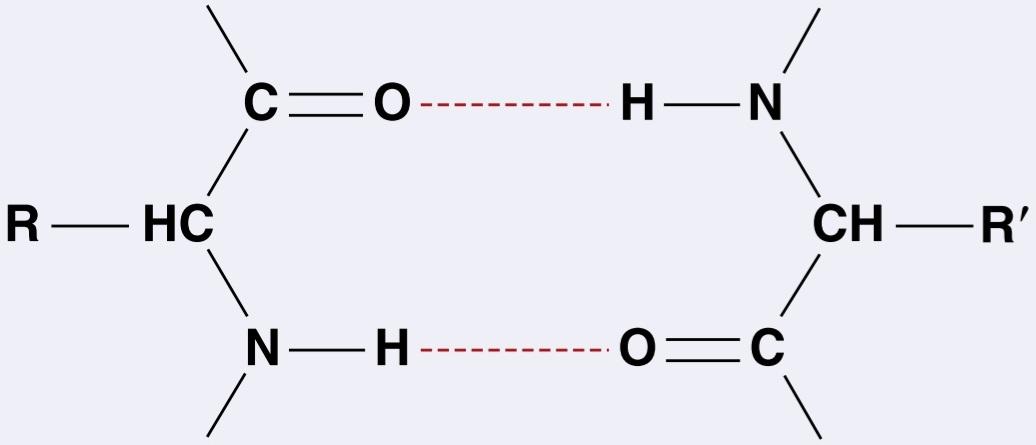
Many peptide chains are coiled or folded, and the successive coils or folds are held in a tight spiral or in other shapes by similar hydrogen bonding and other forces.
بسیاری از زنجیرههای پپتیدی مارپیچ یا تا شده هستند و سیمپیچها یا چینهای متوالی در یک مارپیچ محکم یا به اشکال دیگر توسط پیوند هیدروژنی مشابه و نیروهای دیگر نگه داشته میشوند.

Figure 70-1. Amino acids. The 10 essential amino acids cannot be synthesized in sufficient quantities in the body; these amino acids must be obtained, already formed, from food.
شکل 70-1. اسیدهای آمینه 10 اسید آمینه ضروری را نمی توان به مقدار کافی در بدن سنتز کرد. این آمینو اسیدها باید از مواد غذایی به دست آمده باشند.
Transport and Storage of Amino Acids
Blood Amino Acids
The normal concentration of amino acids in the blood is between 35 and 65 mg/dl, which is an average of about 2 mg/dl for each of the 20 amino acids, although some are present in far greater amounts than are others. Because the amino acids are relatively strong acids, they exist in the blood principally in the ionized state, as a result of the removal of one hydrogen atom from the NH2 radical. They actually account for 2 to 3 milliequivalents of the negative ions in the blood. The precise distribution of the different amino acids in the blood depends to some extent on the types of proteins eaten, but the concentrations of at least some individual amino acids are regulated by selective synthesis in the different cells.
حمل و نقل و نگهداری اسیدهای آمینه
اسیدهای آمینه خون
غلظت طبیعی اسیدهای آمینه در خون بین 35 تا 65 میلی گرم در دسی لیتر است که به طور متوسط برای هر یک از 20 اسید آمینه حدود 2 میلی گرم در دسی لیتر است، اگرچه برخی از آنها در مقادیر بسیار بیشتری نسبت به سایرین وجود دارند. از آنجایی که آمینو اسیدها اسیدهای نسبتاً قوی هستند، در خون عمدتاً در حالت یونیزه وجود دارند که در نتیجه حذف یک اتم هیدروژن از رادیکال NH2 است. آنها در واقع 2 تا 3 میلی اکی والان یون های منفی خون را تشکیل می دهند. توزیع دقیق اسیدهای آمینه مختلف در خون تا حدی به انواع پروتئین های خورده شده بستگی دارد، اما غلظت حداقل برخی از اسیدهای آمینه منفرد با سنتز انتخابی در سلول های مختلف تنظیم می شود.
Fate of Amino Acids Absorbed From the Gastrointestinal Tract. The products of protein digestion and absorption in the gastrointestinal tract are almost entirely amino acids; only rarely are polypeptides or whole protein molecules absorbed from the digestive tract into the blood. Soon after a meal, the amino acid concentration in a person’s blood rises, but the increase is usually only a few milligrams per deciliter, for two reasons: First, protein digestion and absorption are usually extended over 2 to 3 hours, which allows only small quantities of amino acids to be absorbed at a time. Second, after entering the blood, the additional amino acids are absorbed within 5 to 10 minutes by cells throughout the body, especially by the liver. Therefore, large concentrations of amino acids almost never accumulate in the blood and tissue fluids. Nevertheless, the turnover rate of the amino acids is so rapid that many grams of proteins can be carried from one part of the body to another in the form of amino acids each hour.
سرنوشت آمینو اسیدهای جذب شده از دستگاه گوارش. محصولات هضم و جذب پروتئین در دستگاه گوارش تقریباً به طور کامل اسیدهای آمینه هستند. به ندرت پلی پپتیدها یا مولکول های پروتئین کامل از دستگاه گوارش به خون جذب می شوند. بلافاصله پس از صرف غذا، غلظت آمینو اسید در خون فرد افزایش مییابد، اما این افزایش معمولاً تنها چند میلیگرم در دسی لیتر است، به دو دلیل: اول، هضم و جذب پروتئین معمولاً بین 2 تا 3 ساعت طول میکشد، که باعث میشود فقط مقادیر کمی از اسیدهای آمینه در یک زمان جذب شوند. دوم، پس از ورود به خون، اسیدهای آمینه اضافی در عرض 5 تا 10 دقیقه توسط سلول های سراسر بدن، به ویژه توسط کبد، جذب می شوند. بنابراین، غلظت زیادی از اسیدهای آمینه تقریباً هرگز در خون و مایعات بافتی تجمع نمی یابد. با این وجود، سرعت گردش اسیدهای آمینه به قدری سریع است که در هر ساعت می توان بسیاری از گرم پروتئین را به شکل اسیدهای آمینه از یک قسمت از بدن به قسمت دیگر منتقل کرد.
Active Transport of Amino Acids Into the Cells. The molecules of all the amino acids are much too large to diffuse readily through the pores of the cell membranes. Therefore, significant quantities of amino acids can move either inward or outward through the membranes only by facilitated transport or active transport using carrier mechanisms. The nature of some of the carrier mechanisms is not completely understood, but a few are discussed in Chapter 4.
انتقال فعال آمینو اسیدها به داخل سلول. مولکول های تمام آمینو اسیدها بسیار بزرگتر از آن هستند که به راحتی در منافذ غشای سلولی پخش شوند. بنابراین، مقادیر قابل توجهی از اسیدهای آمینه می توانند از طریق غشاها به سمت داخل یا خارج حرکت کنند، تنها با حمل و نقل آسان یا انتقال فعال با استفاده از مکانیسم های حامل. ماهیت برخی از مکانیسم های حامل به طور کامل درک نشده است، اما چند مورد در فصل 4 مورد بحث قرار گرفته است.
Renal Threshold for Amino Acids. In the kidneys, the different amino acids that are filtered by the glomerular capillaries can be reabsorbed through the proximal tubular epithelium by secondary active transport, which returns them to the blood. However, as is true of other active transport mechanisms in the renal tubules, there is an upper limit to the rate at which each type of amino acid can be transported. For this reason, when the concentration of a particular type of amino acid becomes too high in the plasma and glomerular filtrate, the excess that cannot be actively reabsorbed is lost into the urine.
آستانه کلیه برای اسیدهای آمینه. در کلیهها، اسیدهای آمینه مختلف که توسط مویرگهای گلومرولی فیلتر میشوند، میتوانند از طریق اپیتلیوم لولهای پروگزیمال توسط انتقال فعال ثانویه دوباره جذب شوند، که آنها را به خون باز میگرداند. با این حال، همانطور که در مورد سایر مکانیسم های انتقال فعال در لوله های کلیوی صادق است، یک حد بالایی برای سرعت انتقال هر نوع اسید آمینه وجود دارد. به همین دلیل، وقتی غلظت نوع خاصی از اسید آمینه در پلاسما و فیلتر گلومرولی بیش از حد بالا میرود، مقدار اضافی که نمیتواند به طور فعال دوباره جذب شود در ادرار از بین میرود.
Storage of Amino Acids as Proteins in the Cells
After entry into tissue cells, amino acids combine with one another by peptide linkages, under the direction of the cell’s messenger RNA and ribosomal system, to form cellular proteins. Therefore, the concentration of free amino acids inside most cells usually remains low, and storage of large quantities of free amino acids does not occur in the cells; instead, they are stored mainly in the form of actual proteins. However, many of these intracellular proteins can be rapidly decomposed again into amino acids under the influence of intracellular lysosomal digestive enzymes. These amino acids can then be transported back out of the cell into the blood. Special exceptions to this reversal process are the proteins in the chromosomes of the nucleus and the structural proteins such as collagen and muscle contractile proteins. These proteins do not participate significantly in this reverse digestion and transport back out of the cells.
ذخیره اسیدهای آمینه به عنوان پروتئین در سلول ها
پس از ورود به سلول های بافتی، اسیدهای آمینه با پیوندهای پپتیدی، تحت هدایت RNA پیام رسان سلول و سیستم ریبوزومی، با یکدیگر ترکیب می شوند و پروتئین های سلولی را تشکیل می دهند. بنابراین، غلظت آمینو اسیدهای آزاد در داخل اکثر سلول ها معمولاً کم می ماند و ذخیره مقادیر زیادی از اسیدهای آمینه آزاد در سلول ها رخ نمی دهد. در عوض، آنها عمدتاً به شکل پروتئین های واقعی ذخیره می شوند. با این حال، بسیاری از این پروتئینهای درون سلولی میتوانند به سرعت تحت تأثیر آنزیمهای گوارشی لیزوزومی داخل سلولی دوباره به اسیدهای آمینه تجزیه شوند. سپس این آمینو اسیدها می توانند به خارج از سلول به داخل خون منتقل شوند. استثناهای ویژه برای این فرآیند معکوس، پروتئینهای کروموزومهای هسته و پروتئینهای ساختاری مانند کلاژن و پروتئینهای انقباضی عضلانی هستند. این پروتئین ها به طور قابل توجهی در این هضم معکوس شرکت نمی کنند و به خارج از سلول منتقل می شوند.
Some tissues of the body participate in the storage of amino acids to a greater extent than do others. For example, the liver, which is a large organ and has special systems for processing amino acids, can store large quantities of rapidly exchangeable proteins, which is also true of the kidneys and the intestinal mucosa to a lesser extent.
برخی از بافت های بدن به میزان بیشتری نسبت به سایرین در ذخیره اسیدهای آمینه شرکت می کنند. به عنوان مثال، کبد که اندام بزرگی است و دارای سیستمهای ویژهای برای پردازش اسیدهای آمینه است، میتواند مقادیر زیادی پروتئینهای قابل تبادل سریع را ذخیره کند، که این امر در مورد کلیهها و مخاط روده نیز به میزان کمتری صدق میکند.
Amino Acid Release From Cells as a Means of Regulating Plasma Amino Acid Concentration. Whenever plasma amino acid concentrations fall below normal levels, the required amino acids are transported out of the cells to replenish their supply in the plasma. In this way, the plasma concentration of each type of amino acid is maintained at a reasonably constant value. Some of the hormones secreted by the endocrine glands are able to alter the balance between tissue proteins and circulating amino acids. For example, growth hormone and insulin increase the formation of tissue proteins, whereas adrenocortical glucocorticoid hormones increase the concentration of plasma amino acids.
انتشار اسید آمینه از سلول ها به عنوان وسیله ای برای تنظیم غلظت اسید آمینه پلاسما. هر گاه غلظت اسید آمینه پلاسما به زیر سطح نرمال برسد، آمینواسیدهای مورد نیاز به خارج از سلول ها منتقل می شوند تا ذخیره خود را در پلاسما دوباره پر کنند. به این ترتیب غلظت پلاسمایی هر نوع اسید آمینه در مقدار نسبتاً ثابتی حفظ می شود. برخی از هورمون های ترشح شده توسط غدد درون ریز قادرند تعادل بین پروتئین های بافتی و اسیدهای آمینه در گردش را تغییر دهند. به عنوان مثال، هورمون رشد و انسولین تشکیل پروتئین های بافتی را افزایش می دهند، در حالی که هورمون های گلوکوکورتیکوئیدی قشر آدرنال باعث افزایش غلظت اسیدهای آمینه پلاسما می شوند.
Reversible Equilibrium Between the Proteins in Different Parts of the Body. Because cellular proteins in the liver (and, to a much less extent, in other tissues) can be synthesized rapidly from plasma amino acids, and because many of these proteins can be degraded and returned to the plasma almost as rapidly, constant interchange and equilibrium occurs between the plasma amino acids and labile proteins in virtually all cells of the body. For example, if a particular tissue requires proteins, it can synthesize new proteins from amino acids of the blood; in turn, blood amino acids are replenished by degradation of proteins from other cells of the body, especially from liver cells. These effects are particularly noticeable in relation to protein synthesis in cancer cells. Cancer cells are often prolific users of amino acids; therefore, the proteins of the other cells can become markedly depleted.
تعادل برگشت پذیر بین پروتئین ها در قسمت های مختلف بدن. از آنجایی که پروتئین های سلولی در کبد (و به میزان بسیار کمتری در بافت های دیگر) می توانند به سرعت از اسیدهای آمینه پلاسما سنتز شوند، و از آنجا که بسیاری از این پروتئین ها می توانند تقریباً به سرعت تجزیه شده و به پلاسما بازگردانده شوند، تبادل و تعادل ثابت بین اسیدهای آمینه پلاسما در تمام سلول های تقریباً حساس بدن رخ می دهد. به عنوان مثال، اگر بافت خاصی به پروتئین نیاز داشته باشد، می تواند پروتئین های جدیدی را از اسیدهای آمینه خون سنتز کند. به نوبه خود، اسیدهای آمینه خون با تخریب پروتئین های دیگر سلول های بدن، به ویژه از سلول های کبد، دوباره پر می شوند. این اثرات به ویژه در رابطه با سنتز پروتئین در سلول های سرطانی قابل توجه است. سلول های سرطانی اغلب مصرف کنندگان پرکار اسیدهای آمینه هستند. بنابراین، پروتئین های سایر سلول ها می توانند به طور قابل توجهی کاهش پیدا کنند.
Upper Limit for the Storage of Proteins. Each type of cell has an upper limit with regard to the amount of proteins it can store. After all the cells have reached their limits, the excess amino acids still in the circulation are degraded into other products and used for energy, as discussed subsequently, or they are converted to fat or glycogen and stored in these forms.
حد بالایی برای ذخیره پروتئین. هر نوع سلول با توجه به مقدار پروتئینی که می تواند ذخیره کند، حد بالایی دارد. پس از اینکه همه سلولها به محدودیتهای خود رسیدند، اسیدهای آمینه اضافی که هنوز در گردش هستند به محصولات دیگر تجزیه میشوند و برای انرژی استفاده میشوند، همانطور که در ادامه بحث شد، یا به چربی یا گلیکوژن تبدیل میشوند و به این شکلها ذخیره میشوند.
Functional Roles of the Plasma Proteins
The major types of protein present in the plasma are albumin, globulin, and fibrinogen.
نقش های عملکردی پروتئین های پلاسما
انواع اصلی پروتئین موجود در پلاسما آلبومین، گلوبولین و فیبرینوژن هستند.
A major function of albumin is to provide colloid osmotic pressure in the plasma, which prevents plasma loss from the capillaries, as discussed in Chapter 16.
عملکرد اصلی آلبومین ایجاد فشار اسمزی کلوئیدی در پلاسما است که از از دست دادن پلاسما از مویرگ ها جلوگیری می کند، همانطور که در فصل 16 بحث شد.
The globulins perform several enzymatic functions in the plasma, but equally important, they are principally responsible for both the natural and acquired immunity of the body against invading organisms, as discussed in Chapter 35.
گلوبولینها چندین عملکرد آنزیمی را در پلاسما انجام میدهند، اما به همان اندازه مهم، آنها اساساً مسئول ایمنی طبیعی و اکتسابی بدن در برابر ارگانیسمهای مهاجم هستند، همانطور که در فصل 35 بحث شد.
Fibrinogen polymerizes into long fibrin threads during blood coagulation, thereby forming blood clots that help repair leaks in the circulatory system, as discussed in Chapter 37.
فیبرینوژن در طول انعقاد خون به رشتههای فیبرین بلند پلیمریزه میشود و در نتیجه لختههای خونی تشکیل میشود که به ترمیم نشت در سیستم گردش خون کمک میکند، همانطور که در فصل 37 بحث شد.
Formation of the Plasma Proteins. Essentially all the albumin and fibrinogen of the plasma proteins, as well as 50% to 80% of the globulins, are formed in the liver. The remaining globulins, which are formed almost entirely in lymphoid tissues, are mainly gamma globulins that constitute antibodies used in the immune system.
تشکیل پروتئین های پلاسما. اساساً تمام آلبومین و فیبرینوژن پروتئین های پلاسما و همچنین 50 تا 80 درصد گلوبولین ها در کبد تشکیل می شوند. گلوبولین های باقی مانده، که تقریباً به طور کامل در بافت های لنفاوی تشکیل می شوند، عمدتاً گاما گلوبولین هایی هستند که آنتی بادی های مورد استفاده در سیستم ایمنی را تشکیل می دهند.
The rate of plasma protein formation by the liver can be extremely high-as much as 30 g/day. Certain disease conditions cause rapid loss of plasma proteins; for example, severe burns that denude large surface areas of the skin can cause the loss of several liters of plasma through the denuded areas each day. The rapid production of plasma proteins by the liver is valuable in preventing death in such states. Occasionally, a person with severe renal disease loses as much as 20 grams of plasma protein in the urine each day for months, and this plasma protein is continually replaced mainly by liver production of the required proteins.
سرعت تشکیل پروتئین پلاسما توسط کبد می تواند بسیار بالا باشد تا 30 گرم در روز. برخی شرایط بیماری باعث از دست دادن سریع پروتئین های پلاسما می شود. به عنوان مثال، سوختگیهای شدید که سطح وسیعی از پوست را برهنه میکند، میتواند باعث از دست رفتن چندین لیتر پلاسما در روز از طریق مناطق برهنهشده شود. تولید سریع پروتئین های پلاسما توسط کبد برای جلوگیری از مرگ در چنین حالت هایی ارزشمند است. گاهی اوقات، فرد مبتلا به بیماری شدید کلیوی، روزانه 20 گرم پروتئین پلاسما را برای ماه ها در ادرار از دست می دهد و این پروتئین پلاسما به طور مداوم عمدتاً با تولید پروتئین های مورد نیاز کبد جایگزین می شود.
In persons with cirrhosis of the liver, large numbers of liver cells are destroyed and replaced by fibrous tissue, causing a reduction in the liver’s ability to synthesize plasma proteins. As discussed in Chapter 25, liver cirrhosis leads to decreased plasma colloid osmotic pressure, which causes generalized edema.
در افراد مبتلا به سیروز کبدی، تعداد زیادی از سلولهای کبدی تخریب شده و با بافت فیبری جایگزین میشوند و باعث کاهش توانایی کبد در سنتز پروتئینهای پلاسما میشوند. همانطور که در فصل 25 بحث شد، سیروز کبدی منجر به کاهش فشار اسمزی کلوئیدی پلاسما می شود که باعث ادم عمومی می شود.
Plasma Proteins as a Source of Amino Acids for the Tissues. When the tissues become depleted of proteins, the plasma proteins can act as a source of rapid replacement. Indeed, whole plasma proteins can be imbibed in toto by tissue macrophages through the process of pinocytosis; once in these cells, they are split into amino acids that are transported back into the blood and used throughout the body to build cellular proteins wherever they are needed. In this way, the plasma proteins function as a labile protein storage medium and represent a readily available source of amino acids whenever a particular tissue requires them.
پروتئین های پلاسما به عنوان منبع اسیدهای آمینه برای بافت ها. هنگامی که بافت ها از پروتئین خالی می شوند، پروتئین های پلاسما می توانند به عنوان منبع جایگزینی سریع عمل کنند. در واقع، کل پروتئینهای پلاسما را میتوان از طریق فرآیند پینوسیتوز توسط ماکروفاژهای بافتی جذب کرد. هنگامی که در این سلولها قرار میگیرند، به اسیدهای آمینه تقسیم میشوند که به خون منتقل میشوند و در سراسر بدن برای ساختن پروتئینهای سلولی در هر کجا که لازم باشد استفاده میشوند. به این ترتیب، پروتئینهای پلاسما بهعنوان یک محیط ذخیرهسازی پروتئین ناپایدار عمل میکنند و هر زمان که بافت خاصی به آنها نیاز داشته باشد، منبعی از اسیدهای آمینه در دسترس را نشان میدهند.
Reversible Equilibrium Between the Plasma Proteins and the Tissue Proteins. As shown in Figure 70-2, a constant state of equilibrium exists among the plasma proteins, the amino acids of the plasma, and the tissue proteins. On the basis of radioactive tracer studies, it has been estimated that normally about 400 grams of body protein are synthesized and degraded each day as part of the continual state of flux of amino acids, which demonstrates the general principle of reversible exchange of amino acids among the different proteins of the body. Even during starvation or severe debilitating diseases, the ratio of total tissue proteins to total plasma proteins in the body remains relatively constant at about 33:1.
تعادل برگشت پذیر بین پروتئین های پلاسما و پروتئین های بافت. همانطور که در شکل 70-2 نشان داده شده است، یک حالت تعادل ثابت در بین پروتئین های پلاسما، اسیدهای آمینه پلاسما و پروتئین های بافت وجود دارد. بر اساس مطالعات ردیاب رادیواکتیو، تخمین زده شده است که به طور معمول روزانه حدود 400 گرم پروتئین بدن به عنوان بخشی از حالت جریان مداوم اسیدهای آمینه سنتز و تجزیه می شود، که اصل کلی مبادله برگشت پذیر اسیدهای آمینه را بین پروتئین های مختلف بدن نشان می دهد. حتی در زمان گرسنگی یا بیماریهای ناتوانکننده شدید، نسبت کل پروتئینهای بافت به کل پروتئینهای پلاسما در بدن نسبتاً ثابت میماند و در حدود 33:1 است.
Because of this reversible equilibrium between plasma proteins and the other proteins of the body, one of the most effective therapies for severe, acute whole-body protein deficiency is intravenous transfusion of plasma protein. Within a few days, or sometimes within hours, the amino acids of administered protein are distributed throughout the cells of the body to form new proteins as needed.
به دلیل این تعادل برگشتپذیر بین پروتئینهای پلاسما و سایر پروتئینهای بدن، یکی از مؤثرترین درمانها برای کمبود شدید و حاد پروتئین کل بدن، تزریق داخل وریدی پروتئین پلاسما است. در عرض چند روز یا گاهی در عرض چند ساعت، اسیدهای آمینه پروتئین دریافت شده در سراسر سلول های بدن توزیع می شود تا در صورت نیاز پروتئین های جدید تشکیل شود.

Figure 70-2. Reversible equilibrium among the tissue proteins, plasma proteins, and plasma amino acids.
شکل 70-2. تعادل برگشت پذیر بین پروتئین های بافت، پروتئین های پلاسما و اسیدهای آمینه پلاسما.
Essential and Nonessential Amino Acids. Ten of the amino acids normally present in animal proteins can be synthesized in the cells, whereas the other 10 either cannot be synthesized or are synthesized in quantities too small to supply the body’s needs. This second group of amino ac- ids that cannot be synthesized is called the essential amino acids. Use of the word “essential” does not mean that the other 10 “nonessential” amino acids are not required for the formation of proteins but only that the others are not essential in the diet because they can be synthesized in the body.
اسیدهای آمینه ضروری و غیر ضروری. ده آمینو اسیدی که به طور معمول در پروتئین های حیوانی وجود دارد را می توان در سلول ها سنتز کرد، در حالی که 10 اسید آمینه دیگر یا نمی توانند سنتز شوند یا در مقادیر بسیار کم برای تامین نیازهای بدن سنتز می شوند. این گروه دوم از اسیدهای آمینه که نمی توانند سنتز شوند، اسیدهای آمینه ضروری نامیده می شوند. استفاده از کلمه “ضروری” به این معنی نیست که 10 اسید آمینه “غیر ضروری” دیگر برای تشکیل پروتئین مورد نیاز نیستند، بلکه فقط به این معنی است که بقیه در رژیم غذایی ضروری نیستند زیرا می توانند در بدن سنتز شوند.
Synthesis of the nonessential amino acids depends mainly on the formation of appropriate a-keto acids, which are the precursors of the respective amino acids. For ex- ample, pyruvic acid, which is formed in large quantities during the glycolytic breakdown of glucose, is the keto acid precursor of the amino acid alanine. Then, by the process of transamination, an amino radical is transferred to the a-keto acid, and the keto oxygen is transferred to the do- nor of the amino radical. This reaction is shown in Figure 70-3. Note that alanine is formed after the amino radical is transferred to the pyruvic acid from glutamic acid. Amino radicals can be transferred from asparagine, aspartic acid, and glutamine. Glutamine is present in the tissues in large quantities, and one of its principal functions is to serve as an amino radical storehouse.
سنتز اسیدهای آمینه غیر ضروری عمدتاً به تشکیل اسیدهای a-keto مناسب بستگی دارد که پیش ساز اسیدهای آمینه مربوطه هستند. به عنوان مثال، اسید پیروویک، که در مقادیر زیادی در طی تجزیه گلیکولیتیک گلوکز تشکیل می شود، پیش ساز اسید کتو اسید آمینه آلانین است. سپس با فرآیند ترانس آمینو، یک رادیکال آمینه به اسید a-keto و کتواکسیژن به دهنده رادیکال آمینه منتقل می شود. این واکنش در شکل 70-3 نشان داده شده است. توجه داشته باشید که آلانین پس از انتقال رادیکال آمینه به پیروویک اسید از اسید گلوتامیک تشکیل می شود. رادیکال های آمینه را می توان از آسپاراژین، آسپارتیک اسید و گلوتامین منتقل کرد. گلوتامین به مقدار زیاد در بافت ها وجود دارد و یکی از وظایف اصلی آن این است که به عنوان یک انبار رادیکال آمینه عمل کند.
Note also that the reactions are reversible so that trans- fer of amino groups can also occur during degradation of amino acids, as discussed later.
همچنین توجه داشته باشید که واکنشها برگشتپذیر هستند به طوری که انتقال گروههای آمینه نیز میتواند در حین تجزیه اسیدهای آمینه اتفاق بیفتد، همانطور که بعداً بحث شد.
Transamination is promoted by several enzymes, among which are the aminotransferases, which are derivatives of pyridoxine, one of the B vitamins (B6). Without this vitamin, the amino acids are poorly synthesized, and protein formation cannot proceed normally.
ترانس آمینواسیون توسط چندین آنزیم تقویت می شود، از جمله آمینوترانسفرازها، که مشتقات پیریدوکسین، یکی از ویتامین های B (B6) هستند. بدون این ویتامین، اسیدهای آمینه ضعیف سنتز می شوند و تشکیل پروتئین نمی تواند به طور طبیعی انجام شود.

Figure 70-3. Synthesis of alanine from pyruvic acid by transamination.
شکل 70-3. سنتز آلانین از اسید پیروویک با ترانس آمیناسیون.
Use of Proteins for Energy
Once the cells are filled to their limits with stored protein, any additional amino acids in the body fluids are degraded and used for energy or are stored mainly as fat or secondarily as glycogen. This degradation occurs almost entirely in the liver, and it begins with deamination, which is explained in the following section.
استفاده از پروتئین برای انرژی
هنگامی که سلول ها تا حد خود با پروتئین ذخیره شده پر می شوند، هر گونه اسید آمینه اضافی در مایعات بدن تجزیه می شود و برای انرژی استفاده می شود یا عمدتاً به عنوان چربی یا ثانویه به عنوان گلیکوژن ذخیره می شود. این تخریب تقریباً به طور کامل در کبد رخ می دهد و با دآمیناسیون شروع می شود که در قسمت زیر توضیح داده شده است.
Deamination-Removal of Amino Groups From Amino Acids.
Deamination occurs mainly by transamination, which means transfer of the amino group to some acceptor substance. This process is the reverse of the process for synthesis of amino acids, which was previously explained.
دآمیناسیون – حذف گروه های آمینه از اسیدهای آمینه.
دآمیناسیون عمدتاً توسط ترانس آمینواسیون انجام می شود که به معنای انتقال گروه آمینه به برخی از مواد پذیرنده است. این فرآیند برعکس فرآیند سنتز اسیدهای آمینه است که قبلا توضیح داده شد.
The greatest amount of deamination occurs according to the following transamination schema:
بیشترین میزان دآمیناسیون طبق طرح ترانس آمیناسیون زیر رخ می دهد:
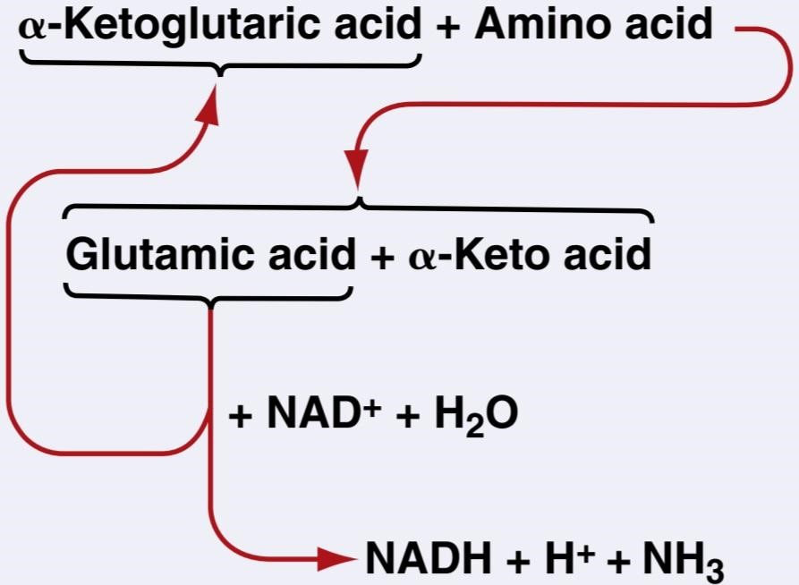
Note from this illustration that the amino group from the amino acid is transferred to a-ketoglutaric acid, which then becomes glutamic acid. The glutamic acid can then transfer the amino group to other substances or release it in the form of ammonia (NH3). In the process of losing the amino group, the glutamic acid once again becomes a-ketoglutaric acid, so the cycle can be repeated again and again. To initiate this process, the excess amino acids in the cells, especially in the liver, induce activation of large quantities of aminotransferases, the enzymes responsible for initiating most deamination.
از این تصویر توجه کنید که گروه آمینه از اسید آمینه به اسید a-ketoglutaric منتقل می شود که سپس به اسید گلوتامیک تبدیل می شود. سپس اسید گلوتامیک می تواند گروه آمینه را به مواد دیگر منتقل کند یا آن را به شکل آمونیاک (NH3) آزاد کند. در فرآیند از دست دادن گروه آمینه، اسید گلوتامیک یک بار دیگر به اسید a-ketoglutaric تبدیل می شود، بنابراین چرخه می تواند بارها و بارها تکرار شود. برای شروع این فرآیند، اسیدهای آمینه اضافی در سلول ها، به ویژه در کبد، باعث فعال شدن مقادیر زیادی آمینوترانسفرازها می شود، آنزیم هایی که مسئول بیشتر دآمیناسیون هستند.
Urea Formation by the Liver. The ammonia released during deamination of amino acids is removed from the blood almost entirely by conversion into urea. Two molecules of ammonia (NH3) and one molecule of carbon di- oxide (CO2) combine in the following net reaction:
تشکیل اوره توسط کبد. آمونیاک آزاد شده در طی دآمیناسیون اسیدهای آمینه تقریباً به طور کامل با تبدیل به اوره از خون خارج می شود. دو مولکول آمونیاک (NH3) و یک مولکول دی اکسید کربن (CO2) در واکنش خالص زیر ترکیب می شوند:

Essentially all urea formed in the human body is synthesized in the liver. In the absence of the liver or in persons with serious liver disease, ammonia accumulates in the blood. This accumulation of ammonia is extremely toxic, especially to the brain, and can lead to a state called hepatic coma. The stages in the formation of urea are essentially the following:
اساساً تمام اوره تشکیل شده در بدن انسان در کبد سنتز می شود. در غیاب کبد یا در افراد مبتلا به بیماری جدی کبد، آمونیاک در خون تجمع می یابد. این تجمع آمونیاک به خصوص برای مغز بسیار سمی است و می تواند منجر به حالتی به نام کمای کبدی شود. مراحل تشکیل اوره اساساً به شرح زیر است:
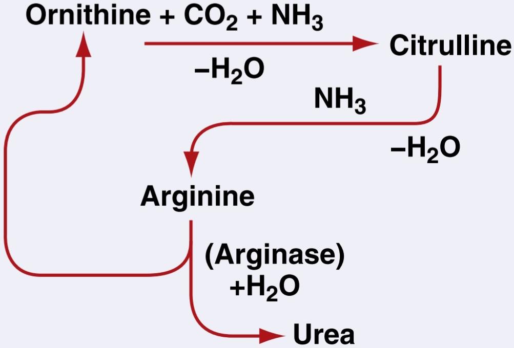
After its formation, the urea diffuses from the liver cells into the body fluids and is excreted by the kidneys.
پس از تشکیل، اوره از سلول های کبد به مایعات بدن منتشر شده و از طریق کلیه ها دفع می شود.
Oxidation of Deaminated Amino Acids. Once amino acids have been deaminated, the resulting keto acids can, in most cases, be oxidized to release energy for metabolic purposes. This oxidation usually involves two successive processes:
اکسیداسیون اسیدهای آمینه دآمینه شده. هنگامی که اسیدهای آمینه دآمینه شدند، کتواسیدهای حاصل می توانند در اکثر موارد اکسید شوند تا انرژی برای اهداف متابولیکی آزاد شود. این اکسیداسیون معمولاً شامل دو فرآیند متوالی است:
1. The keto acid is changed into an appropriate chemical substance that can enter the citric acid cycle.
1. کتو اسید به یک ماده شیمیایی مناسب تبدیل می شود که می تواند وارد چرخه اسید سیتریک شود.
2. This substance is degraded by the cycle and used for energy in the same manner that acetyl coenzyme A (acetyl-CoA) derived from carbohydrate and lipid metabolism is used, as explained in Chapters 68 and 69.
2. این ماده در چرخه تجزیه می شود و برای انرژی استفاده می شود، همانطور که در فصل های 68 و 69 توضیح داده شد، همانطور که از استیل کوآنزیم A (استیل کوآ) مشتق شده از متابولیسم کربوهیدرات و لیپید استفاده می شود.
In general, the amount of adenosine triphosphate formed for each gram of protein that is oxidized is slightly less than that formed for each gram of glucose that is oxidized.
به طور کلی، مقدار آدنوزین تری فسفات تشکیل شده برای هر گرم پروتئین اکسید شده اندکی کمتر از مقدار تشکیل شده برای هر گرم گلوکز اکسید شده است.
Gluconeogenesis and Ketogenesis. Certain deaminated amino acids are similar to the substrates normally used by the cells, mainly the liver cells, to synthesize glucose or fatty acids. For example, deaminated alanine is pyruvic acid, which can be converted into either glucose or glycogen. Alternatively, it can be converted into acetyl-CoA, which can then be polymerized into fatty acids. Also, two molecules of acetyl-CoA can condense to form acetoacetic acid, which is one of the ketone bodies, as explained in Chapter 69.
گلوکونئوژنز و کتوژنز. برخی از اسیدهای آمینه دآمینه شده مشابه سوبستراهایی هستند که معمولاً توسط سلول ها، عمدتاً سلول های کبد، برای سنتز گلوکز یا اسیدهای چرب استفاده می شود. به عنوان مثال، آلانین دآمینه، اسید پیروویک است که می تواند به گلوکز یا گلیکوژن تبدیل شود. از طرف دیگر، می توان آن را به استیل-CoA تبدیل کرد، که سپس می تواند به اسیدهای چرب پلیمریزه شود. همچنین، همانطور که در فصل 69 توضیح داده شد، دو مولکول استیل کوآ می توانند متراکم شوند و اسید استواستیک را تشکیل دهند که یکی از اجسام کتون است.
The conversion of amino acids into glucose or glycogen is called gluconeogenesis, and the conversion of amino acids into keto acids or fatty acids is called ketogenesis. Of the 20 deaminated amino acids, 18 have chemical structures that allow them to be converted into glucose, and 19 of them can be converted into fatty acids.
تبدیل اسیدهای آمینه به گلوکز یا گلیکوژن را گلوکونئوژنز و تبدیل اسیدهای آمینه به اسیدهای کتو یا اسیدهای چرب را کتوژنز می نامند. از 20 اسید آمینه دآمینه شده، 18 اسید آمینه دارای ساختار شیمیایی هستند که به آنها اجازه می دهد به گلوکز تبدیل شوند و 19 مورد از آنها می توانند به اسیدهای چرب تبدیل شوند.
Obligatory Degradation of Proteins
When a person eats no proteins, a certain proportion of body proteins is degraded into amino acids and then deaminated and oxidized. This process involves 20 to 30 grams of protein each day, which is called the obligatory loss of proteins. There- fore, to prevent net loss of protein from the body, the average person must ingest a minimum of 20 to 30 grams of protein each day, although this amount depends on multiple factors, including muscle mass, activity, and age; to be on the safe side, a minimum of 60 to 75 grams is usually recommended.
تخریب اجباری پروتئین ها
وقتی فردی پروتئین نمی خورد، نسبت مشخصی از پروتئین های بدن به اسیدهای آمینه تجزیه می شود و سپس دآمینه و اکسید می شود. این فرآیند شامل 20 تا 30 گرم پروتئین در روز است که به آن از دست دادن اجباری پروتئین گفته می شود. بنابراین، برای جلوگیری از از دست دادن خالص پروتئین از بدن، فرد عادی باید حداقل 20 تا 30 گرم پروتئین در روز مصرف کند، اگرچه این مقدار به عوامل متعددی از جمله توده عضلانی، فعالیت و سن بستگی دارد. برای ایمن بودن، حداقل 60 تا 75 گرم معمولا توصیه می شود.
The ratios of the different amino acids in the dietary protein must be about the same as the ratios in the body tissues if the entire dietary protein is to be fully usable to form new tissue proteins. If one particular type of essential amino acid is low in concentration, the others become unusable because cells synthesize either whole proteins or none at all, as explained in Chapter 3 in relation to protein synthesis. The unusable amino acids are deaminated and oxidized. A protein that has a ratio of amino acids different from that of the average body protein is called a partial protein or an incomplete protein, and such a protein is less valuable for nutrition than is a complete protein.
نسبت آمینواسیدهای مختلف در پروتئین جیره باید تقریباً با نسبتهای موجود در بافتهای بدن یکسان باشد تا بتوان از کل پروتئین رژیم غذایی برای تشکیل پروتئینهای بافت جدید استفاده کرد. اگر یک نوع خاص از اسید آمینه ضروری غلظت کمی داشته باشد، بقیه غیرقابل استفاده می شوند زیرا سلول ها یا پروتئین کامل را سنتز می کنند یا اصلاً پروتئین را سنتز نمی کنند، همانطور که در فصل 3 در رابطه با سنتز پروتئین توضیح داده شد. اسیدهای آمینه غیر قابل استفاده دآمینه و اکسید می شوند. پروتئینی که نسبت اسیدهای آمینه آن با نسبت پروتئین متوسط بدن متفاوت باشد، پروتئین جزئی یا پروتئین ناقص نامیده می شود و چنین پروتئینی برای تغذیه ارزش کمتری نسبت به پروتئین کامل دارد.
Effect of Starvation on Protein Degradation. Except for the 20 to 30 grams of obligatory protein degradation each day, the body uses almost entirely carbohydrates or fats for energy, as long as they are available. However, after several weeks of starvation, when the quantities of stored carbohydrates and fats begin to run out, the amino acids of the blood are rapidly deaminated and oxidized for energy. From this point on, the proteins of the tissues degrade rapidly-as much as 125 grams daily-and, as a result, cellular functions deteriorate precipitously. Because carbohydrate and fat utilization for energy normally occurs in preference to protein utilization, carbohydrates and fats are called protein sparers.
تاثیر گرسنگی بر تخریب پروتئین. به جز 20 تا 30 گرم تجزیه اجباری پروتئین در روز، بدن تقریباً به طور کامل از کربوهیدرات ها یا چربی ها برای انرژی استفاده می کند، تا زمانی که در دسترس هستند. با این حال، پس از چندین هفته گرسنگی، زمانی که مقادیر کربوهیدراتها و چربیهای ذخیره شده شروع به تمام شدن میکنند، اسیدهای آمینه خون به سرعت دآمینه شده و برای تولید انرژی اکسید میشوند. از این مرحله به بعد، پروتئینهای بافتها به سرعت تجزیه میشوند – به میزان 125 گرم در روز – و در نتیجه عملکرد سلولی بهسرعت بدتر میشود. از آنجایی که استفاده از کربوهیدرات و چربی برای انرژی معمولاً در ترجیح استفاده از پروتئین اتفاق میافتد، کربوهیدراتها و چربیها را ذخیرهکننده پروتئین مینامند.
Hormonal Regulation of Protein Metabolism
Growth Hormone Increases the Synthesis of Cellular Proteins. Growth hormone causes the tissue proteins to increase. The precise mechanism by which this increase occurs is not fully understood, but it is believed to result mainly from increased transport of amino acids through the cell membranes, acceleration of the DNA and RNA transcription and translation processes for protein synthesis, and decreased oxidation of tissue proteins.
تنظیم هورمونی متابولیسم پروتئین
هورمون رشد سنتز پروتئین های سلولی را افزایش می دهد. هورمون رشد باعث افزایش پروتئین های بافت می شود. مکانیسم دقیقی که توسط آن این افزایش رخ می دهد به طور کامل شناخته نشده است، اما اعتقاد بر این است که عمدتاً ناشی از افزایش انتقال اسیدهای آمینه از طریق غشای سلولی، تسریع در رونویسی DNA و RNA و فرآیندهای ترجمه برای سنتز پروتئین، و کاهش اکسیداسیون پروتئین های بافتی است.
Insulin Is Necessary for Protein Synthesis. Total lack of insulin reduces protein synthesis to almost zero. Insulin accelerates transport of some amino acids into cells, which could be the stimulus for protein synthesis. Also, insulin reduces protein degradation and increases the availability of glucose to the cells, so the need for amino acids for energy is correspondingly reduced.
انسولین برای سنتز پروتئین ضروری است. کمبود کامل انسولین سنتز پروتئین را تقریباً به صفر می رساند. انسولین انتقال برخی از اسیدهای آمینه را به داخل سلول ها تسریع می کند، که می تواند محرک سنتز پروتئین باشد. همچنین، انسولین تجزیه پروتئین را کاهش می دهد و دسترسی گلوکز به سلول ها را افزایش می دهد، بنابراین نیاز به اسیدهای آمینه برای انرژی به ترتیب کاهش می یابد.
Glucocorticoids Increase Breakdown of Most Tissue Proteins.
Glucocorticoids secreted by the adrenal cortex decrease the quantity of protein in most tissues while increasing amino acid concentration in the plasma, as well as increasing liver proteins and plasma proteins. Glucocorticoids increase the rate of breakdown of extrahepatic proteins, thereby making increased quantities of amino acids available in the body fluids. This allows the liver to synthesize increased quantities of hepatic cellular proteins and plasma proteins.
گلوکوکورتیکوئیدها تجزیه اکثر پروتئین های بافت را افزایش می دهند.
گلوکوکورتیکوئیدهای ترشح شده از قشر آدرنال باعث کاهش مقدار پروتئین در اکثر بافت ها و در عین حال افزایش غلظت اسید آمینه در پلاسما و همچنین افزایش پروتئین های کبد و پروتئین های پلاسما می شود. گلوکوکورتیکوئیدها سرعت تجزیه پروتئین های خارج کبدی را افزایش می دهند و در نتیجه مقادیر بیشتری از آمینو اسیدها را در مایعات بدن در دسترس قرار می دهند. این به کبد اجازه می دهد تا مقادیر بیشتری از پروتئین های سلولی کبدی و پروتئین های پلاسما را سنتز کند.
Testosterone Increases Protein Deposition in Tissues. Testosterone, the male sex hormone, causes increased deposition of protein in tissues throughout the body, especially the contractile proteins of the muscles (a 30%-50% increase). The mechanism of this effect is unknown, but it is definitely different from the effect of growth hormone, in the following way: Growth hormone causes tissues to continue growing almost indefinitely, whereas testosterone causes the muscles and, to a much lesser extent, some other protein tissues to enlarge for only several months. Once the muscles and other protein tissues have reached a maximum, despite continued administration of testosterone, further protein deposition ceases.
تستوسترون رسوب پروتئین را در بافت ها افزایش می دهد. تستوسترون، هورمون جنسی مردانه، باعث افزایش رسوب پروتئین در بافتهای سراسر بدن، بهویژه پروتئینهای انقباضی ماهیچهها میشود (30 تا 50 درصد افزایش). مکانیسم این اثر ناشناخته است، اما به طور قطع با اثر هورمون رشد به روش زیر متفاوت است: هورمون رشد باعث می شود بافت ها تقریباً به طور نامحدود به رشد خود ادامه دهند، در حالی که تستوسترون باعث می شود عضلات و به میزان بسیار کمتری برخی از بافت های پروتئینی دیگر تنها برای چند ماه بزرگ شوند. هنگامی که ماهیچه ها و سایر بافت های پروتئینی به حداکثر می رسند، با وجود تجویز مداوم تستوسترون، رسوب بیشتر پروتئین متوقف می شود.
Estrogen. Estrogen, the principal female sex hormone, also causes some deposition of protein, but the effect of estrogen is much less compared with that of testosterone.
استروژن. استروژن، هورمون جنسی اصلی زنانه نیز باعث رسوب پروتئین می شود، اما تاثیر استروژن در مقایسه با تستوسترون بسیار کمتر است.
Thyroxine Increases Metabolism of Cells. Thyroxine in- directly affects protein metabolism by increasing metabolism of the cells. If insufficient carbohydrates and fats are available for energy, thyroxine causes rapid degradation of proteins and uses them for energy. Conversely, if adequate quantities of carbohydrates and fats are available and excess amino acids are also available in the extracellular fluid, thyroxine can actually increase the rate of protein synthesis. In growing animals or human beings, deficiency of thyroxine causes growth to be greatly inhibited because of lack of protein synthesis. In essence, it is believed that thyroxine has little specific effect on protein metabolism but does have an important general effect by increasing the rates of both normal anabolic and normal catabolic protein reactions.
تیروکسین متابولیسم سلول ها را افزایش می دهد. تیروکسین به طور مستقیم بر متابولیسم پروتئین با افزایش متابولیسم سلول ها تأثیر می گذارد. اگر کربوهیدرات و چربی کافی برای انرژی در دسترس نباشد، تیروکسین باعث تخریب سریع پروتئین ها شده و از آنها برای انرژی استفاده می کند. برعکس، اگر مقادیر کافی کربوهیدرات و چربی در دسترس باشد و اسیدهای آمینه اضافی نیز در مایع خارج سلولی موجود باشد، تیروکسین در واقع می تواند سرعت سنتز پروتئین را افزایش دهد. در حیوانات یا انسان های در حال رشد، کمبود تیروکسین باعث می شود که به دلیل عدم سنتز پروتئین، رشد تا حد زیادی مهار شود. در اصل، اعتقاد بر این است که تیروکسین اثر خاص کمی بر متابولیسم پروتئین دارد، اما با افزایش سرعت واکنشهای پروتئین آنابولیک طبیعی و پروتئین کاتابولیک طبیعی، اثر کلی مهمی دارد.
کلیک کنید «Bibliography: فهرست کتب مربوطه»
Bröer S, Fairweather SJ: Amino acid transport across the mammalian intestine. Compr Physiol 9:343, 2018.
Bröer S, Bröer A: Amino acid homeostasis and signalling in mamma- lian cells and organisms. Biochem J 474:1935, 2017.
Finn PF, Dice JF: Proteolytic and lipolytic responses to starvation. Nu- trition 22:830, 2006.
Hawley JA, Burke LM, Phillips SM, Spriet LL: Nutritional modulation of training-induced skeletal muscle adaptations. J Appl Physiol 110:834, 2011.
Kandasamy P, Gyimesi G, Kanai Y, Hediger MA: Amino acid trans- porters revisited: new views in health and disease. Trends Biochem Sci 43:752, 2018.
Kaur J, Debnath J: Autophagy at the crossroads of catabolism and anabolism. Nat Rev Mol Cell Biol 16:461, 2015.
Mann GE, Yudilevich DL, Sobrevia L: Regulation of amino acid and glucose transporters in endothelial and smooth muscle cells. Phys- iol Rev 83:183, 2003.
Pencharz PB, Elango R, Wolfe RR: Recent developments in under- standing protein needs – how much and what kind should we eat? Appl Physiol Nutr Metab 41:577, 2016.
Rossetti ML, Steiner JL, Gordon BS: Androgen-mediated regulation of skeletal muscle protein balance. Mol Cell Endocrinol 447:35, 2017.
Tavernarakis N: Ageing and the regulation of protein synthesis: a bal- ancing act? Trends Cell Biol 18:228, 2008.
Vandenberg RJ, Ryan RM: Mechanisms of glutamate transport. Phys- iol Rev 93:1621, 2013.
Wolfe RR: The 2017 Sir David P Cuthbertson lecture. Amino acids and muscle protein metabolism in critical care. Clin Nutr 37:1093, 2018.
Wolfe RR, Cifelli AM, Kostas G, Kim IY: Optimizing protein intake in adults: Interpretation and application of the recommended dietary allowance compared with the acceptable macronutrient distribu- tion range. Adv Nutr 8:266, 2017.
🚀 با ما همراه شوید!
تازهترین مطالب و آموزشهای مغز و اعصاب را از دست ندهید. با فالو کردن کانال تلگرام آیندهنگاران مغز، از ما حمایت کنید!



