
فیزیولوژی پزشکی گایتون و هال؛ متابولیسم لیپید


دعای مطالعه [ نمایش ]
بِسْمِ الله الرَّحْمنِ الرَّحیمِ
اَللّهُمَّ اَخْرِجْنى مِنْ ظُلُماتِ الْوَهْمِ
خدايا مرا بيرون آور از تاريكىهاى وهم،
وَ اَكْرِمْنى بِنُورِ الْفَهْمِ
و به نور فهم گرامى ام بدار،
اَللّهُمَّ افْتَحْ عَلَيْنا اَبْوابَ رَحْمَتِكَ
خدايا درهاى رحمتت را به روى ما بگشا،
وَانْشُرْ عَلَيْنا خَزائِنَ عُلُومِكَ بِرَحْمَتِكَ يا اَرْحَمَ الرّاحِمينَ
و خزانههاى علومت را بر ما باز كن به امید رحمتت اى مهربانترين مهربانان.
کتاب «فیزیولوژی پزشکی گایتون و هال» بهعنوان یکی از جامعترین و معتبرترین منابع در حوزه علوم پزشکی، همچنان مرجع کلیدی برای درک عملکرد پیچیده بدن انسان است. این اثر با تکیه بر تازهترین پژوهشها و توضیحات دقیق از سازوکارهای فیزیولوژیک، پلی میان علوم پایه پزشکی و کاربردهای بالینی ایجاد میکند و نقشی بیبدیل در آموزش، پژوهش و ارتقای دانش سلامت ایفا مینماید.
ترجمه دقیق و علمی این شاهکار توسط برند علمی آیندهنگاران مغز به مدیریت داریوش طاهری، دسترسی فارسیزبانان به مرزهای نوین دانش فیزیولوژی را ممکن ساخته و رسالتی علمی برای ارتقای آموزش پزشکی، فهم عمیقتر سازوکارهای بدن و توسعه روشهای نوین در حوزه سلامت فراهم آورده است.
» Guyton and Hall Textbook of Medical Physiology
»» CHAPTER 69: Lipid Metabolism

Several chemical compounds in food and in the body are classified as lipids, including the following: (1) neutral fat, also known as triglycerides; (2) phospholipids; (3) cholesterol; and (4) a few others of less importance. Chemically, the basic lipid moiety of triglycerides and phospholipids is fatty acids, which are long-chain hydrocarbon organic acids. A typical fatty acid, palmitic acid, is the following: CH3(CH2)14COOH.
چندین ترکیب شیمیایی موجود در غذا و بدن به عنوان لیپید طبقه بندی می شوند، از جمله: (1) چربی خنثی، همچنین به عنوان تری گلیسیرید شناخته می شود. (2) فسفولیپیدها؛ (3) کلسترول؛ و (4) چند مورد دیگر که اهمیت کمتری دارند. از نظر شیمیایی، بخش لیپیدی پایه تری گلیسیریدها و فسفولیپیدها اسیدهای چرب هستند که اسیدهای آلی هیدروکربنی با زنجیره بلند هستند. یک اسید چرب معمولی، اسید پالمیتیک، به شرح زیر است: CH3(CH2)14COOH.
Although cholesterol does not contain fatty acid, its sterol nucleus is synthesized from portions of fatty acid molecules, thus giving it many of the physical and chemical properties of other lipids.
اگرچه کلسترول حاوی اسید چرب نیست، هسته استرول آن از بخشهایی از مولکولهای اسید چرب سنتز میشود، بنابراین بسیاری از خواص فیزیکی و شیمیایی چربیهای دیگر را به آن میدهد.
The triglycerides are used in the body mainly to provide energy for the different metabolic processes, a function they share almost equally with carbohydrates. However, some lipids, especially cholesterol, phosphor lipids, and small amounts of triglycerides, are used to form the membranes of all cells of the body and to perform other essential cellular functions.
تری گلیسیریدها در بدن عمدتاً برای تأمین انرژی برای فرآیندهای متابولیک مختلف استفاده می شوند، عملکردی که تقریباً به طور مساوی با کربوهیدرات ها تقسیم می شوند. با این حال، برخی از لیپیدها، به ویژه کلسترول، لیپیدهای فسفر و مقادیر کمی تری گلیسیرید، برای تشکیل غشای تمام سلولهای بدن و انجام سایر عملکردهای ضروری سلولی استفاده میشوند.
BASIC CHEMICAL STRUCTURE OF TRIGLYCERIDES (NEUTRAL FAT)
Because most of this chapter deals with utilization of triglycerides for energy, the following typical structure of the triglyceride molecule should be understood:
ساختار شیمیایی پایه تری گلیسریدها (چربی خنثی)
از آنجایی که بیشتر این فصل به استفاده از تری گلیسیرید برای انرژی می پردازد، ساختار معمولی زیر مولکول تری گلیسیرید باید درک شود:

Note that three long-chain fatty acid molecules are bound with one molecule of glycerol. The three fatty acids most commonly present in the triglycerides of the human body are as follows: (1) stearic acid (shown in the tristearin example), which has an 18-carbon chain and is fully saturated with hydrogen atoms; (2) oleic acid, which also has an 18-carbon chain but has one double bond in the middle of the chain; and (3) palmitic acid, which has 16 carbon atoms and is fully saturated.
توجه داشته باشید که سه مولکول اسید چرب با زنجیره بلند با یک مولکول گلیسرول پیوند دارند. سه اسید چرب که بیشتر در تری گلیسیریدهای بدن انسان وجود دارند به شرح زیر است: (1) اسید استئاریک (در مثال تری استئارین نشان داده شده است)، که دارای یک زنجیره 18 کربنی است و کاملاً با اتم های هیدروژن اشباع شده است. (2) اسید اولئیک، که همچنین دارای یک زنجیره 18 کربنی است اما دارای یک پیوند دوگانه در وسط زنجیره است. و (3) اسید پالمیتیک که 16 اتم کربن دارد و کاملاً اشباع است.
TRANSPORT OF LIPIDS IN THE BODY FLUIDS
TRANSPORT OF TRIGLYCERIDES AND OTHER LIPIDS FROM THE GASTRO- INTESTINAL TRACT BY LYMPH-THE CHYLOMICRONS
As explained in Chapter 66, almost all the fats in the diet, with the principal exception of a few short-chain fatty acids, are absorbed from the intestines into the intestinal lymph. During digestion, most triglycerides are split into monoglycerides and fatty acids. Then, while passing through the intestinal epithelial cells, the monoglycerides and fatty acids are resynthesized into new molecules of triglycerides that enter the lymph as minute, dispersed droplets called chylomicrons (Figure 69-1), whose diameters are between 0.08 and 0.6 micron. A small amount of apolipoprotein, mainly apolipoprotein B, is adsorbed to the outer surfaces of the chylomicrons. The remainder of the protein molecules project into the surrounding water and thereby increase the suspension stability of the chylomicrons in the lymph fluid and prevent their adherence to the lymphatic vessel walls.
انتقال لیپیدها در مایعات بدن
انتقال تری گلیسیریدها و سایر لیپیدها از دستگاه گوارش و روده توسط لنف – شیلومیکرون ها
همانطور که در فصل 66 توضیح داده شد، تقریباً تمام چربی های رژیم غذایی، به استثنای چند اسید چرب با زنجیره کوتاه، از روده ها به لنف روده جذب می شوند. در طول هضم، بیشتر تری گلیسیریدها به مونوگلیسرید و اسیدهای چرب تقسیم می شوند. سپس، در حین عبور از سلول های اپیتلیال روده، مونوگلیسریدها و اسیدهای چرب به مولکول های جدید تری گلیسیرید تبدیل می شوند که به صورت قطرات ریز و پراکنده به نام شیلومیکرون وارد لنف می شوند (شکل 69-1)، که قطر آنها بین 0.08 تا 0.6 میکرون است. مقدار کمی آپولیپوپروتئین، عمدتا آپولیپوپروتئین B، به سطوح بیرونی شیلومیکرون ها جذب می شود. باقی مانده مولکول های پروتئین به داخل آب اطراف پیش می روند و در نتیجه پایداری سوسپانسیون شیلومیکرون ها در مایع لنفاوی را افزایش می دهند و از چسبیدن آنها به دیواره های عروق لنفاوی جلوگیری می کنند.
Most of the cholesterol and phospholipids absorbed from the gastrointestinal tract enter the chylomicrons. Thus, although the chylomicrons are composed principally of triglycerides, they also contain about 9% phosphor lipids, 3% cholesterol, and 1% apolipoproteins. The chylomicrons are then transported upward through the thoracic duct and emptied into the circulating venous blood at the juncture of the jugular and subclavian veins.
بیشتر کلسترول و فسفولیپیدهای جذب شده از دستگاه گوارش وارد شیلومیکرون ها می شوند. بنابراین، اگرچه شیلومیکرون ها عمدتاً از تری گلیسیریدها تشکیل شده اند، اما حاوی حدود 9٪ لیپید فسفر، 3٪ کلسترول و 1٪ آپولیپوپروتئین هستند. سپس شیلومیکرون ها از طریق مجرای قفسه سینه به سمت بالا منتقل می شوند و در محل اتصال وریدهای ژوگولار و ساب کلاوین به داخل خون وریدی در گردش تخلیه می شوند.

Figure 69-1. Chylomicrons are lipoprotein particles that consist of phospholipids and apolipoproteins on the outer surface and triglycerides and cholesterol inside.
شکل 69-1. شیلومیکرون ها ذرات لیپوپروتئینی هستند که از فسفولیپیدها و آپولیپوپروتئین ها در سطح بیرونی و تری گلیسیرید و کلسترول در داخل تشکیل شده اند.
REMOVAL OF THE CHYLOMICRONS FROM THE BLOOD
About 1 hour after a meal containing large quantities of fat, the chylomicron concentration in the plasma may rise to 1% to 2% of the total plasma, and because of the large size of the chylomicrons, the plasma appears turbid and sometimes yellow. However, the chylomicrons have a half-life of less than 1 hour, so the plasma becomes clear again within a few hours. The fat of the chylomicrons is removed mainly in the following way.
حذف شیلومیکرون ها از خون
حدود 1 ساعت پس از یک وعده غذایی حاوی مقادیر زیادی چربی، غلظت شیلومیکرون در پلاسما ممکن است به 1٪ تا 2٪ از کل پلاسما افزایش یابد و به دلیل اندازه بزرگ شیلومیکرون ها، پلاسما کدر و گاهی اوقات زرد به نظر می رسد. با این حال، شیلومیکرون ها نیمه عمر کمتر از 1 ساعت دارند، بنابراین پلاسما در عرض چند ساعت دوباره شفاف می شود. چربی شیلومیکرون ها عمدتاً به روش زیر حذف می شود.
Chylomicron Triglycerides Are Hydrolyzed by Lipoprotein Lipase, and Fat Is Stored in Adipose Tissue. Most of the chylomicrons are removed from the circulating blood as they pass through the capillaries of various tissues, especially adipose tissue, skeletal muscle, and heart. These tissues synthesize the enzyme lipoprotein lipase, which is transported to the surface of capillary endothelial cells, where it hydrolyzes the triglycerides of chylomicrons as they come in contact with the endothelial wall, thus releasing fatty acids and glycerol (Figure 69-2).
تری گلیسیریدهای شیلومیکرون توسط لیپوپروتئین لیپاز هیدرولیز می شوند و چربی در بافت چربی ذخیره می شود. بیشتر شیلومیکرون ها با عبور از مویرگ های بافت های مختلف به ویژه بافت چربی، ماهیچه های اسکلتی و قلب از خون در گردش خارج می شوند. این بافت ها آنزیم لیپوپروتئین لیپاز را سنتز می کنند که به سطح سلول های اندوتلیال مویرگی منتقل می شود و در آنجا تری گلیسیرید شیلومیکرون ها را در تماس با دیواره اندوتلیال هیدرولیز می کند و در نتیجه اسیدهای چرب و گلیسرول آزاد می کند (شکل 69-2).
The fatty acids released from the chylomicrons, being highly miscible with the membranes of the cells, diffuse into the fat cells of the adipose tissue and muscle cells. Once inside these cells, the fatty acids can be used for fuel or again synthesized into triglycerides, with new glycerol being supplied by the metabolic processes of the storage cells, as discussed later in the chapter. The lipase also causes hydrolysis of phospholipids, which also releases fatty acids to be stored in the cells in the same way.
اسیدهای چرب آزاد شده از شیلومیکرون ها که به شدت با غشای سلول ها قابل اختلاط هستند، در سلول های چربی بافت چربی و سلول های ماهیچه ای پخش می شوند. هنگامی که در داخل این سلول ها قرار می گیرند، اسیدهای چرب می توانند برای سوخت استفاده شوند یا دوباره به تری گلیسیرید سنتز شوند، با گلیسرول جدید که توسط فرآیندهای متابولیکی سلول های ذخیره سازی تامین می شود، همانطور که بعداً در این فصل بحث شد. لیپاز همچنین باعث هیدرولیز فسفولیپیدها می شود که اسیدهای چرب را نیز آزاد می کند تا به همان روش در سلول ها ذخیره شوند.
After the triglycerides are removed from the chylomicrons, the cholesterol-enriched chylomicron remnants are rapidly cleared from the plasma. The chylomicron remnants bind to receptors on endothelial cells in the liver sinusoids. Apolipoprotein-E on the surface of the chylomicron remnants and secreted by liver cells also plays an important role in initiating clearance of these plasma lipoproteins.
پس از حذف تری گلیسیرید از شیلومیکرون ها، بقایای شیلومیکرون غنی شده با کلسترول به سرعت از پلاسما پاک می شوند. بقایای شیلومیکرون به گیرنده های سلول های اندوتلیال در سینوسوئیدهای کبد متصل می شوند. آپولیپوپروتئین-E بر روی سطح بقایای شیلومیکرون و ترشح شده توسط سلول های کبدی نیز نقش مهمی در آغاز پاکسازی این لیپوپروتئین های پلاسما ایفا می کند.
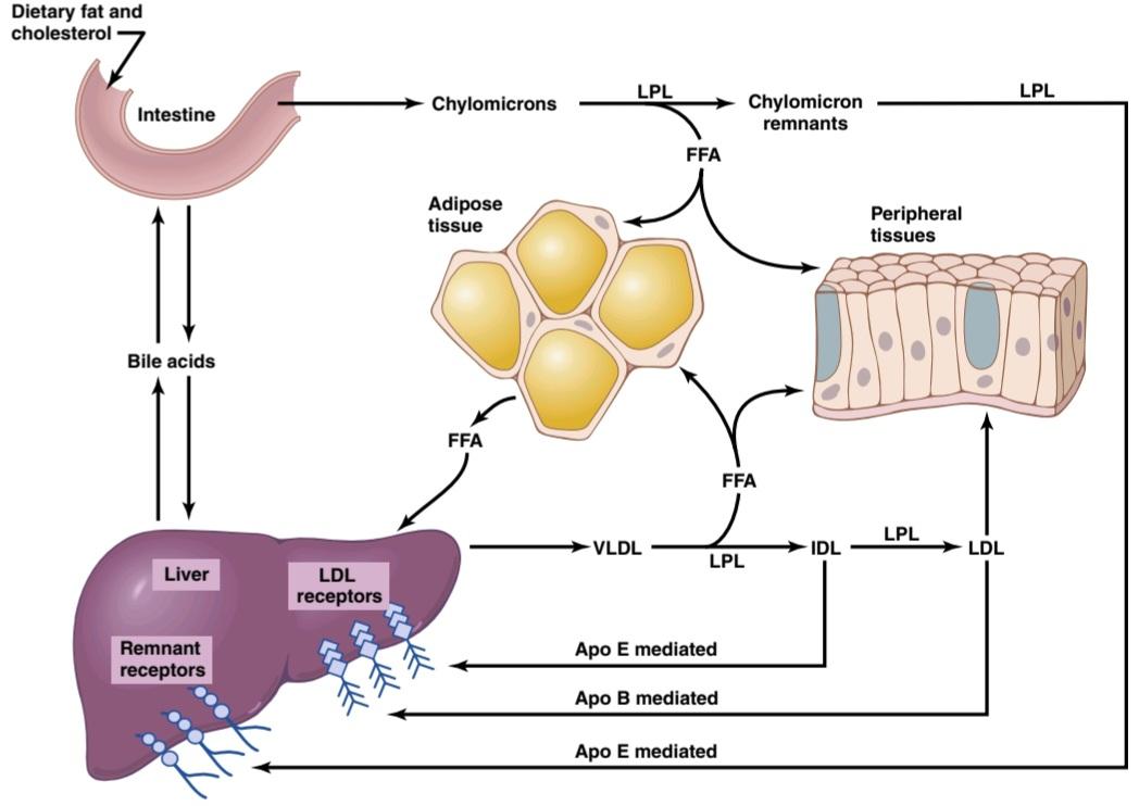
Figure 69-2. Summary of major pathways for metabolism of chylomicrons synthesized in the intestine and very low density
شکل 69-2. خلاصه ای از مسیرهای اصلی متابولیسم شیلومیکرون های سنتز شده در روده با چگالی بسیار کم
“Free Fatty Acids” Are Transported in the Blood in Combination With Albumin
When fat that has been stored in the adipose tissue is to be used elsewhere in the body to provide energy, it must first be transported from the adipose tissue to the other tissue. It is transported mainly in the form of free fatty acids. This transport is achieved by hydrolysis of the triglycerides back into fatty acids and glycerol.
“اسیدهای چرب آزاد” در ترکیب با آلبومین در خون منتقل می شوند
وقتی قرار است چربی ذخیره شده در بافت چربی در جای دیگری از بدن برای تامین انرژی استفاده شود، ابتدا باید از بافت چربی به بافت دیگر منتقل شود. عمدتاً به شکل اسیدهای چرب آزاد منتقل می شود. این انتقال با هیدرولیز تری گلیسیریدها به اسیدهای چرب و گلیسرول حاصل می شود.
At least two classes of stimuli play important roles in promoting this hydrolysis. First, when the amount of glucose available to the fat cell is inadequate, one of the glucose breakdown products, a-glycerophosphate, is also available in insufficient quantities. Because this substance is required to maintain the glycerol portion of triglycerides, the result is hydrolysis of triglycerides. Second, a hormone-sensitive cellular lipase can be activated by several hormones from the endocrine glands, and this also promotes rapid hydrolysis of triglycerides. This topic is discussed later in the chapter.
حداقل دو دسته از محرک ها نقش مهمی در ترویج این هیدرولیز دارند. اول اینکه، زمانی که مقدار گلوکز موجود در سلول چربی ناکافی است، یکی از محصولات تجزیه گلوکز، a-glycerophosphate، نیز به مقدار ناکافی در دسترس است. از آنجا که این ماده برای حفظ بخش گلیسرول تری گلیسیرید مورد نیاز است، نتیجه هیدرولیز تری گلیسیرید است. دوم، یک لیپاز سلولی حساس به هورمون می تواند توسط چندین هورمون از غدد درون ریز فعال شود، و این نیز باعث هیدرولیز سریع تری گلیسیرید می شود. این موضوع بعداً در فصل مورد بحث قرار می گیرد.
Upon leaving fat cells, fatty acids ionize strongly in the plasma and the ionic portion combines immediately with albumin molecules of the plasma proteins. Fatty acids bound in this manner are called free fatty acids or nonesterified fatty acids, to distinguish them from other fatty acids in the plasma that exist in the form of (1) esters of glycerol, (2) cholesterol, or (3) other substances.
با خروج از سلول های چربی، اسیدهای چرب به شدت در پلاسما یونیزه می شوند و بخش یونی بلافاصله با مولکول های آلبومین پروتئین های پلاسما ترکیب می شود. اسیدهای چرب متصل به این روش، اسیدهای چرب آزاد یا اسیدهای چرب غیر استری شده نامیده می شوند تا آنها را از سایر اسیدهای چرب موجود در پلاسما که به شکل (1) استرهای گلیسرول، (2) کلسترول، یا (3) مواد دیگر وجود دارند، متمایز کنند.
The concentration of free fatty acids in the plasma under resting conditions is about 15 mg/dl, which is a total of only 0.45 gram of fatty acids in the entire circulatory system. Even this small amount accounts for almost all the transport of fatty acids from one part of the body to another for the following reasons:
غلظت اسیدهای چرب آزاد در پلاسما در شرایط استراحت حدود 15 میلی گرم در دسی لیتر است که در کل فقط 45/0 گرم اسیدهای چرب در کل سیستم گردش خون است. حتی همین مقدار ناچیز تقریباً تمام انتقال اسیدهای چرب از یک قسمت بدن به قسمت دیگر را به دلایل زیر تشکیل می دهد:
1. Despite the minute amount of free fatty acid in the blood, its rate of “turnover” is extremely rapid: half the plasma fatty acid is replaced by new fatty acid every 2 to 3 minutes. One can calculate that at this rate, almost all the normal energy requirements of the body can be provided by the oxidation of transported free fatty acids, without using any carbohydrates or proteins for energy.
1. علیرغم مقدار کمی اسید چرب آزاد در خون، سرعت “تبدیل” آن بسیار سریع است: هر 2 تا 3 دقیقه نیمی از اسید چرب پلاسما با اسید چرب جدید جایگزین می شود. می توان محاسبه کرد که با این سرعت، تقریباً تمام انرژی طبیعی مورد نیاز بدن را می توان از طریق اکسیداسیون اسیدهای چرب آزاد منتقل شده، بدون استفاده از هیچ کربوهیدرات یا پروتئینی برای انرژی تأمین کرد.
2. Conditions that increase the rate of utilization of fat for cellular energy also increase the free fatty acid concentration in the blood. In fact, the concentration sometimes increases fivefold to eightfold. Such a large increase occurs especially in cases of starvation and in diabetes mellitus; in both these conditions, the person derives little or no metabolic energy from carbohydrates.
2. شرایطی که میزان استفاده از چربی برای انرژی سلولی را افزایش می دهد، غلظت اسیدهای چرب آزاد در خون را نیز افزایش می دهد. در واقع، غلظت آن گاهی پنج برابر تا هشت برابر می شود. چنین افزایش زیادی به ویژه در موارد گرسنگی و دیابت رخ می دهد. در هر دو این شرایط، فرد انرژی متابولیکی کمی از کربوهیدرات ها دریافت می کند یا اصلا انرژی متابولیک دریافت نمی کند.
Under normal conditions, only about 3 molecules of fatty acid combine with each molecule of albumin, but as many as 30 fatty acid molecules can combine with a single albumin molecule when the need for fatty acid transport is extreme. This shows how variable the rate of lipid transport can be under different physiological conditions.
در شرایط عادی، تنها حدود 3 مولکول اسید چرب با هر مولکول آلبومین ترکیب میشوند، اما 30 مولکول اسید چرب میتوانند با یک مولکول آلبومین ترکیب شوند که نیاز به انتقال اسید چرب بسیار زیاد باشد. این نشان می دهد که نرخ انتقال چربی در شرایط مختلف فیزیولوژیکی چقدر می تواند متغیر باشد.
lipoprotein (VLDL) synthesized in the liver. Apo B, Apolipoprotein B; Apo E, apolipoprotein E; FFA, free fatty acids; IDL, intermediate-density lipoprotein; LDL, low- density lipoprotein; LPL, lipoprotein lipase.
لیپوپروتئین (VLDL) در کبد سنتز می شود. Apo B، آپولیپوپروتئین B; Apo E، آپولیپوپروتئین E; FFA، اسیدهای چرب آزاد؛ IDL، لیپوپروتئین با چگالی متوسط؛ LDL، لیپوپروتئین با چگالی کم؛ LPL، لیپوپروتئین لیپاز.
Lipoproteins-Their Special Function in Transporting Cholesterol and Phospholipids
In the postabsorptive state, after all the chylomicrons have been removed from the blood, more than 95% of all the lipids in the plasma are in the form of lipoprotein. These lipids are small particles-much smaller than chylomicrons, but qualitatively similar in composition-containing triglycerides, cholesterol, phospholipids, and protein. The total concentration of lipoproteins in the plasma averages about 700 mg per 100 ml of plasma-that is, 700 mg/dl-and can be broken down into the following individual lipoprotein constituents:
لیپوپروتئین ها – عملکرد ویژه آنها در انتقال کلسترول و فسفولیپیدها
در حالت پسابجذب، پس از حذف تمام شیلومیکرون ها از خون، بیش از 95 درصد از کل لیپیدهای پلاسما به شکل لیپوپروتئین هستند. این لیپیدها ذرات کوچکی هستند – بسیار کوچکتر از شیلومیکرون ها، اما از نظر کیفی از نظر تری گلیسریدها، کلسترول، فسفولیپیدها و پروتئین حاوی ترکیب مشابه هستند. غلظت کل لیپوپروتئین ها در پلاسما به طور متوسط حدود 700 میلی گرم در 100 میلی لیتر پلاسما است – یعنی 700 میلی گرم در دسی لیتر – و می تواند به اجزای لیپوپروتئین فردی زیر تقسیم شود:

Types of Lipoproteins. Aside from the chylomicrons, which are very large lipoproteins, there are four major types of lipoproteins, classified by their densities as measured in the ultracentrifuge: (1) very low density lipoproteins (VLDLs), which contain high concentrations of triglycerides and moderate concentrations of both cholesterol and phospholipids; (2) intermediate-density lipoproteins (IDLs), which are VLDLS from which a share of the triglycerides has been removed, so the concentrations of cholesterol and phospholipids are increased; (3) low- density lipoproteins (LDLs), which are derived from IDLs by the removal of almost all the triglycerides, leaving an especially high concentration of cholesterol and a moderately high concentration of phospholipids; and (4) high-density lipoproteins (HDLs), which contain a high concentration of protein (~50%) but much smaller concentrations of cholesterol and phospholipids.
انواع لیپوپروتئین ها. جدای از شیلومیکرون ها که لیپوپروتئین های بسیار بزرگی هستند، چهار نوع اصلی لیپوپروتئین وجود دارد که بر اساس چگالی آنها در دستگاه اولتراسانتریفیوژ طبقه بندی می شوند: (1) لیپوپروتئین های با چگالی بسیار کم (VLDL)، که حاوی غلظت بالایی از تری گلیسیرید و غلظت متوسطی از کلسترول و فولیکول هستند. (2) لیپوپروتئینهای با چگالی متوسط (IDL)، که VLDLS هستند که بخشی از تری گلیسیریدها از آن حذف شده است، بنابراین غلظت کلسترول و فسفولیپیدها افزایش مییابد. (3) لیپوپروتئین های با چگالی کم (LDL)، که از IDL ها با حذف تقریباً تمام تری گلیسیریدها به دست می آیند و غلظت بالایی از کلسترول و غلظت نسبتاً بالایی از فسفولیپیدها را بر جای می گذارند. و (4) لیپوپروتئین های با چگالی بالا (HDL)، که حاوی غلظت بالایی از پروتئین (~50٪) اما غلظت بسیار کمتری از کلسترول و فسفولیپیدها هستند.
Formation and Function of Lipoproteins. Almost all the lipoproteins are formed in the liver, which is also where most of the plasma cholesterol, phospholipids, and triglycerides are synthesized. In addition, small quantities of HDLs are synthesized in the intestinal epithelium during absorption of fatty acids from the intestines.
تشکیل و عملکرد لیپوپروتئین ها. تقریباً تمام لیپوپروتئینها در کبد تشکیل میشوند، جایی که بیشتر کلسترول پلاسما، فسفولیپیدها و تری گلیسیریدها نیز در آن سنتز میشوند. علاوه بر این، مقادیر کمی HDL در اپیتلیوم روده در طی جذب اسیدهای چرب از روده سنتز می شود.
The primary function of the lipoproteins is to transport their lipid components in the blood. The VLDLs transport triglycerides synthesized in the liver mainly to the adipose tissue. The other lipoproteins are especially important in different stages of phospholipid and cholesterol transport from the liver to the peripheral tissues or from the periphery back to the liver. Later in the chapter, we discuss in more detail special problems of cholesterol transport in relation to the disease atherosclerosis, which is associated with the development of fatty lesions on the insides of arterial walls.
وظیفه اصلی لیپوپروتئین ها انتقال اجزای لیپیدی آنها در خون است. VLDL ها تری گلیسیریدهای سنتز شده در کبد را عمدتاً به بافت چربی منتقل می کنند. سایر لیپوپروتئین ها به ویژه در مراحل مختلف انتقال فسفولیپید و کلسترول از کبد به بافت های محیطی یا از حاشیه به کبد اهمیت دارند. در ادامه این فصل، مشکلات ویژه انتقال کلسترول در رابطه با بیماری آترواسکلروز که با ایجاد ضایعات چربی در داخل دیوارههای شریان همراه است، با جزئیات بیشتری مورد بحث قرار میدهیم.
Fat Deposits
Large quantities of fat are stored in two major tissues of the body, the adipose tissue and the liver. The adipose tissue is usually called fat deposits, or simply tissue fat.
رسوبات چربی
مقدار زیادی چربی در دو بافت اصلی بدن یعنی بافت چربی و کبد ذخیره می شود. بافت چربی معمولاً رسوبات چربی یا به سادگی چربی بافت نامیده می شود.
Adipose Tissue
A major function of adipose tissue is storage of triglycerides until they are needed to provide energy elsewhere in the body. Additional functions are to provide heat insulation for the body, as discussed in Chapter 74, and secretion of hormones, such as leptin and adiponectin, which affect multiple body functions, including appetite and energy expenditure, as discussed in Chapter 72.
بافت چربی
عملکرد اصلی بافت چربی ذخیره تری گلیسیرید است تا زمانی که برای تامین انرژی در سایر نقاط بدن مورد نیاز باشد. کارکردهای اضافی عبارتند از ایجاد عایق حرارتی برای بدن، همانطور که در فصل 74 بحث شد، و ترشح هورمونهایی مانند لپتین و آدیپونکتین، که بر عملکردهای متعدد بدن، از جمله اشتها و مصرف انرژی، که در فصل 72 بحث شد، تأثیر میگذارند.
Fat Cells (Adipocytes) Store Triglycerides. The fat cells (adipocytes) of adipose tissue are modified fibroblasts that store almost pure triglycerides in quantities as great as 80% to 95% of the entire cell volume. Triglycerides inside the fat cells are generally in a liquid form. When the tissues are exposed to prolonged cold, the fatty acid chains of the cell triglycerides, over a period of weeks, become either shorter or more unsaturated to decrease their melting point, there- by always allowing the fat to remain in a liquid state. This characteristic is particularly important because only liquid fat can be hydrolyzed and transported from the cells.
سلول های چربی (آدیپوسیت ها) تری گلیسیرید را ذخیره می کنند. سلول های چربی (آدیپوسیت ها) بافت چربی فیبروبلاست های اصلاح شده ای هستند که تری گلیسیریدهای تقریباً خالص را در مقادیری به اندازه 80 تا 95 درصد از کل حجم سلول ذخیره می کنند. تری گلیسیرید درون سلول های چربی عموما به شکل مایع هستند. هنگامی که بافت ها در معرض سرمای طولانی مدت قرار می گیرند، زنجیره های اسید چرب تری گلیسیرید سلولی، در طی چند هفته، کوتاه تر یا غیراشباع تر می شوند تا نقطه ذوب خود را کاهش دهند و در نتیجه همیشه به چربی اجازه می دهند در حالت مایع باقی بماند. این ویژگی به ویژه مهم است زیرا فقط چربی مایع می تواند هیدرولیز شده و از سلول ها منتقل شود.
Fat cells can synthesize very small amounts of fatty acids and triglycerides from carbohydrates; this function supplements the synthesis of fat in the liver, as discussed later in the chapter.
سلول های چربی می توانند مقادیر بسیار کمی اسیدهای چرب و تری گلیسیرید را از کربوهیدرات ها سنتز کنند. این عملکرد مکمل سنتز چربی در کبد است، همانطور که بعدا در فصل مورد بحث قرار گرفت.
Tissue Lipases Permit Exchange of Fat Between Adipose Tissue and the Blood. As discussed earlier, large quantities of lipases are present in adipose tissue. Some of these enzymes catalyze the deposition of cell triglycerides from the chylomicrons and lipoproteins. Others, when activated by hormones, cause splitting of the triglycerides of the fat cells to release free fatty acids. Because of the rapid exchange of fatty acids, the triglycerides in fat cells are renewed about once every 2 to 3 weeks, which means that the fat stored in the tissues today is not the same fat that was stored last month, thus emphasizing the dynamic state of storage fat.
لیپازهای بافت اجازه تبادل چربی بین بافت چربی و خون را می دهند. همانطور که قبلاً گفته شد، مقادیر زیادی لیپاز در بافت چربی وجود دارد. برخی از این آنزیم ها رسوب تری گلیسیرید سلولی را از شیلومیکرون ها و لیپوپروتئین ها کاتالیز می کنند. برخی دیگر، هنگامی که توسط هورمون ها فعال می شوند، باعث تقسیم تری گلیسیرید سلول های چربی برای آزاد شدن اسیدهای چرب آزاد می شوند. به دلیل تبادل سریع اسیدهای چرب، تری گلیسیرید موجود در سلول های چربی تقریباً هر 2 تا 3 هفته یک بار تجدید می شود، به این معنی که چربی ذخیره شده در بافت های امروزی همان چربی ذخیره شده در ماه گذشته نیست، بنابراین بر وضعیت پویای چربی ذخیره شده تأکید می شود.
Liver Lipids
The principal functions of the liver in lipid metabolism are to (1) degrade fatty acids into small compounds that can be used for energy; (2) synthesize triglycerides, mainly from carbohydrates, but to a lesser extent from proteins as well; and (3) synthesize other lipids from fatty acids, especially cholesterol and phospholipids.
لیپیدهای کبد
کارکردهای اصلی کبد در متابولیسم لیپیدها عبارتند از (1) تجزیه اسیدهای چرب به ترکیبات کوچکی که می توانند برای انرژی استفاده شوند. (2) سنتز تری گلیسیریدها، عمدتاً از کربوهیدرات ها، اما به میزان کمتری از پروتئین ها نیز. و (3) سنتز چربی های دیگر از اسیدهای چرب، به ویژه کلسترول و فسفولیپیدها.
Large quantities of triglycerides appear in the liver during (1) the early stages of starvation, (2) in diabetes mellitus, and (3) in any other condition in which fat instead of carbohydrates is being used for energy. In these conditions, large quantities of triglycerides are mobilized from the adipose tissue, transported as free fatty acids in the blood, and redeposited as triglycerides in the liver, where the initial stages of much of fat degradation begin. Thus, under normal physiological conditions, the total amount of triglycerides in the liver is determined to a great extent by the overall rate at which lipids are being used for energy.
مقادیر زیادی تری گلیسیرید در کبد (1) در مراحل اولیه گرسنگی، (2) در دیابت قندی، و (3) در هر شرایط دیگری که در آن از چربی به جای کربوهیدرات برای انرژی استفاده می شود، ظاهر می شود. در این شرایط، مقادیر زیادی تری گلیسیرید از بافت چربی به حرکت در می آید، به عنوان اسیدهای چرب آزاد در خون منتقل می شود، و به صورت تری گلیسیرید در کبد، جایی که مراحل اولیه تخریب بیشتر چربی آغاز می شود، دوباره رسوب می کنند. بنابراین، در شرایط طبیعی فیزیولوژیکی، مقدار کل تری گلیسیرید در کبد تا حد زیادی با میزان کلی استفاده از لیپیدها برای انرژی تعیین می شود.
The liver may also store large amounts of lipids in people who are obese or have lipodystrophy, a condition characterized by atrophy or genetic deficiency of adipocytes. In both of these conditions, excess fat that cannot be stored in adipose tissue accumulates in the liver and, to a lesser extent, in other tissues that normally store minimal amounts of lipids.
کبد همچنین ممکن است مقادیر زیادی لیپید را در افراد چاق یا لیپودیستروفی ذخیره کند، وضعیتی که با آتروفی یا کمبود ژنتیکی سلولهای چربی مشخص میشود. در هر دوی این شرایط، چربی اضافی که نمیتواند در بافت چربی ذخیره شود، در کبد و به میزان کمتری در بافتهای دیگر که معمولاً مقادیر حداقلی لیپید را ذخیره میکنند، تجمع مییابد.
The liver cells, in addition to containing triglycerides, contain large quantities of phospholipids and cholesterol, which are continually synthesized by the liver. Also, the liver cells are much more capable of desaturating fatty ac- ids than are other tissues, and thus liver triglycerides normally are much more unsaturated than the triglycerides of adipose tissue. This capability of the liver to desaturate fatty acids is functionally important to all tissues of the body because many structural elements of all cells contain reasonable quantities of unsaturated fats, and their principal source is the liver. This desaturation is accomplished by a dehydrogenase in the liver cells.
سلول های کبدی علاوه بر اینکه حاوی تری گلیسیرید هستند، حاوی مقادیر زیادی فسفولیپید و کلسترول هستند که به طور مداوم توسط کبد سنتز می شوند. همچنین سلولهای کبدی نسبت به سایر بافتها توانایی غیراشباعکردن اسیدهای چرب را دارند و بنابراین تریگلیسریدهای کبد معمولاً غیراشباعتر از تریگلیسریدهای بافت چربی هستند. این توانایی کبد برای غیراشباع کردن اسیدهای چرب از نظر عملکردی برای تمام بافتهای بدن مهم است، زیرا بسیاری از عناصر ساختاری همه سلولها حاوی مقادیر معقولی از چربیهای غیراشباع هستند و منبع اصلی آنها کبد است. این غیراشباع شدن توسط یک دهیدروژناز در سلول های کبد انجام می شود.
Use of Triglycerides for Energy: Formation of Adenosine Triphosphate
The dietary intake of fat varies considerably in persons of different cultures, averaging as little as 10% to 15% of caloric intake in some Asian populations to as much as 35% to 50% of the calories in many Western populations. For many persons the use of fats for energy is therefore as important as the use of carbohydrates. In addition, many of the carbohydrates ingested with each meal are converted into triglycerides, stored, and used later in the form of fatty acids released from the triglycerides for energy.
استفاده از تری گلیسیرید برای انرژی: تشکیل آدنوزین تری فسفات
مصرف چربی در رژیم غذایی در افراد با فرهنگ های مختلف به طور قابل توجهی متفاوت است، به طور متوسط بین 10 تا 15 درصد کالری دریافتی در برخی از جمعیت های آسیایی تا 35 تا 50 درصد کالری در بسیاری از جمعیت های غربی است. بنابراین برای بسیاری از افراد استفاده از چربی ها برای انرژی به اندازه استفاده از کربوهیدرات ها مهم است. علاوه بر این، بسیاری از کربوهیدرات های مصرف شده با هر وعده غذایی به تری گلیسیرید تبدیل می شوند، ذخیره می شوند و بعداً به شکل اسیدهای چرب آزاد شده از تری گلیسیرید برای انرژی استفاده می شوند.
Hydrolysis of Triglycerides Into Fatty Acids and Glycerol. The first stage in using triglycerides for energy is their hydrolysis into fatty acids and glycerol. Then, both the fatty acids and the glycerol are transported in the blood to the active tissues, where they will be oxidized to give energy. Almost all cells with some exceptions, such as brain tissue and red blood cells-can use fatty acids for energy.
هیدرولیز تری گلیسیرید به اسیدهای چرب و گلیسرول. اولین مرحله در استفاده از تری گلیسیریدها برای انرژی، هیدرولیز آنها به اسیدهای چرب و گلیسرول است. سپس، هم اسیدهای چرب و هم گلیسرول در خون به بافتهای فعال منتقل میشوند و در آنجا اکسید میشوند تا انرژی بدهند. تقریباً همه سلولها به استثنای بافت مغز و گلبولهای قرمز میتوانند از اسیدهای چرب برای انرژی استفاده کنند.
Glycerol, upon entering the active tissue, is immediately changed by intracellular enzymes into glycerol-3- phosphate, which enters the glycolytic pathway for glucose breakdown and is thus used for energy. Before the fatty ac- ids can be used for energy, they must be processed further in the mitochondria.
گلیسرول، پس از ورود به بافت فعال، بلافاصله توسط آنزیم های داخل سلولی به گلیسرول-3- فسفات تبدیل می شود که برای تجزیه گلوکز وارد مسیر گلیکولیتیک می شود و بنابراین برای انرژی استفاده می شود. قبل از اینکه اسیدهای چرب برای انرژی مورد استفاده قرار گیرند، باید بیشتر در میتوکندری پردازش شوند.
Entry of Fatty Acids Into Mitochondria. Degradation and oxidation of fatty acids occur only in the mitochondria. Therefore, the first step for the use of fatty acids is their transport into the mitochondria using carnitine as a carrier. Once inside the mitochondria, fatty acids split away from carnitine and are degraded and oxidized.
ورود اسیدهای چرب به میتوکندری. تخریب و اکسیداسیون اسیدهای چرب فقط در میتوکندری اتفاق می افتد. بنابراین، اولین قدم برای استفاده از اسیدهای چرب، انتقال آنها به داخل میتوکندری با استفاده از کارنیتین به عنوان یک حامل است. هنگامی که در داخل میتوکندری قرار می گیرند، اسیدهای چرب از کارنیتین جدا می شوند و تجزیه و اکسید می شوند.
Degradation of Fatty Acids to Acetyl Coenzyme A by Beta-Oxidation. Fatty acids are degraded in the mitochondria by progressive release of two-carbon segments in the form of acetyl coenzyme A (acetyl-CoA). This degradation process, which is shown in Figure 69-3, is called beta- oxidation of fatty acids.
تجزیه اسیدهای چرب به استیل کوآنزیم A توسط بتا اکسیداسیون. اسیدهای چرب در میتوکندری با آزادسازی تدریجی بخشهای دو کربنه به شکل استیل کوآنزیم A (استیل کوآ) تجزیه میشوند. این فرآیند تخریب، که در شکل 69-3 نشان داده شده است، اکسیداسیون اسیدهای چرب بتا نامیده می شود.
To understand the essential steps in the beta-oxidation process, note that in Equation 1 in Figure 69-3, the first step is combination of the fatty acid molecule with coenzyme A (COA) to form fatty acyl-CoA. In Equations 2, 3, and 4, the beta carbon (the second carbon from the right) of the fatty acyl-CoA binds with an oxygen molecule-that is, the beta carbon becomes oxidized.
برای درک مراحل ضروری در فرآیند بتا اکسیداسیون، توجه داشته باشید که در معادله 1 در شکل 69-3، اولین مرحله ترکیب مولکول اسید چرب با کوآنزیم A (COA) برای تشکیل آسیل-CoA چرب است. در معادلات 2، 3 و 4، کربن بتا (کربن دوم از سمت راست) آسیل-CoA چرب با یک مولکول اکسیژن متصل می شود، یعنی کربن بتا اکسید می شود.
Then, in Equation 5, the right-hand two-carbon portion of the molecule is split off to release acetyl-CoA into the cell fluid. At the same time, another CoA molecule binds at the end of the remaining portion of the fatty acid molecule, and thus a new fatty acyl-CoA molecule is formed; this time, however, the molecule is two carbon atoms shorter because of the loss of the first acetyl-CoA from its terminal end.
سپس، در معادله 5، بخش دو کربنه سمت راست مولکول جدا می شود تا استیل-CoA در مایع سلولی آزاد شود. در همان زمان، یک مولکول CoA دیگر در انتهای بخش باقی مانده از مولکول اسید چرب متصل می شود و بنابراین یک مولکول آسیل-CoA چرب جدید تشکیل می شود. با این حال، این بار، مولکول دو اتم کربن کوتاهتر است، زیرا اولین استیل-CoA از انتهای انتهایی آن از بین میرود.
Next, this shorter fatty acyl-CoA enters into Equation 2 and progresses through Equations 3, 4, and 5 to release another acetyl-CoA molecule, thus shortening the original fatty acid molecule by another two carbons. In addition to the released acetyl-CoA molecules, four atoms of hydrogen are released from the fatty acid molecule at the same time, entirely separate from the acetyl-CoA.
در مرحله بعد، این آسیل کوآ چرب کوتاهتر وارد معادله 2 میشود و از طریق معادلات 3، 4 و 5 پیش میرود تا مولکول استیل کوآ دیگری آزاد شود، بنابراین مولکول اسید چرب اولیه توسط دو کربن دیگر کوتاه میشود. علاوه بر مولکول های آزاد شده استیل کوآ، چهار اتم هیدروژن به طور همزمان از مولکول اسید چرب آزاد می شود که کاملاً جدا از استیل کوآ است.
Oxidation of Acetyl-CoA. The acetyl-CoA molecules formed by beta-oxidation of fatty acids in the mitochondria enter immediately into the citric acid cycle (see Chapter 68), combining first with oxaloacetic acid to form citric acid, which then is degraded into carbon dioxide and hydrogen atoms. The hydrogen is subsequently oxidized by the chemiosmotic oxidative system of the mitochondria, which was also explained in Chapter 68. The net reaction in the citric acid cycle for each molecule of acetyl-CoA is the following:
اکسیداسیون استیل کوآ. مولکولهای استیل کوآ که از بتا اکسیداسیون اسیدهای چرب در میتوکندری تشکیل میشوند، بلافاصله وارد چرخه اسید سیتریک میشوند (به فصل 68 مراجعه کنید)، ابتدا با اسید اگزالواستیک ترکیب میشوند و اسید سیتریک را تشکیل میدهند که سپس به اتمهای دی اکسید کربن و هیدروژن تجزیه میشود. متعاقباً هیدروژن توسط سیستم اکسیداتیو شیمیایی میتوکندری اکسید می شود که در فصل 68 نیز توضیح داده شد. واکنش خالص در چرخه اسید سیتریک برای هر مولکول استیل-CoA به شرح زیر است:

Thus, after initial degradation of fatty acids to acetyl- CoA, their final breakdown is precisely the same as that of the acetyl-CoA formed from pyruvic acid during the metabolism of glucose. The extra hydrogen atoms are also oxidized by the same chemiosmotic oxidative system of the mitochondria that is used in carbohydrate oxidation, liberating large amounts of adenosine triphosphate (ATP).
بنابراین، پس از تجزیه اولیه اسیدهای چرب به استیل-CoA، تجزیه نهایی آنها دقیقاً مشابه استیل-CoA است که از پیروویک اسید در طی متابولیسم گلوکز تشکیل شده است. اتم های هیدروژن اضافی نیز توسط همان سیستم اکسیداتیو شیمیایی میتوکندری که در اکسیداسیون کربوهیدرات استفاده می شود، اکسید می شوند و مقادیر زیادی آدنوزین تری فسفات (ATP) آزاد می کنند.
Large Amounts of ATP Are Formed by Oxidation of Fatty Acids. Note in Figure 69-3 that the four separate hydrogen atoms released each time a molecule of acetyl-CoA is split from the fatty acid chain are released in the forms reduced flavin adenine dinucleotide (FADH2), reduced nicotinamide adenine dinucleotide (NADH), and H+. There- fore, for every stearic fatty acid molecule that is split to form 9 acetyl-CoA molecules, 32 extra hydrogen atoms are removed. In addition, for each of the 9 molecules of acetyl- CoA that are subsequently degraded by the citric acid cycle, 8 more hydrogen atoms are removed, making another 72 hydrogen atoms. Thus a total of 104 hydrogen atoms are eventually released by the degradation of each stearic acid molecule. Of this group, 34 are removed from the degrading fatty acids by flavoproteins and 70 are removed by nicotinamide adenine dinucleotide (NAD+) as NADH and H+.
مقادیر زیادی ATP از اکسیداسیون اسیدهای چرب تشکیل می شود. در شکل 69-3 توجه داشته باشید که چهار اتم هیدروژن مجزای آزاد شده هر بار که یک مولکول استیل-CoA از زنجیره اسیدهای چرب جدا می شود به شکل های فلاوین آدنین دی نوکلئوتید احیا شده (FADH2)، نیکوتین آمید آدنین دی نوکلئوتید احیا شده (NADH) و H+ آزاد می شوند. بنابراین، به ازای هر مولکول اسید چرب استئاریک که برای تشکیل 9 مولکول استیل کوآ تقسیم می شود، 32 اتم هیدروژن اضافی حذف می شود. علاوه بر این، برای هر یک از 9 مولکول استیل CoA که متعاقباً توسط چرخه اسید سیتریک تجزیه میشوند، 8 اتم هیدروژن دیگر حذف میشود و 72 اتم هیدروژن دیگر ایجاد میشود. بنابراین در مجموع 104 اتم هیدروژن در نهایت با تجزیه هر مولکول اسید استئاریک آزاد می شود. از این گروه، 34 مورد توسط فلاووپروتئین ها از اسیدهای چرب تجزیه کننده و 70 توسط نیکوتین آمید آدنین دی نوکلئوتید (+NAD) به صورت NADH و +H حذف می شوند.
These two groups of hydrogen atoms are oxidized in the mitochondria, as discussed in Chapter 68, but they enter the oxidative system at different points. Therefore, 1 molecule of ATP is synthesized for each of the 34 flavoprotein hydrogens, and 1.5 molecules of ATP are synthesized for each of the 70 NADH and H+ hydrogens. This makes 34 plus 105, or a total of 139 molecules of ATP formed by the oxidation of hydrogen derived from each molecule of stearic acid. Another 9 molecules of ATP are formed in the citric acid cycle itself (separate from the ATP released by the oxidation of hydrogen), 1 for each of the nine acetyl-CoA molecules metabolized. Thus, a total of 148 molecules of ATP are formed during the complete oxidation of 1 molecule of stearic acid. However, 2 high-energy bonds are consumed in the initial combination of CoA with the stearic acid molecule, making a net gain of 146 molecules of ATP.
این دو گروه از اتم های هیدروژن همانطور که در فصل 68 بحث شد در میتوکندری اکسید می شوند، اما در نقاط مختلف وارد سیستم اکسیداتیو می شوند. بنابراین، 1 مولکول ATP برای هر یک از 34 هیدروژن فلاووپروتئین و 1.5 مولکول ATP برای هر یک از 70 هیدروژن NADH و +H سنتز می شود. این باعث می شود 34 به علاوه 105 یا در مجموع 139 مولکول ATP از اکسیداسیون هیدروژن حاصل از هر مولکول اسید استئاریک تشکیل شود. 9 مولکول دیگر ATP در خود چرخه اسید سیتریک (جدا از ATP آزاد شده توسط اکسیداسیون هیدروژن) تشکیل می شود، 1 مولکول برای هر یک از 9 مولکول استیل کوآ متابولیزه شده. بنابراین، در مجموع 148 مولکول ATP در طی اکسیداسیون کامل 1 مولکول اسید استئاریک تشکیل می شود. با این حال، 2 پیوند پرانرژی در ترکیب اولیه CoA با مولکول اسید استئاریک مصرف میشود و سود خالص 146 مولکول ATP را ایجاد میکند.
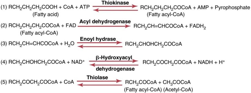
Figure 69-3. Beta oxidation of fatty acids to yield acetyl coenzyme A.
شکل 69-3. اکسیداسیون بتا اسیدهای چرب برای تولید استیل کوآنزیم A.
Formation of Acetoacetic Acid in the Liver and Its Transport in the Blood
A large share of the initial degradation of fatty acids occurs in the liver, especially when large amounts of lipids are being used for energy. However, the liver uses only a small proportion of the fatty acids for its own intrinsic metabolic processes. Instead, when the fatty acid chains have been split into acetyl-CoA, two molecules of acetyl-CoA condense to form one molecule of acetoacetic acid, which is then transported in the blood to the other cells throughout the body, where it is used for energy. The following chemical processes occur:
تشکیل اسید استواستیک در کبد و انتقال آن در خون
بخش بزرگی از تخریب اولیه اسیدهای چرب در کبد اتفاق می افتد، به ویژه زمانی که مقادیر زیادی از لیپیدها برای انرژی استفاده می شود. با این حال، کبد فقط از نسبت کمی از اسیدهای چرب برای فرآیندهای متابولیک ذاتی خود استفاده می کند. در عوض، زمانی که زنجیرههای اسید چرب به استیل کوآ تقسیم میشوند، دو مولکول استیل کوآ متراکم میشوند و یک مولکول اسید استواستیک را تشکیل میدهند، که سپس در خون به سلولهای دیگر در سراسر بدن منتقل میشود و در آنجا برای انرژی استفاده میشود. فرآیندهای شیمیایی زیر رخ می دهد:

Part of the acetoacetic acid is also converted into ẞ- hydroxybutyric acid, and minute quantities are converted into acetone in accord with the following reactions:
بخشی از اسید استواستیک نیز به اسید هیدروکسی بوتیریک تبدیل میشود و مقادیر بسیار کمی مطابق با واکنشهای زیر به استون تبدیل میشوند:

The acetoacetic acid, ẞ-hydroxybutyric acid, and acetone diffuse freely through the liver cell membranes and are transported by the blood to the peripheral tissues. Here they again diffuse into the cells, where reverse reactions occur, and acetyl-CoA molecules are formed. These molecules in turn enter the citric acid cycle and are oxidized for energy, as already explained.
اسید استواستیک، ẞ-هیدروکسی بوتیریک اسید و استون آزادانه از طریق غشاهای سلولی کبد منتشر می شوند و توسط خون به بافت های محیطی منتقل می شوند. در اینجا دوباره به داخل سلول ها پخش می شوند، جایی که واکنش های معکوس رخ می دهد و مولکول های استیل کوآ تشکیل می شوند. این مولکول ها به نوبه خود وارد چرخه اسید سیتریک می شوند و همانطور که قبلاً توضیح داده شد برای انرژی اکسید می شوند.
Normally, the acetoacetic acid and ẞ-hydroxybutyric acid that enter the blood are transported so rapidly to the tissues that their combined concentration in the plasma seldom rises above 3 mg/dl. Yet, despite this small con- centration in the blood, large quantities are actually trans- ported, as is also true for free fatty acid transport. The rapid transport of both these substances results from their high solubility in the membranes of the target cells, which allows almost instantaneous diffusion into the cells.
به طور معمول، اسید استواستیک و اسید هیدروکسی بوتیریک که وارد خون می شوند، به قدری سریع به بافت ها منتقل می شوند که غلظت ترکیبی آنها در پلاسما به ندرت از 3 میلی گرم در دسی لیتر بالاتر می رود. با این حال، علیرغم این غلظت کم در خون، مقادیر زیادی در واقع منتقل می شود، همانطور که برای انتقال اسیدهای چرب آزاد نیز صادق است. انتقال سریع هر دوی این مواد ناشی از حلالیت بالای آنها در غشای سلول های هدف است که امکان انتشار تقریباً آنی به داخل سلول ها را فراهم می کند.
Ketosis in Starvation, Diabetes, and Other Diseases.
The concentrations of acetoacetic acid, ẞ-hydroxybutyric acid, and acetone occasionally rise to levels many times normal in the blood and interstitial fluids; this condition is called ketosis because acetoacetic acid is a keto acid. The three compounds are called ketone bodies. Ketosis occurs especially as a consequence of starvation, in persons with diabetes mellitus, and sometimes even when a person’s diet is composed almost entirely of fat. In all these states, essentially no carbohydrates are metabolized-in starvation and with a high-fat diet because carbohydrates are not available, and in diabetes because insulin is not available to cause glucose transport into the cells.
کتوز در گرسنگی، دیابت و سایر بیماری ها.
غلظت استواستیک اسید، ẞ-هیدروکسی بوتیریک اسید و استون گهگاه در خون و مایعات بینابینی تا حد طبیعی افزایش می یابد. این وضعیت کتوز نامیده می شود زیرا اسید استواستیک یک کتو اسید است. این سه ترکیب اجسام کتون نامیده می شوند. کتوز به ویژه در نتیجه گرسنگی، در افراد مبتلا به دیابت رخ می دهد، و گاهی اوقات حتی زمانی که رژیم غذایی فرد تقریباً به طور کامل از چربی تشکیل شده باشد. در همه این حالت ها، اساساً هیچ کربوهیدراتی متابولیزه نمی شود – در گرسنگی و با رژیم غذایی پرچرب به دلیل در دسترس نبودن کربوهیدرات ها، و در دیابت به دلیل اینکه انسولین برای انتقال گلوکز به سلول ها در دسترس نیست.
When carbohydrates are not used for energy, almost all the energy of the body must come from fat metabolism. We shall see later in the chapter that the unavailability of carbohydrates automatically increases the rate of removal of fatty acids from adipose tissues. In addition, several hormonal factors such as increased secretion of glucocorticoids by the adrenal cortex, increased secretion of glucagon by the pancreas, and decreased secretion of insulin by the pancreas-further enhance removal of fatty acids from the fat tissues. As a result, large quantities of fatty acids become available (1) to the peripheral tissue cells to be used for energy and (2) to the liver cells, where much of the fatty acid is converted to ketone bodies.
وقتی از کربوهیدرات ها برای انرژی استفاده نمی شود، تقریبا تمام انرژی بدن باید از متابولیسم چربی تامین شود. بعداً در این فصل خواهیم دید که در دسترس نبودن کربوهیدرات ها به طور خودکار سرعت حذف اسیدهای چرب از بافت های چربی را افزایش می دهد. علاوه بر این، چندین فاکتور هورمونی مانند افزایش ترشح گلوکوکورتیکوئیدها توسط قشر آدرنال، افزایش ترشح گلوکاگون توسط لوزالمعده و کاهش ترشح انسولین توسط پانکراس – حذف اسیدهای چرب از بافت های چربی را افزایش می دهد. در نتیجه، مقادیر زیادی اسیدهای چرب (1) در دسترس سلول های بافت محیطی قرار می گیرند تا برای انرژی استفاده شوند و (2) برای سلول های کبد، جایی که بیشتر اسید چرب به اجسام کتون تبدیل می شود.
The ketone bodies pour out of the liver to be carried to the cells. For several reasons, the cells are limited in the amount of ketone bodies that can be oxidized. The most important reason for this limitation is that one of the products of carbohydrate metabolism is the oxaloacetate that is required to bind with acetyl-CoA before it can be processed in the citric acid cycle. Therefore, deficiency of oxaloacetate derived from carbohydrates limits the entry of acetyl-CoA into the citric acid cycle, and when a simultaneous outpouring of large quantities of acetoacetic acid and other ketone bodies from the liver occurs, the blood concentrations of acetoacetic acid and ẞ-hydroxybutyric acid sometimes rise to as high as 20 times normal, thus leading to extreme acidosis, as explained in Chapter 31.
اجسام کتونی از کبد بیرون می ریزند تا به سلول ها منتقل شوند. به چند دلیل، سلول ها در مقدار اجسام کتون که می توانند اکسید شوند محدود هستند. مهمترین دلیل این محدودیت این است که یکی از محصولات متابولیسم کربوهیدرات اگزالواستات است که قبل از پردازش در چرخه اسید سیتریک به استیل کوآ نیاز دارد. بنابراین، کمبود اگزالواستات به دست آمده از کربوهیدراتها، ورود استیل کوآ به چرخه اسید سیتریک را محدود میکند و هنگامی که همزمان مقادیر زیادی اسید استواستیک و سایر اجسام کتون از کبد خارج میشود، غلظت بالای استواستیک اسید و اسید هیدریک در خون گاهی اوقات تا 20 برابر طبیعی افزایش مییابد. اسیدوز همانطور که در فصل 31 توضیح داده شد.
The acetone that is formed during ketosis is a volatile substance, some of which is blown off in small quantities in the expired air of the lungs, thus giving the breath an acetone smell that is frequently used to diagnose ketosis.
استونی که در طی کتوز ایجاد میشود، یک ماده فرار است که مقدار کمی از آن در هوای منقضی شده ریهها خارج میشود، بنابراین بوی استون به نفس میدهد که اغلب برای تشخیص کتوز استفاده میشود.
Adaptation to a High-Fat Diet. When changing slowly from a carbohydrate diet to a diet almost entirely of fat, a person’s body adapts to use far more acetoacetic acid than usual, and in this case, ketosis normally does not occur. For instance, in the Inuit (Eskimos), who sometimes live mainly on a fat diet, ketosis does not develop. Undoubtedly, several factors, none of which is clear, enhance the rate of acetoacetic acid metabolism by the cells. After a few weeks, even the brain cells, which normally derive almost all their energy from glucose, can derive 50% to 75% of their energy from fats.
سازگاری با رژیم غذایی پرچرب. هنگامی که به آرامی از یک رژیم کربوهیدراتی به یک رژیم غذایی تقریباً تمام چربی تغییر میکند، بدن فرد برای استفاده از اسید استواستیک بسیار بیشتر از حد معمول سازگار میشود و در این حالت، کتوز معمولاً رخ نمیدهد. به عنوان مثال، در اینوئیت ها (اسکیموها)، که گاهی اوقات عمدتاً با رژیم غذایی چرب زندگی می کنند، کتوز ایجاد نمی شود. بدون شک عوامل متعددی که هیچ کدام مشخص نیستند، سرعت متابولیسم اسید استواستیک توسط سلول ها را افزایش می دهند. بعد از چند هفته، حتی سلول های مغزی که به طور معمول تقریباً تمام انرژی خود را از گلوکز می گیرند، می توانند 50 تا 75 درصد انرژی خود را از چربی ها به دست آورند.
Synthesis of Triglycerides From Carbohydrates
Whenever a greater quantity of carbohydrates enters the body than can be used immediately for energy or can be stored in the form of glycogen, the excess is rapidly converted into triglycerides and stored in this form in the adipose tissue.
سنتز تری گلیسیرید از کربوهیدرات ها
هر گاه مقدار بیشتری از کربوهیدرات ها وارد بدن شود که بتوان بلافاصله از آن برای انرژی استفاده کرد یا به شکل گلیکوژن ذخیره کرد، مقدار اضافی آن به سرعت به تری گلیسیرید تبدیل شده و به این شکل در بافت چربی ذخیره می شود.
In humans, most triglyceride synthesis occurs in the liver, but minute quantities are also synthesized in the adipose tissue. The triglycerides formed in the liver are trans- ported mainly in VLDLS to the adipose tissue, where they are stored.
در انسان، بیشتر سنتز تری گلیسیرید در کبد اتفاق می افتد، اما مقادیر کمی نیز در بافت چربی سنتز می شود. تری گلیسیریدهای تشکیل شده در کبد عمدتاً در VLDLS به بافت چربی منتقل می شوند و در آنجا ذخیره می شوند.
Conversion of Acetyl-CoA Into Fatty Acids. The first step in the synthesis of triglycerides is conversion of carbohydrates into acetyl-CoA. As explained in Chapter 68, this conversion occurs during the normal degradation of glucose by the glycolytic system. Because fatty acids are actually large polymers of acetic acid, it is easy to understand how acetyl-CoA can be converted into fatty acids. However, the synthesis of fatty acids from acetyl-CoA is not achieved by simply reversing the oxidative degradation described earlier. Instead, this occurs by the two-step process shown in Figure 69-4, using malonyl-CoA and reduced nicotinamide adenine dinucleotide phosphate (NADPH) as the principal intermediates in the polymerization process.
تبدیل استیل کوآ به اسیدهای چرب. اولین مرحله در سنتز تری گلیسیرید، تبدیل کربوهیدرات ها به استیل کوآ است. همانطور که در فصل 68 توضیح داده شد، این تبدیل در طی تجزیه طبیعی گلوکز توسط سیستم گلیکولیتیک اتفاق می افتد. از آنجایی که اسیدهای چرب در واقع پلیمرهای بزرگ اسید استیک هستند، درک چگونگی تبدیل استیل کوآ به اسیدهای چرب آسان است. با این حال، سنتز اسیدهای چرب از استیل کوآ به سادگی با معکوس کردن تخریب اکسیداتیو که قبلا توضیح داده شد به دست نمی آید. در عوض، این با فرآیند دو مرحله ای نشان داده شده در شکل 69-4، با استفاده از مالونیل-CoA و نیکوتین آمید آدنین دی نوکلئوتید فسفات احیا شده (NADPH) به عنوان واسطه های اصلی در فرآیند پلیمریزاسیون رخ می دهد.
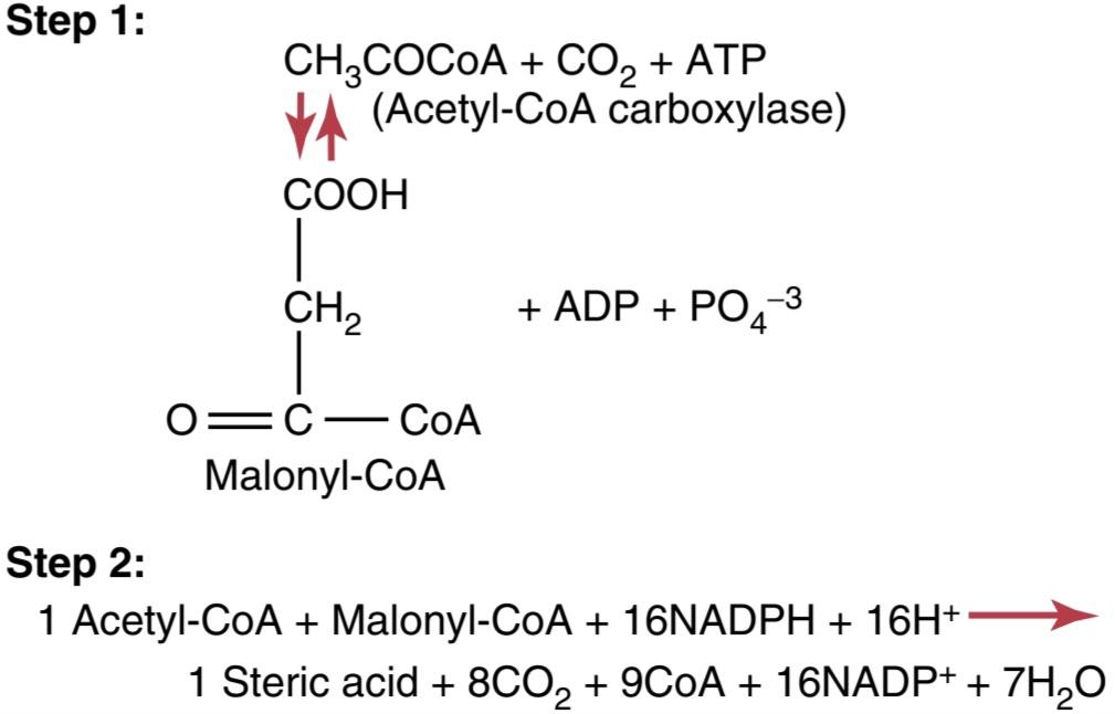
Figure 69-4. Synthesis of fatty acids.
شکل 69-4. سنتز اسیدهای چرب.
Combination of Fatty Acids With a-Glycerophosphate to Form Triglycerides
Once the synthesized fatty acid chains have grown to contain 14 to 18 carbon atoms, they bind with glycerol to form triglycerides. The enzymes that cause this conversion are highly specific for fatty acids, with chain lengths of 14 carbon atoms or greater, a factor that controls the physical quality of the triglycerides stored in the body.
ترکیب اسیدهای چرب با گلیسروفسفات برای تشکیل تری گلیسیرید
هنگامی که زنجیره های اسیدهای چرب سنتز شده رشد کرده و حاوی 14 تا 18 اتم کربن هستند، با گلیسرول متصل می شوند و تری گلیسیرید تشکیل می دهند. آنزیم هایی که باعث این تبدیل می شوند برای اسیدهای چرب بسیار خاص هستند، با طول زنجیره ای 14 اتم کربن یا بیشتر، عاملی که کیفیت فیزیکی تری گلیسیریدهای ذخیره شده در بدن را کنترل می کند.
As shown in Figure 69-5, the glycerol portion of triglycerides is furnished by a-glycerophosphate, which is another product derived from the glycolytic scheme of glucose degradation. This mechanism is discussed in Chapter 68.
همانطور که در شکل 69-5 نشان داده شده است، بخش گلیسرول تری گلیسیرید توسط a-glycerophosphate که محصول دیگری است که از طرح گلیکولیتیک تجزیه گلوکز به دست می آید، ارائه می شود. این مکانیسم در فصل 68 مورد بحث قرار گرفته است.
Efficiency of Carbohydrate Conversion Into Fat. During triglyceride synthesis, only about 15% of the original energy in the glucose is lost in the form of heat; the remaining 85% is transferred to the stored triglycerides.
کارایی تبدیل کربوهیدرات به چربی. در طول سنتز تری گلیسیرید، تنها حدود 15٪ از انرژی اولیه در گلوکز به شکل گرما از دست می رود. 85 درصد باقیمانده به تری گلیسیریدهای ذخیره شده منتقل می شود.
Importance of Fat Synthesis and Storage. Fat synthesis from carbohydrates is especially important for two reasons:
اهمیت سنتز و ذخیره چربی. سنتز چربی از کربوهیدرات ها به دو دلیل مهم است:
1. The ability of the different cells of the body to store carbohydrates in the form of glycogen is generally slight; a maximum of only a few hundred grams of glycogen can be stored in the liver, the skeletal muscles, and all other tissues of the body put together. In contrast, many kilograms of fat can be stored in adipose tissue. Therefore, fat synthesis provides a means by which the energy of excess ingested carbohydrates (and proteins) can be stored for later use. Indeed, the average person has almost 150 times as much energy stored in the form of fat as stored in the form of carbohydrate.
1. توانایی سلول های مختلف بدن برای ذخیره کربوهیدرات ها به شکل گلیکوژن به طور کلی ناچیز است. حداکثر تنها چند صد گرم گلیکوژن می تواند در کبد، ماهیچه های اسکلتی و سایر بافت های بدن در کنار هم ذخیره شود. در مقابل، بسیاری از کیلوگرم چربی را می توان در بافت چربی ذخیره کرد. بنابراین، سنتز چربی وسیلهای را فراهم میکند که به وسیله آن انرژی کربوهیدراتها (و پروتئینهای دریافتی) اضافی را میتوان برای استفاده بعدی ذخیره کرد. در واقع، یک فرد متوسط تقریباً 150 برابر انرژی ذخیره شده به شکل چربی نسبت به کربوهیدرات ذخیره شده دارد.
2. Each gram of fat contains almost two and a half times the calories of energy contained by each gram of glycogen. Therefore, for a given weight gain, a person can store several times as much energy in the form of fat as in the form of carbohydrate, which is exceedingly important when an animal must be highly motile to survive.
2. هر گرم چربی تقریباً دو و نیم برابر کالری انرژی موجود در هر گرم گلیکوژن دارد. بنابراین، برای افزایش وزن معین، یک فرد می تواند چندین برابر بیشتر از کربوهیدرات انرژی را به شکل چربی ذخیره کند، که زمانی که یک حیوان برای زنده ماندن باید تحرک بالایی داشته باشد، بسیار مهم است.
Failure to Synthesize Fats From Carbohydrates in the Absence of Insulin. When insufficient insulin is available, as occurs in persons with serious cases of diabetes mellitus, fats are poorly synthesized, if at all, for the following reasons. First, when insulin is not available, glucose does not enter the fat and liver cells satisfactorily, so little of the acetyl-CoA and NADPH needed for fat synthesis can be derived from glucose. Second, lack of glucose in the fat cells greatly reduces the availability of a-glycerophosphate, which also makes it difficult for the tissues to form triglycerides.
عدم سنتز چربی ها از کربوهیدرات ها در غیاب انسولین. هنگامی که انسولین کافی در دسترس نیست، همانطور که در افراد مبتلا به موارد جدی دیابت رخ می دهد، چربی ها به دلایل زیر ضعیف سنتز می شوند. اول اینکه وقتی انسولین در دسترس نیست، گلوکز به طور رضایت بخشی وارد سلول های چربی و کبد نمی شود، بنابراین مقدار کمی از استیل-CoA و NADPH مورد نیاز برای سنتز چربی را می توان از گلوکز به دست آورد. دوم، کمبود گلوکز در سلولهای چربی، دسترسی به گلیسروفسفات را تا حد زیادی کاهش میدهد، که همچنین تشکیل تری گلیسیرید را برای بافتها دشوار میکند.

Figure 69-5. Overall schema for synthesis of triglycerides from glucose.
شکل 69-5. طرح کلی برای سنتز تری گلیسیرید از گلوکز.
Synthesis of Triglycerides From Proteins
Many amino acids can be converted into acetyl-CoA, as discussed in Chapter 70. The acetyl-CoA can then be synthesized into triglycerides. Therefore, when people have more proteins in their diets than their tissues can use as proteins, a large share of the excess is stored as fat.
سنتز تری گلیسیرید از پروتئین ها
بسیاری از اسیدهای آمینه می توانند به استیل-CoA تبدیل شوند، همانطور که در فصل 70 مورد بحث قرار گرفت. سپس استیل-CoA می تواند به تری گلیسیرید سنتز شود. بنابراین، زمانی که افراد در رژیم غذایی خود پروتئین بیشتری نسبت به بافت هایشان به عنوان پروتئین دارند، سهم زیادی از مازاد آن به عنوان چربی ذخیره می شود.
Regulation of Energy Release From Triglycerides
Carbohydrates Are Preferred Over Fats for Energy When Excess Carbohydrates Are Available. When excess carbohydrates are available in the body, carbohydrates are used preferentially over triglycerides for energy. Several reasons exist for this “fat-sparing” effect of carbohydrates.
تنظیم آزاد شدن انرژی از تری گلیسیرید
وقتی کربوهیدراتهای اضافی در دسترس هستند، کربوهیدراتها بر چربیها ترجیح داده میشوند. زمانی که کربوهیدرات های اضافی در بدن موجود باشد، کربوهیدرات ها ترجیحاً نسبت به تری گلیسیرید برای انرژی استفاده می شوند. دلایل متعددی برای این اثر «چربیدهی» کربوهیدراتها وجود دارد.
First, fats in adipose tissue cells are present in two forms: stored triglycerides and small quantities of free fatty acids. They are in constant equilibrium with each other. When excess a-glycerophosphate is present (which occurs when excess carbohydrates are available), the excess a-glycerophosphate binds the free fatty acids in the form of stored triglycerides. As a result, the equilibrium between free fatty acids and triglycerides shifts toward the stored triglycerides; consequently, only minute quantities of fatty acids are available to be used for energy. Because a-glycerophosphate is an important product of glucose metabolism, the availability of large amounts of glucose automatically inhibits the use of fatty acids for energy.
اول، چربی ها در سلول های بافت چربی به دو شکل وجود دارند: تری گلیسیریدهای ذخیره شده و مقادیر کمی اسیدهای چرب آزاد. آنها در تعادل دائمی با یکدیگر هستند. هنگامی که a-glycerophosphate اضافی وجود داشته باشد (که زمانی رخ می دهد که کربوهیدرات اضافی در دسترس باشد)، a-glycerophosphate اضافی به اسیدهای چرب آزاد به شکل تری گلیسیرید ذخیره شده متصل می شود. در نتیجه، تعادل بین اسیدهای چرب آزاد و تری گلیسیریدها به سمت تری گلیسیریدهای ذخیره شده تغییر می کند. در نتیجه، فقط مقادیر بسیار کمی از اسیدهای چرب برای مصرف انرژی در دسترس هستند. از آنجایی که a-glycerophosphate محصول مهم متابولیسم گلوکز است، در دسترس بودن مقادیر زیادی گلوکز به طور خودکار استفاده از اسیدهای چرب برای انرژی را مهار می کند.
Second, when carbohydrates are available in excess, fatty acids are synthesized more rapidly than they are de- graded. This effect is caused partially by the large quantities of acetyl-CoA formed from the carbohydrates and by the low concentration of free fatty acids in the adipose tissue, thus creating conditions appropriate for the conversion of acetyl-CoA into fatty acids.
ثانیاً، وقتی کربوهیدراتها بیش از حد در دسترس باشند، اسیدهای چرب سریعتر از تجزیه آنها سنتز میشوند. این اثر تا حدی به دلیل مقادیر زیاد استیل-CoA تشکیل شده از کربوهیدرات ها و غلظت کم اسیدهای چرب آزاد در بافت چربی ایجاد می شود، بنابراین شرایط مناسب برای تبدیل استیل-CoA به اسیدهای چرب ایجاد می شود.
An even more important effect that promotes conversion of carbohydrates to fats is the following: The first step, which is the rate-limiting step, in the synthesis of fatty acids is carboxylation of acetyl-CoA to form malonyl-CoA. The rate of this reaction is controlled primarily by the enzyme acetyl-CoA carboxylase, the activity of which is accelerated in the presence of intermediates of the citric acid cycle. When excess carbohydrates are being used, these intermediates increase, automatically causing increased synthesis of fatty acids.
یک اثر مهم تر که تبدیل کربوهیدرات ها به چربی ها را افزایش می دهد موارد زیر است: مرحله اول که مرحله محدود کننده سرعت است در سنتز اسیدهای چرب کربوکسیلاسیون استیل-CoA برای تشکیل مالونیل-CoA است. سرعت این واکنش در درجه اول توسط آنزیم استیل کوآ کربوکسیلاز کنترل می شود که فعالیت آن در حضور واسطه های چرخه اسید سیتریک تسریع می شود. هنگامی که کربوهیدرات های اضافی استفاده می شود، این واسطه ها افزایش می یابد و به طور خودکار باعث افزایش سنتز اسیدهای چرب می شود.
Thus, an excess of carbohydrates in the diet not only acts as a fat-sparer but also increases fat stores. In fact, all the excess carbohydrates not used for energy or stored in the small glycogen deposits of the body are converted to fat for storage.
بنابراین، کربوهیدرات بیش از حد در رژیم غذایی نه تنها به عنوان یک کاهش دهنده چربی عمل می کند، بلکه باعث افزایش ذخایر چربی می شود. در واقع، تمام کربوهیدراتهای اضافی که برای انرژی استفاده نمیشوند یا در ذخایر کوچک گلیکوژن بدن ذخیره نمیشوند، برای ذخیره به چربی تبدیل میشوند.
Acceleration of Fat Utilization for Energy in the Absence of Carbohydrates. All the fat-sparing effects of carbohydrates are lost and actually reversed when carbohydrates are not available. The equilibrium shifts in the opposite direction, and fat is mobilized from adipose cells and used for energy in place of carbohydrates.
تسریع استفاده از چربی برای انرژی در غیاب کربوهیدرات. وقتی کربوهیدراتها در دسترس نباشند، تمام اثرات چربیدهی کربوهیدراتها از بین میروند و در واقع معکوس میشوند. تعادل در جهت مخالف تغییر می کند و چربی از سلول های چربی بسیج می شود و به جای کربوهیدرات ها برای انرژی استفاده می شود.
Also important are several hormonal changes that take place to promote rapid fatty acid mobilization from adipose tissue. Among the most important of these hormonal changes is a marked decrease in pancreatic secretion of insulin caused by the absence of carbohydrates. This decrease in insulin not only reduces the rate of glucose utilization by the tissues but also decreases fat storage, which further shifts the equilibrium in favor of fat metabolism in place of carbohydrates.
همچنین تغییرات هورمونی متعددی مهم است که برای تحریک سریع اسیدهای چرب از بافت چربی رخ می دهد. از جمله مهمترین این تغییرات هورمونی، کاهش قابل توجه ترشح انسولین از پانکراس است که به دلیل عدم وجود کربوهیدرات ایجاد می شود. این کاهش انسولین نه تنها میزان استفاده از گلوکز توسط بافت ها را کاهش می دهد، بلکه ذخیره چربی را نیز کاهش می دهد، که باعث تغییر بیشتر تعادل به نفع متابولیسم چربی به جای کربوهیدرات ها می شود.
Hormonal Regulation of Fat Utilization. At least seven of the hormones secreted by the endocrine glands have significant effects on fat utilization. Some important hormonal effects on fat metabolism, in addition to lack of insulin, are noted here.
تنظیم هورمونی مصرف چربی. حداقل هفت هورمون ترشح شده توسط غدد درون ریز تأثیرات قابل توجهی بر مصرف چربی دارند. برخی از اثرات مهم هورمونی بر متابولیسم چربی، علاوه بر کمبود انسولین، در اینجا ذکر شده است.
Probably the most dramatic increase that occurs in fat utilization is that observed during heavy exercise. This in- crease results almost entirely from release of epinephrine and norepinephrine by the adrenal medullae during exercise, as a result of sympathetic stimulation. These two hormones directly activate hormone-sensitive triglyceride lipase, which is present in abundance in the fat cells, and this activation causes rapid breakdown of triglycerides and mobilization of fatty acids. Sometimes the free fatty acid concentration in the blood of an exercising person rises as much as eightfold, and the use of these fatty acids by the muscles for energy is correspondingly increased. Other types of stress that activate the sympathetic nervous system can also increase fatty acid mobilization and utilization in a similar manner.
احتمالاً چشمگیرترین افزایشی که در مصرف چربی رخ می دهد، افزایشی است که در طول ورزش سنگین مشاهده می شود. این افزایش تقریباً به طور کامل ناشی از آزاد شدن اپی نفرین و نوراپی نفرین توسط بصل الکلیف آدرنال در حین ورزش در نتیجه تحریک سمپاتیک است. این دو هورمون مستقیماً لیپاز تری گلیسیرید حساس به هورمون را فعال می کنند که به وفور در سلول های چربی وجود دارد و این فعال شدن باعث تجزیه سریع تری گلیسیرید و تحرک اسیدهای چرب می شود. گاهی اوقات غلظت اسیدهای چرب آزاد در خون یک فرد ورزشکار تا 8 برابر افزایش می یابد و مصرف این اسیدهای چرب توسط ماهیچه ها برای انرژی به همان نسبت افزایش می یابد. انواع دیگر استرس که سیستم عصبی سمپاتیک را فعال میکنند نیز میتوانند حرکت و استفاده از اسیدهای چرب را به روشی مشابه افزایش دهند.
Stress also causes large quantities of adrenocorticotropic hormone (ACTH) to be released by the anterior pituitary gland, which causes the adrenal cortex to secrete extra quantities of glucocorticoids. Both ACTH and glucocorticoids activate either the same hormone-sensitive triglyceride lipase as that activated by epinephrine and norepinephrine or a similar lipase. When ACTH and glucocorticoids are secreted in excessive amounts for long periods, as occurs in the endocrine condition called Cushing’s syndrome, fats are frequently mobilized to such a great extent that ketosis results. ACTH and glucocorticoids are then said to have a ketogenic effect. Growth hormone has an effect similar to but weaker than that of ACTH and glucocorticoids in activating hormone-sensitive lipase. Therefore, growth hormone can also have a mild ketogenic effect.
استرس همچنین باعث می شود مقادیر زیادی هورمون آدرنوکورتیکوتروپیک (ACTH) توسط غده هیپوفیز قدامی ترشح شود که باعث می شود قشر آدرنال مقادیر اضافی گلوکوکورتیکوئید ترشح کند. هم ACTH و هم گلوکوکورتیکوئیدها همان لیپاز تری گلیسیرید حساس به هورمون را فعال می کنند که توسط اپی نفرین و نوراپی نفرین یا لیپاز مشابه فعال می شود. هنگامی که ACTH و گلوکوکورتیکوئیدها در مقادیر بیش از حد برای دورههای طولانی ترشح میشوند، همانطور که در شرایط غدد درون ریز به نام سندرم کوشینگ رخ میدهد، چربیها اغلب بهقدری زیاد حرکت میکنند که منجر به کتوز میشود. سپس گفته می شود که ACTH و گلوکوکورتیکوئیدها دارای اثر کتوژنیک هستند. هورمون رشد اثری مشابه اما ضعیف تر از ACTH و گلوکوکورتیکوئیدها در فعال کردن لیپاز حساس به هورمون دارد. بنابراین، هورمون رشد نیز می تواند اثر کتوژنیک خفیفی داشته باشد.
Thyroid hormone indirectly causes rapid mobilization of fat by increasing overall rate of energy metabolism in all cells of the body under the influence of this hormone. The resulting reduction in acetyl-CoA and other intermediates of both fat and carbohydrate metabolism in the cells is a stimulus to fat mobilization.
هورمون تیروئید به طور غیرمستقیم با افزایش سرعت کلی متابولیسم انرژی در تمام سلولهای بدن تحت تأثیر این هورمون باعث تحرک سریع چربی میشود. کاهش حاصل در استیل کوآ و سایر واسطه های متابولیسم چربی و کربوهیدرات در سلول ها محرکی برای بسیج چربی است.
The effects of the different hormones on metabolism are discussed further in the chapters dealing with each hormone.
اثرات هورمون های مختلف بر متابولیسم بیشتر در فصل های مربوط به هر هورمون مورد بحث قرار می گیرد.
Obesity-Excess Deposition of Fat
Obesity is discussed in Chapter 72 in relation to dietary balances, but briefly, it is caused by the ingestion of greater amounts of food than can be used by the body for energy. The excess food, whether fats, carbohydrates, or proteins, is then stored almost entirely as fat in the adipose tissue, to be used later for energy. The ability of humans to store excess energy in adipose tissue is tremendous, with some people achieving body weights in excess of 500 kilograms (1100 pounds), mainly as a result of fat accumulation.
چاقی- رسوب بیش از حد چربی
چاقی در فصل 72 در رابطه با تعادل رژیم غذایی مورد بحث قرار گرفته است، اما به طور خلاصه، این چاقی ناشی از مصرف مقادیر بیشتری از مواد غذایی است که بدن می تواند برای انرژی از آن استفاده کند. غذای اضافی، اعم از چربی، کربوهیدرات یا پروتئین، تقریباً به طور کامل به عنوان چربی در بافت چربی ذخیره می شود تا بعداً برای انرژی استفاده شود. توانایی انسان برای ذخیره انرژی اضافی در بافت چربی فوق العاده است، به طوری که برخی از افراد به وزن بیش از 500 کیلوگرم (1100 پوند) عمدتاً در نتیجه تجمع چربی می رسند.
Several strains of rodents have been found in which hereditary obesity occurs. In at least one of these strains, the obesity is caused by ineffective mobilization of fat from the adipose tissue by tissue lipase, while synthesis and storage of fat continue normally. Such a one-way process causes progressive enhancement of the fat stores, resulting in severe obesity. Multiple genetic factors that influence brain feeding centers or pathways that control energy expenditure or that alter energy storage can also cause hereditary obesity in humans. However, monogenic (single gene) causes of human obesity are rare, as discussed in Chapter 72.
چندین گونه از جوندگان یافت شده است که در آنها چاقی ارثی رخ می دهد. حداقل در یکی از این سویه ها، چاقی ناشی از حرکت بی اثر چربی از بافت چربی توسط لیپاز بافت است، در حالی که سنتز و ذخیره چربی به طور طبیعی ادامه می یابد. چنین فرآیند یک طرفه باعث افزایش تدریجی ذخایر چربی و در نتیجه چاقی شدید می شود. عوامل ژنتیکی متعددی که بر مراکز تغذیه مغز یا مسیرهایی که مصرف انرژی را کنترل میکنند یا ذخیره انرژی را تغییر میدهند، تأثیر میگذارند، همچنین میتوانند باعث چاقی ارثی در انسان شوند. با این حال، علل تک ژنی (تک ژنی) چاقی انسان، همانطور که در فصل 72 مورد بحث قرار گرفت، نادر است.
Phospholipids and Cholesterol
Phospholipids
The major types of body phospholipids are lecithins, cephalins, and sphingomyelin; their typical chemical formulas are shown in Figure 69-6. Phospholipids always contain one or more fatty acid molecules and one phosphoric acid radical, and they usually contain a nitrogenous base. Although the chemical structures of phospholipids are somewhat variant, their physical properties are similar because they are all lipid soluble, transported in lipoproteins, and used throughout the body for various structural purposes, such as in cell membranes and intracellular membranes.
فسفولیپیدها و کلسترول
فسفولیپیدها
انواع عمده فسفولیپیدهای بدن لسیتین ها، سفالین ها و اسفنگومیلین هستند. فرمول های شیمیایی معمول آنها در شکل 69-6 نشان داده شده است. فسفولیپیدها همیشه حاوی یک یا چند مولکول اسید چرب و یک رادیکال اسید فسفریک هستند و معمولاً حاوی یک باز نیتروژن هستند. اگرچه ساختارهای شیمیایی فسفولیپیدها تا حدودی متفاوت است، خواص فیزیکی آنها مشابه است زیرا همه آنها محلول در چربی هستند، در لیپوپروتئین ها منتقل می شوند و در سراسر بدن برای اهداف ساختاری مختلف مانند غشای سلولی و غشای داخل سلولی استفاده می شوند.

Figure 69-6. Typical phospholipids.
شکل 69-6. فسفولیپیدهای معمولی
Formation of Phospholipids. Phospholipids are synthesized in essentially all cells of the body, although certain cells have a special ability to form great quantities of them. Probably 90% of phospholipids are formed in liver cells; substantial quantities are also formed by the intestinal epithelial cells during lipid absorption from the gut.
تشکیل فسفولیپیدها. فسفولیپیدها اساساً در تمام سلولهای بدن سنتز میشوند، اگرچه سلولهای خاصی توانایی خاصی برای تشکیل مقادیر زیادی از آنها دارند. احتمالاً 90 درصد فسفولیپیدها در سلول های کبدی تشکیل می شوند. مقادیر قابل توجهی نیز توسط سلول های اپیتلیال روده در طول جذب لیپید از روده تشکیل می شود.
The rate of phospholipid formation is governed to some extent by the usual factors that control the overall rate of fat metabolism because, when triglycerides are deposited in the liver, the rate of phospholipid formation increases. Also, specific chemical substances are needed for the formation of some phospholipids. For instance, choline, either obtained in the diet or synthesized in the body, is necessary for formation of lecithin, because choline is the nitrogenous base of the lecithin molecule. In addition, inositol is necessary for formation of some cephalins.
سرعت تشکیل فسفولیپید تا حدی توسط عوامل معمولی کنترل می شود که میزان کلی متابولیسم چربی را کنترل می کنند، زیرا وقتی تری گلیسیرید در کبد رسوب می کند، سرعت تشکیل فسفولیپید افزایش می یابد. همچنین برای تشکیل برخی از فسفولیپیدها به مواد شیمیایی خاصی نیاز است. به عنوان مثال، کولین، یا در رژیم غذایی به دست می آید یا در بدن سنتز می شود، برای تشکیل لسیتین ضروری است، زیرا کولین پایه نیتروژنی مولکول لسیتین است. علاوه بر این، اینوزیتول برای تشکیل برخی از سفالین ها ضروری است.
Specific Uses of Phospholipids. Phospholipids have several functions, including the following:
کاربردهای خاص فسفولیپیدها .فسفولیپیدها عملکردهای مختلفی دارند، از جمله:
1. Phospholipids are an important constituent of lipoproteins in the blood and are essential for formation and function of most of these lipoproteins; in the absence of phospholipids, serious abnormalities of transport of cholesterol and other lipids can occur.
1. فسفولیپیدها جزء مهم لیپوپروتئین های خون هستند و برای تشکیل و عملکرد بیشتر این لیپوپروتئین ها ضروری هستند. در غیاب فسفولیپیدها، ناهنجاری های جدی انتقال کلسترول و سایر لیپیدها ممکن است رخ دهد.
2. Thromboplastin, which is necessary to initiate the clotting process, is composed mainly of one of the cephalins.
2. ترومبوپلاستین که برای شروع فرآیند لخته شدن ضروری است، عمدتاً از یکی از سفالین ها تشکیل شده است.
3. Large quantities of sphingomyelin are present in the nervous system; this substance acts as an electrical insulator in the myelin sheath around nerve fibers.
3. مقادیر زیادی اسفنگومیلین در سیستم عصبی وجود دارد. این ماده به عنوان یک عایق الکتریکی در غلاف میلین اطراف رشته های عصبی عمل می کند.
4. Phospholipids are donors of phosphate radicals when these radicals are necessary for different chemical reactions in the tissues.
4. فسفولیپیدها اهداکننده رادیکال های فسفات هستند که این رادیکال ها برای واکنش های شیمیایی مختلف در بافت ها ضروری باشند.
5. One of the most important functions of phospholipids is participation in formation of structural elements- mainly membranes-in cells throughout the body, as discussed in the next section of this chapter in connection with a similar function for cholesterol.
5. یکی از مهم ترین عملکردهای فسفولیپیدها، مشارکت در تشکیل عناصر ساختاری- عمدتاً غشاء- در سلول ها در سراسر بدن است، همانطور که در بخش بعدی این فصل در رابطه با عملکرد مشابه کلسترول مورد بحث قرار گرفت.
Cholesterol
Cholesterol, the formula of which is shown in Figure 69-7, is present in the normal diet, and it can be absorbed slowly from the gastrointestinal tract into the intestinal lymph. It is highly fat soluble but only slightly soluble in water. It is specifically capable of forming esters with fatty acids. Indeed, about 70% of the cholesterol in the lipoproteins of the plasma is in the form of cholesterol esters.
کلسترول
کلسترول که فرمول آن در شکل 69-7 نشان داده شده است، در رژیم غذایی معمولی وجود دارد و می تواند به آرامی از دستگاه گوارش به داخل لنف روده جذب شود. بسیار محلول در چربی است اما کمی در آب حل می شود. به طور خاص قادر به تشکیل استر با اسیدهای چرب است. در واقع، حدود 70 درصد کلسترول موجود در لیپوپروتئین های پلاسما به شکل استرهای کلسترول است.

Figure 69-7. Cholesterol.
شکل 69-7. کلسترول
Formation of Cholesterol. Besides the cholesterol ab- sorbed each day from the gastrointestinal tract, which is called exogenous cholesterol, an even greater quantity is formed in the cells of the body, called endogenous cholesterol. Essentially all the endogenous cholesterol that circulates in the lipoproteins of the plasma is formed by the liver, but all other cells of the body form at least some cholesterol, which is consistent with the fact that many of the membra- nous structures of all cells are partially composed of this substance.
تشکیل کلسترول. علاوه بر کلسترولی که هر روز از دستگاه گوارش جذب می شود، که به آن کلسترول اگزوژن می گویند، مقدار بیشتری نیز در سلول های بدن تشکیل می شود که به آن کلسترول درون زا می گویند. اساساً تمام کلسترول درون زا که در لیپوپروتئینهای پلاسما گردش میکند توسط کبد تشکیل میشود، اما تمام سلولهای دیگر بدن حداقل مقداری کلسترول را تشکیل میدهند که با این واقعیت مطابقت دارد که بسیاری از ساختارهای غشایی همه سلولها تا حدی از این ماده تشکیل شدهاند.
The basic structure of cholesterol is a sterol nucleus, which is synthesized entirely from multiple molecules of acetyl-CoA. In turn, the sterol nucleus can be modified by various side chains to form (1) cholesterol; (2) cholic acid, which is the basis of the bile acids formed in the liver; and (3) many important steroid hormones secreted by the adrenal cortex, the ovaries, and the testes (these hormones are discussed in later chapters).
ساختار اصلی کلسترول یک هسته استرول است که به طور کامل از چندین مولکول استیل-CoA سنتز می شود. به نوبه خود، هسته استرول را می توان با زنجیره های جانبی مختلف تغییر داد تا (1) کلسترول را تشکیل دهد. (2) اسید کولیک، که اساس اسیدهای صفراوی تشکیل شده در کبد است. و (3) بسیاری از هورمونهای استروئیدی مهم که از قشر آدرنال، تخمدانها و بیضهها ترشح میشوند (این هورمونها در فصلهای بعدی مورد بحث قرار خواهند گرفت).
Factors That Affect Plasma Cholesterol Concentration- Feedback Control of Body Cholesterol. Among the important factors that affect plasma cholesterol concentration are the following:
عوامل موثر بر غلظت کلسترول پلاسما- کنترل بازخورد کلسترول بدن. از جمله عوامل مهمی که بر غلظت کلسترول پلاسما تأثیر می گذارد می توان به موارد زیر اشاره کرد:
1. An increase in the amount of cholesterol ingested each day may increase the plasma concentration slightly. However, when cholesterol is ingested, the rising concentration of cholesterol inhibits the most essential enzyme for endogenous synthesis of cholesterol, 3-hydroxy-3-methylglutaryl CoA reductase, thus pro- viding an intrinsic feedback control system to prevent an excessive increase in plasma cholesterol concentration. As a result, plasma cholesterol concentration usually is not changed upward or downward more than +15% by altering the amount of cholesterol in the diet, although the response of individuals differs markedly.
1. افزایش مقدار کلسترول مصرفی در هر روز ممکن است غلظت پلاسما را کمی افزایش دهد. با این حال، هنگامی که کلسترول خورده می شود، افزایش غلظت کلسترول، ضروری ترین آنزیم برای سنتز درون زا کلسترول، 3-هیدروکسی-3- متیل گلوتاریل CoA ردوکتاز را مهار می کند، بنابراین یک سیستم کنترل بازخورد ذاتی برای جلوگیری از افزایش بیش از حد غلظت پلاسمایی کلسترول فراهم می کند. در نتیجه، غلظت کلسترول پلاسما معمولاً با تغییر مقدار کلسترول در رژیم غذایی بیش از 15% به سمت بالا یا پایین تغییر نمی کند، اگرچه پاسخ افراد به طور قابل توجهی متفاوت است.
2. A diet high in saturated fat increases blood cholesterol concentration 15% to 25%, especially when this diet is associated with excess weight gain and obesity. This increase in blood cholesterol results from increased fat deposition in the liver, which then provides increased quantities of acetyl-CoA in the liver cells for the production of cholesterol. Therefore, to decrease the blood cholesterol concentration, maintaining a normal body weight and a diet low in saturated fat are even more important than maintaining a diet low in cholesterol.
2. رژیم غذایی سرشار از چربی اشباع شده، غلظت کلسترول خون را 15 تا 25 درصد افزایش می دهد، به خصوص زمانی که این رژیم با اضافه وزن و چاقی همراه باشد. این افزایش در کلسترول خون ناشی از افزایش رسوب چربی در کبد است که پس از آن مقادیر بیشتری از استیل کوآ را در سلول های کبد برای تولید کلسترول فراهم می کند. بنابراین، برای کاهش غلظت کلسترول خون، حفظ وزن طبیعی بدن و رژیم غذایی کم چربی اشباع شده حتی مهمتر از حفظ یک رژیم غذایی کم کلسترول است.
3. Ingestion of fat containing highly unsaturated fatty acids usually depresses the blood cholesterol concentration a slight to moderate amount. The mechanism of this effect is unknown, despite the fact that this observation is the basis of much present-day dietary strategy.
3. مصرف چربی حاوی اسیدهای چرب غیراشباع بالا معمولاً غلظت کلسترول خون را کمی تا متوسط کاهش می دهد. مکانیسم این اثر ناشناخته است، علیرغم این واقعیت که این مشاهدات اساس بسیاری از استراتژی های رژیم غذایی امروزی است.
4. Lack of insulin or thyroid hormone increases the blood cholesterol concentration, whereas excess thyroid hormone decreases the concentration. These effects are probably caused mainly by changes in the degree of activation of specific enzymes responsible for the metabolism of lipids and overall metabolic rate.
4. کمبود انسولین یا هورمون تیروئید باعث افزایش غلظت کلسترول خون می شود در حالی که هورمون تیروئید اضافی باعث کاهش غلظت می شود. این اثرات احتمالاً عمدتاً ناشی از تغییر در درجه فعال شدن آنزیم های خاص مسئول متابولیسم لیپیدها و میزان متابولیسم کلی است.
5. Genetic disorders of cholesterol metabolism may greatly increase plasma cholesterol levels. For example, mutations of the LDL receptor gene prevent the liver from adequately removing the cholesterol-rich LDLs from the plasma. As discussed later, this phenomenon causes the liver to produce excessive amounts of cholesterol. Mutations of the gene that encodes apolipoprotein B, the part of the LDL that binds to the receptor, also cause excessive cholesterol production by the liver.
5. اختلالات ژنتیکی متابولیسم کلسترول ممکن است سطح کلسترول پلاسما را تا حد زیادی افزایش دهد. به عنوان مثال، جهش ژن گیرنده LDL مانع از حذف کافی LDL های غنی از کلسترول از پلاسما توسط کبد می شود. همانطور که بعداً بحث شد، این پدیده باعث می شود کبد مقادیر زیادی کلسترول تولید کند. جهش ژنی که آپولیپوپروتئین B را کد می کند، بخشی از LDL که به گیرنده متصل می شود، همچنین باعث تولید بیش از حد کلسترول توسط کبد می شود.
Specific Uses of Cholesterol in the Body. By far the most abundant non-membranous use of cholesterol in the body is to form cholic acid in the liver. As much as 80% of cholesterol is converted into cholic acid. As explained in Chapter 71, cholic acid is conjugated with other substances to form bile salts, which promote digestion and absorption of fats.
کاربردهای خاص کلسترول در بدن. بیشترین کاربرد غیر غشایی کلسترول در بدن، تشکیل اسید کولیک در کبد است. 80 درصد کلسترول به اسید کولیک تبدیل می شود. همانطور که در فصل 71 توضیح داده شد، اسید کولیک با مواد دیگری ترکیب می شود تا نمک های صفراوی را تشکیل دهد که هضم و جذب چربی ها را تقویت می کند.
A small quantity of cholesterol is used by (1) the adrenal glands to form adrenocortical hormones, (2) the ovaries to form progesterone and estrogen, and (3) the testes to form testosterone. These glands can also synthesize their own sterols and then form hormones from them, as discussed in the chapters on endocrinology.
مقدار کمی از کلسترول توسط (1) غدد فوق کلیوی برای تشکیل هورمون های قشر آدرنال، (2) تخمدان ها برای تشکیل پروژسترون و استروژن، و (3) بیضه ها برای تشکیل تستوسترون استفاده می شود. این غدد همچنین می توانند استرول های خود را سنتز کنند و سپس هورمون هایی از آنها تشکیل دهند، همانطور که در فصل های غدد درون ریز مورد بحث قرار گرفت.
A large amount of cholesterol is precipitated in the corneum of the skin. This cholesterol, along with other lipids, makes the skin highly resistant to the absorption of water-soluble substances and to the action of many chemical agents because cholesterol and the other skin lipids are highly inert to acids and to many solvents that might other- wise easily penetrate the body. Also, these lipid substances help prevent water evaporation from the skin; without this protection, the amount of evaporation can be 5 to 10 liters per day (as occurs in patients with burns who have lost their skin) instead of the usual 300 to 400 milliliters.
مقدار زیادی کلسترول در قرنیه پوست رسوب می کند. این کلسترول همراه با سایر لیپیدها باعث می شود که پوست در برابر جذب مواد محلول در آب و در برابر عمل بسیاری از عوامل شیمیایی مقاوم باشد زیرا کلسترول و سایر چربی های پوست نسبت به اسیدها و بسیاری از حلال ها که در غیر این صورت به راحتی به بدن نفوذ می کنند بسیار بی اثر هستند. همچنین، این مواد لیپیدی به جلوگیری از تبخیر آب از پوست کمک می کنند. بدون این محافظ، میزان تبخیر می تواند 5 تا 10 لیتر در روز (همانطور که در بیماران سوختگی که پوست خود را از دست داده اند) به جای 300 تا 400 میلی لیتر معمول باشد.
Cellular Structural Functions of Phospholipids and Cholesterol Especially for Membranes. The previously mentioned uses of phospholipids and cholesterol are of only minor importance in comparison with their function of forming specialized structures, mainly membranes, in all cells of the body. In Chapter 2, it was pointed out that large quantities of phospholipids and cholesterol are present in both the cell membrane and the membranes of the internal organelles of all cells. It is also known that the ratio of membrane cholesterol to phospholipids is especially important in determining the fluidity of cell membranes.
عملکردهای ساختاری سلولی فسفولیپیدها و کلسترول به ویژه برای غشاء. کاربردهای ذکر شده قبلی از فسفولیپیدها و کلسترول در مقایسه با عملکرد آنها برای تشکیل ساختارهای تخصصی، عمدتاً غشاء، در تمام سلول های بدن اهمیت ناچیزی دارند. در فصل 2 اشاره شد که مقادیر زیادی فسفولیپید و کلسترول هم در غشای سلولی و هم در غشای اندامک های داخلی همه سلول ها وجود دارد. همچنین مشخص شده است که نسبت کلسترول غشایی به فسفولیپیدها در تعیین سیالیت غشاهای سلولی اهمیت ویژه ای دارد.
For membranes to be formed, substances that are not soluble in water must be available. In general, the only substances in the body that are not soluble in water (be- sides the inorganic substances of bone) are the lipids and some proteins. Thus, the physical integrity of cells everywhere in the body is based mainly on phospholipids, cholesterol, and certain insoluble proteins. The polar charges on the phospholipids also reduce the interfacial tension between the cell membranes and the surrounding fluids.
برای تشکیل غشاها، موادی که در آب محلول نیستند باید در دسترس باشند. به طور کلی، تنها موادی در بدن که در آب حل نمی شوند (علاوه بر مواد معدنی استخوان)، لیپیدها و برخی پروتئین ها هستند. بنابراین، یکپارچگی فیزیکی سلول ها در همه جای بدن عمدتاً بر اساس فسفولیپیدها، کلسترول و برخی پروتئین های نامحلول است. بارهای قطبی روی فسفولیپیدها همچنین کشش سطحی بین غشای سلولی و مایعات اطراف را کاهش می دهد.
Another fact that indicates the importance of phospholipids and cholesterol for the formation of structural elements of the cells is the slow turnover rates of these substances in most non-hepatic tissues-turnover rates measured in months or years. For instance, their function in brain cells to provide memory processes is related mainly to their indestructible physical properties.
واقعیت دیگری که نشاندهنده اهمیت فسفولیپیدها و کلسترول برای تشکیل عناصر ساختاری سلولها است، نرخ گردش آهسته این مواد در اکثر بافتهای غیر کبدی است – نرخ گردش مالی که در ماهها یا سالها اندازهگیری میشود. به عنوان مثال، عملکرد آنها در سلول های مغز برای ارائه فرآیندهای حافظه عمدتاً به خواص فیزیکی تخریب ناپذیر آنها مربوط می شود.
Atherosclerosis
Atherosclerosis is a disease of the large and intermediatesized arteries in which fatty lesions called atheromatous plaques develop on the inside surfaces of the arterial walls. Arteriosclerosis, in contrast, is a general term that refers to thickened and stiffened blood vessels of all sizes.
آترواسکلروز
آترواسکلروز یک بیماری شریان های بزرگ و متوسط است که در آن ضایعات چربی به نام پلاک های آتروماتوز در سطوح داخلی دیواره شریان ایجاد می شود. در مقابل، تصلب شرایین یک اصطلاح عمومی است که به عروق خونی ضخیم و سفت شده در اندازههای مختلف اشاره دارد.
One abnormality that can be measured very early in blood vessels that later become atherosclerotic is damage to the vascular endothelium. This damage, in turn, increases the expression of adhesion molecules on endothelial cells and decreases their ability to release nitric oxide and other substances that help prevent adhesion of macromolecules, platelets, and monocytes to the endothelium. After damage to the vascular endothelium occurs, circulating monocytes and lipids (mostly LDLs) begin to accumulate at the site of injury (Figure 69-8A). The monocytes cross the endothelium, enter the intima of the vessel wall, and differentiate to become macrophages, which then ingest and oxidize the accumulated lipoproteins, giving the macrophages a foam- like appearance. These macrophage foam cells then aggregate on the blood vessel and form a visible fatty streak.
یکی از ناهنجاریهایی که میتوان خیلی زود در عروق خونی که بعداً آترواسکلروتیک میشوند اندازهگیری کرد، آسیب به اندوتلیوم عروقی است. این آسیب، به نوبه خود، بیان مولکولهای چسبنده را بر روی سلولهای اندوتلیال افزایش میدهد و توانایی آنها را برای آزاد کردن اکسید نیتریک و سایر موادی که به جلوگیری از چسبندگی ماکرومولکولها، پلاکتها و مونوسیتها به اندوتلیوم کمک میکنند، کاهش میدهد. پس از آسیب به اندوتلیوم عروقی، مونوسیت ها و لیپیدهای در حال گردش (بیشتر LDL) شروع به تجمع در محل آسیب می کنند (شکل 69-8A). مونوسیت ها از اندوتلیوم عبور می کنند، وارد انتیما دیواره عروق می شوند و به ماکروفاژ تبدیل می شوند که سپس لیپوپروتئین های انباشته شده را می بلعند و اکسید می کنند و به ماکروفاژها ظاهری شبیه کف می دهند. این سلول های کف ماکروفاژ سپس روی رگ خونی جمع می شوند و یک رگه چربی قابل مشاهده را تشکیل می دهند.
With time, the fatty streaks grow larger and coalesce, and the surrounding fibrous and smooth muscle tissues proliferate to form larger and larger plaques (see Figure 69-8B). Also, the macrophages release substances that cause inflammation and further proliferation of smooth muscle and fibrous tissue on the inside surfaces of the arterial wall. The lipid deposits plus the cellular proliferation can become so large that the plaque bulges into the lumen of the artery and greatly reduces blood flow, sometimes completely occluding the vessel. Even without occlusion, the fibroblasts of the plaque eventually deposit extensive amounts of dense connective tissue; sclerosis (fibrosis) becomes so great that the arteries become stiff. Still later, calcium salts often precipitate with the cholesterol and other lipids of the plaques, leading to bony-hard calcifications that can make the arteries rigid tubes. Both of these later stag- es of the disease are called “hardening of the arteries.”
با گذشت زمان، رگه های چربی بزرگتر می شوند و به هم می پیوندند، و بافت های فیبری و ماهیچه های صاف اطراف تکثیر می شوند تا پلاک های بزرگتر و بزرگتری تشکیل دهند (شکل 69-8B را ببینید). همچنین، ماکروفاژها موادی را آزاد می کنند که باعث التهاب و تکثیر بیشتر عضلات صاف و بافت فیبری در سطوح داخلی دیواره شریان می شود. رسوبات لیپیدی به علاوه تکثیر سلولی می تواند به قدری بزرگ شود که پلاک در مجرای شریان برآمده شده و جریان خون را تا حد زیادی کاهش می دهد و گاهی رگ را کاملاً مسدود می کند. حتی بدون انسداد، فیبروبلاستهای پلاک در نهایت مقادیر زیادی از بافت همبند متراکم را رسوب میکنند. اسکلروز (فیبروز) آنقدر زیاد می شود که شریان ها سفت می شوند. هنوز هم بعداً، نمکهای کلسیم اغلب با کلسترول و سایر چربیهای پلاکها رسوب میکنند و منجر به کلسیفیکاسیونهای استخوانی میشوند که میتوانند شریانها را لولههای سفت کنند. هر دوی این مراحل بعدی بیماری “سخت شدن شریان ها” نامیده می شوند.
Atherosclerotic arteries lose most of their distensibility, and because of the degenerative areas in their walls, they are easily ruptured. Also, where the plaques protrude into the flowing blood, their rough surfaces can cause blood clots to develop, with resultant thrombus or embolus formation (see Chapter 37), leading to a sudden blockage of all blood flow in the artery.
شریان های آترواسکلروتیک بیشتر قابلیت انبساط خود را از دست می دهند و به دلیل نواحی دژنراتیو در دیواره آنها، به راحتی پاره می شوند. همچنین، در جایی که پلاکها به داخل خون جاری بیرون میزنند، سطوح خشن آنها میتواند باعث ایجاد لختههای خونی شود که در نتیجه ترومبوز یا آمبولی ایجاد میشود (به فصل 37 مراجعه کنید)، که منجر به انسداد ناگهانی تمام جریان خون در شریان میشود.
Almost half of all deaths in the United States and Europe are due to vascular disease. About two-thirds of these deaths are caused by thrombosis of one or more coronary arteries. The remaining one-third are caused by thrombosis or hemorrhage of vessels in other organs of the body, especially the brain (causing strokes), but also the kidneys, liver, gastrointestinal tract, limbs, and so forth.
تقریبا نیمی از مرگ و میرها در ایالات متحده و اروپا به دلیل بیماری های عروقی است. حدود دو سوم از این مرگها ناشی از ترومبوز یک یا چند شریان کرونر است. یک سوم باقیمانده در اثر ترومبوز یا خونریزی عروق در سایر اندام های بدن، به ویژه مغز (باعث سکته مغزی) و همچنین کلیه ها، کبد، دستگاه گوارش، اندام ها و غیره ایجاد می شود.

Figure 69-8. Development of atherosclerotic plaque. A, Attachment of a monocyte to an adhesion molecule on a damaged endothelial cell of an artery. The monocyte then migrates through the endothelium into the intimal layer of the arterial wall and is transformed into a macrophage. The macrophage then ingests and oxidizes lipoprotein molecules, becoming a macrophage foam cell. The foam cells release substances that cause inflammation and growth of the intimal layer. B, Additional accumulation of macrophages and growth of the intima cause the plaque to grow larger and accumulate lipids. Eventually, the plaque may occlude the vessel or rupture, causing the blood in the artery to coagulate and form a thrombus. (Modified from Libby P: Inflammation in atherosclerosis. Nature 420:868, 2002.)
شکل 69-8. ایجاد پلاک آترواسکلروتیک. الف، اتصال یک مونوسیت به یک مولکول چسبنده روی یک سلول اندوتلیال آسیب دیده یک شریان. سپس مونوسیت از طریق اندوتلیوم به لایه داخلی دیواره شریانی مهاجرت می کند و به ماکروفاژ تبدیل می شود. سپس ماکروفاژ مولکول های لیپوپروتئین را می خورد و اکسید می کند و به سلول کف ماکروفاژ تبدیل می شود. سلول های فوم موادی را آزاد می کنند که باعث التهاب و رشد لایه انتیما می شود. ب، تجمع اضافی ماکروفاژها و رشد انتیما باعث بزرگتر شدن پلاک و تجمع لیپیدها می شود. در نهایت، پلاک ممکن است رگ را مسدود کند یا پاره شود و باعث انعقاد خون در شریان و تشکیل ترومبوز شود. (اصلاح شده از Libby P: Inflammation in atherosclerosis. Nature 420:868، 2002.)
Roles of Cholesterol and Lipoproteins in Atherosclerosis
Increased Low-Density Lipoproteins. An important factor in causing atherosclerosis is a high blood plasma concentration of cholesterol in the form of LDLs. The plasma concentration of these high-cholesterol LDLs is increased by several factors, especially by eating highly saturated fat in the daily diet, obesity, and physical inactivity. To a much lesser extent, eating excess cholesterol may also raise plasma levels of LDLs.
نقش کلسترول و لیپوپروتئین در آترواسکلروز
افزایش لیپوپروتئین های کم چگالی. یکی از عوامل مهم در ایجاد آترواسکلروز، غلظت بالای کلسترول خون در پلاسمای خون به شکل LDL است. غلظت پلاسمایی این LDL های کلسترول بالا توسط عوامل متعددی افزایش می یابد، به ویژه با خوردن چربی های بسیار اشباع شده در رژیم غذایی روزانه، چاقی و عدم فعالیت بدنی. به میزان بسیار کمتری، خوردن کلسترول اضافی ممکن است سطح LDL پلاسما را افزایش دهد.
Familial Hypercholesterolemia. In 80% to 90% of patients with familial hypercholesterolemia, the person inherits defective genes for the formation of LDL receptors on the membrane surfaces of the body’s cells. In the absence of these receptors, the liver cannot absorb either IDL or LDL. Without this absorption, the cholesterol machinery of the liver cells goes on a rampage, producing new cholesterol;it is no longer responsive to the feedback inhibition of too much plasma cholesterol. As a result, the number of VLDLs released by the liver into the plasma increases immensely.
هیپرکلسترولمی خانوادگی. در 80 تا 90 درصد از بیماران مبتلا به هیپرکلسترولمی خانوادگی، فرد ژن های معیوب برای تشکیل گیرنده های LDL در سطوح غشایی سلول های بدن را به ارث می برد. در غیاب این گیرنده ها، کبد نمی تواند IDL یا LDL را جذب کند. بدون این جذب، دستگاه کلسترول سلول های کبدی به شدت شروع می شود و کلسترول جدیدی تولید می کند؛ این دستگاه دیگر به مهار بازخورد کلسترول بیش از حد پلاسما پاسخ نمی دهد. در نتیجه، تعداد VLDL های آزاد شده توسط کبد در پلاسما به شدت افزایش می یابد.
A much smaller percentage (~2%) of persons with familial hypercholesterolemia have gain of function mutations of the PCSK9 gene that encodes the enzyme proprotein convertase subtilisin/kexin type 9; this enzyme binds to the LDL receptor and induces a conformational change that leads to its destruction, thereby reducing LDL absorption by the liver and other cells and increasing plasma cholesterol markedly. Drugs that inhibit PCSK9 are now available to treat familial hypercholesterolemia, although they are still very expensive and not widely used.
درصد بسیار کمتری (~2%) از افراد مبتلا به هیپرکلسترولمی خانوادگی دارای جهش های عملکردی ژن PCSK9 هستند که آنزیم پروپروتئین کانورتاز سوبتیلیسین/ککسین نوع 9 را کد می کند. این آنزیم به گیرنده LDL متصل می شود و تغییر ساختاری را القا می کند که منجر به تخریب آن می شود و در نتیجه جذب LDL توسط کبد و سایر سلول ها را کاهش می دهد و کلسترول پلاسما را به میزان قابل توجهی افزایش می دهد. داروهایی که PCSK9 را مهار می کنند اکنون برای درمان هیپرکلسترولمی خانوادگی در دسترس هستند، اگرچه هنوز بسیار گران هستند و به طور گسترده مورد استفاده قرار نمی گیرند.
Patients with full-blown familial hypercholesterolemia may have blood cholesterol concentrations of 600 to 1000 mg/dl, levels that are four to six times normal. If untreated, many of these people die before age 30 years because of myocardial infarction or other sequelae of atherosclerotic blockage of blood vessels throughout the body.
بیماران مبتلا به هیپرکلسترولمی خانوادگی کامل ممکن است غلظت کلسترول خون 600 تا 1000 میلی گرم در دسی لیتر داشته باشند، سطوحی که چهار تا شش برابر طبیعی است. در صورت عدم درمان، بسیاری از این افراد قبل از 30 سالگی به دلیل انفارکتوس میوکارد یا سایر عواقب انسداد آترواسکلروتیک عروق خونی در سراسر بدن می میرند.
Heterozygous familial hypercholesterolemia is relatively common and occurs in about 1 in 500 people. The more severe form of this disorder caused by homozygous mutations is much rarer, occurring in only about one of every million births on average.
هیپرکلسترولمی خانوادگی هتروزیگوت نسبتا شایع است و در حدود 1 نفر از هر 500 نفر رخ می دهد. شکل شدیدتر این اختلال ناشی از جهش های هموزیگوت بسیار نادرتر است و به طور متوسط تنها در یک تولد از هر میلیون تولد رخ می دهد.
Role of High-Density Lipoproteins in Preventing Atherosclerosis. Much less is known about the function of HDLS compared with that of LDLs. It is believed that HDLs can actually absorb cholesterol crystals that are be- ginning to be deposited in arterial walls. Animal experiments also suggest that HDL may have other actions that protect against atherosclerosis, such as inhibition of oxidative stress and prevention of inflammation in blood vessels. Whether or not these mechanisms are true, epidemiological studies indicate that when a person has a high ratio of high-density to low-density lipoproteins, the likelihood of developing atherosclerosis is greatly reduced. Yet, clinical studies with drugs that increase HDL levels have failed to demonstrate decreased risk for cardiovascular disease. These discrepant results indicate the need for additional research on the basic mechanisms by which HDL may influence atherosclerosis.
نقش لیپوپروتئین های با چگالی بالا در پیشگیری از آترواسکلروز. در مورد عملکرد HDLS در مقایسه با عملکرد LDL بسیار کمتر شناخته شده است. اعتقاد بر این است که HDLها در واقع می توانند کریستال های کلسترول را که در دیواره های شریانی رسوب می کنند جذب کنند. آزمایشهای حیوانی همچنین نشان میدهد که HDL ممکن است اقدامات دیگری مانند مهار استرس اکسیداتیو و جلوگیری از التهاب در عروق خونی داشته باشد که از تصلب شرایین محافظت میکند. خواه این مکانیسم ها درست باشند یا نه، مطالعات اپیدمیولوژیک نشان می دهد که وقتی فردی دارای نسبت بالایی از لیپوپروتئین های با چگالی بالا به کم چگالی باشد، احتمال ابتلا به تصلب شرایین بسیار کاهش می یابد. با این حال، مطالعات بالینی با داروهایی که سطح HDL را افزایش می دهند نتوانسته اند کاهش خطر بیماری قلبی عروقی را نشان دهند. این نتایج متناقض نشان دهنده نیاز به تحقیقات بیشتر در مورد مکانیسم های اساسی است که HDL ممکن است بر آترواسکلروز تأثیر بگذارد.
Other Major Risk Factors for Atherosclerosis
In some people with perfectly normal levels of cholesterol and lipoproteins, atherosclerosis still develops. Some of the factors that are known to predispose to atherosclerosis are (1) physical inactivity and obesity, (2) diabetes mellitus, (3) hypertension, (4) hyperlipidemia, and (5) cigarette smoking.
سایر عوامل خطر اصلی برای آترواسکلروز
در برخی از افراد با سطوح کاملاً طبیعی کلسترول و لیپوپروتئین ها، آترواسکلروز همچنان ایجاد می شود. برخی از عواملی که مستعد ابتلا به آترواسکلروز هستند عبارتند از: (1) کم تحرکی و چاقی، (2) دیابت شیرین، (3) فشار خون بالا، (4) چربی خون بالا، و (5) کشیدن سیگار.
Hypertension, for example, increases the risk for atherosclerotic coronary artery disease by at least twofold. Likewise, persons with diabetes mellitus have, on average, more than a twofold increased risk of developing coronary artery disease. When hypertension and diabetes mellitus occur together, the risk for coronary artery disease is increased by more than eightfold. When hypertension, diabetes mellitus, and hyperlipidemia are all present, the risk for atherosclerotic coronary artery disease is increased almost 20-fold, suggesting that these factors interact in a synergistic manner to increase the risk of developing atherosclerosis. In many overweight and obese patients, these three risk factors do occur together, greatly increasing their risk for atherosclerosis, which in turn may lead to heart attack, stroke, and kidney disease.
به عنوان مثال، فشار خون بالا، خطر ابتلا به بیماری آترواسکلروتیک عروق کرونر را حداقل تا دو برابر افزایش می دهد. به همین ترتیب، افراد مبتلا به دیابت به طور متوسط بیش از دو برابر بیشتر در معرض خطر ابتلا به بیماری عروق کرونر هستند. هنگامی که فشار خون و دیابت با هم اتفاق می افتند، خطر ابتلا به بیماری عروق کرونر بیش از هشت برابر افزایش می یابد. هنگامی که فشار خون، دیابت و چربی خون همه وجود دارند، خطر ابتلا به بیماری آترواسکلروتیک شریان کرونر تقریباً 20 برابر افزایش می یابد، که نشان می دهد این عوامل به شیوه ای هم افزایی برای افزایش خطر ابتلا به تصلب شرایین در تعامل هستند. در بسیاری از بیماران دارای اضافه وزن و چاق، این سه عامل خطر با هم اتفاق میافتند و خطر تصلب شرایین را در آنها افزایش میدهد که به نوبه خود ممکن است منجر به حمله قلبی، سکته مغزی و بیماری کلیوی شود.
In early and middle adulthood, men are more likely to develop atherosclerosis than are women of comparable age, suggesting that male sex hormones might be atherogenic or, conversely, that female sex hormones might be protective.
در اوایل و اواسط بزرگسالی، احتمال ابتلای مردان به تصلب شرایین بیشتر از زنان در سنین مشابه است، که نشان میدهد هورمونهای جنسی مردانه ممکن است آتروژنیک باشند یا برعکس، هورمونهای جنسی زنانه ممکن است محافظ باشند.
Some of these factors cause atherosclerosis by increasing the concentration of LDLs in the plasma. Others, such as hypertension, lead to atherosclerosis by causing damage to the vascular endothelium and other changes in the vascular tissues that predispose to cholesterol deposition.
برخی از این عوامل با افزایش غلظت LDL در پلاسما باعث آترواسکلروز می شوند. برخی دیگر، مانند فشار خون بالا، با ایجاد آسیب به اندوتلیوم عروقی و سایر تغییرات در بافتهای عروقی که مستعد رسوب کلسترول هستند، منجر به تصلب شرایین میشوند.
To add to the complexity of atherosclerosis, experimental studies suggest that excess blood levels of iron can lead to atherosclerosis, perhaps by forming free radicals in the blood that damage the vessel walls. About one-quarter of all people have a special type of LDL called lipoprotein(a), containing an additional protein, apolipoprotein(a), that almost doubles the incidence of atherosclerosis. The precise mechanisms of these atherogenic effects have yet to be discovered.
برای افزودن به پیچیدگی آترواسکلروز، مطالعات تجربی نشان میدهد که سطوح اضافی آهن در خون میتواند منجر به تصلب شرایین شود، شاید با تشکیل رادیکالهای آزاد در خون که به دیوارههای عروق آسیب میزند. حدود یک چهارم افراد دارای نوع خاصی از LDL به نام لیپوپروتئین (a) هستند که حاوی یک پروتئین اضافی به نام آپولیپوپروتئین (a) است که تقریباً بروز تصلب شرایین را دو برابر می کند. مکانیسم های دقیق این اثرات آتروژنیک هنوز کشف نشده است.
Prevention of Atherosclerosis
The most important measures to protect against the development of atherosclerosis and its progression to serious vascular disease are (1) maintaining a healthy weight, being physically active, and eating a diet that contains mainly unsaturated fat with a low cholesterol content; (2) preventing hypertension by maintaining a healthy diet and being physically active, or effectively controlling blood pressure with antihypertensive drugs if hypertension does develop; (3) effectively controlling blood glucose with insulin treatment or other drugs if diabetes develops; and (4) avoiding cigarette smoking.
پیشگیری از آترواسکلروز
مهمترین اقدامات برای محافظت در برابر پیشرفت آترواسکلروز و پیشرفت آن به سمت بیماری های عروقی جدی عبارتند از (1) حفظ وزن سالم، فعالیت بدنی و خوردن رژیم غذایی که عمدتاً حاوی چربی غیراشباع با محتوای کلسترول پایین است. (2) پیشگیری از فشار خون بالا با حفظ یک رژیم غذایی سالم و فعالیت بدنی، یا کنترل موثر فشار خون با داروهای ضد فشار خون در صورت بروز فشار خون بالا. (3) کنترل مؤثر گلوکز خون با درمان انسولین یا سایر داروها در صورت بروز دیابت. و (4) اجتناب از کشیدن سیگار.
Several types of drugs that lower plasma lipids and cholesterol have proved to be valuable in preventing atherosclerosis. Most of the cholesterol formed in the liver is converted into bile acids and secreted in this form into the duodenum; then, more than 90% of these same bile acids is reabsorbed in the terminal ileum and used over and over again in the bile. Therefore, any agent that combines with the bile acids in the gastrointestinal tract and prevents their reabsorption into the circulation can decrease the total bile acid pool in the circulating blood. As a result, far more of the liver cholesterol is converted into new bile acids. Thus, simply eating oat bran, which binds bile acids and is a constituent of many breakfast cereals, increases the proportion of liver cholesterol that forms new bile acids rather than forming new LDLs and atherogenic plaques. Resin agents can also be used to bind bile acids in the gut and increase their fecal excretion, thereby reducing cholesterol synthesis by the liver.
چندین نوع دارو که چربی و کلسترول پلاسما را کاهش می دهند، در پیشگیری از تصلب شرایین ارزشمند هستند. بیشتر کلسترول تشکیل شده در کبد به اسیدهای صفراوی تبدیل می شود و به این شکل به دوازدهه ترشح می شود. سپس بیش از 90 درصد از همین اسیدهای صفراوی در ایلئوم انتهایی بازجذب می شوند و بارها و بارها در صفرا استفاده می شوند. بنابراین، هر عاملی که با اسیدهای صفراوی موجود در دستگاه گوارش ترکیب شود و از جذب مجدد آنها به گردش خون جلوگیری کند، می تواند مخزن کل اسید صفراوی را در خون در گردش کاهش دهد. در نتیجه، مقدار بیشتری از کلسترول کبد به اسیدهای صفراوی جدید تبدیل می شود. بنابراین، صرف خوردن سبوس جو دوسر، که اسیدهای صفراوی را به هم متصل می کند و جزء بسیاری از غلات صبحانه است، نسبت کلسترول کبد را افزایش می دهد که اسیدهای صفراوی جدید را به جای تشکیل LDL و پلاک های آتروژنیک جدید افزایش می دهد. از عوامل رزینی نیز می توان برای اتصال اسیدهای صفراوی در روده و افزایش دفع مدفوع آنها استفاده کرد و در نتیجه سنتز کلسترول توسط کبد را کاهش داد.
Another group of drugs called statins competitively inhibits hydroxy methyl glutaryl-coenzyme A (HMG-CoA) reductase, a rate-limiting enzyme in the synthesis of cholesterol. This inhibition decreases cholesterol synthesis and increases LDL receptors in the liver, usually causing a 25% to 50% reduction in plasma levels of LDLs. The statins may also have other beneficial effects that help prevent atherosclerosis, such as attenuating vascular inflammation. These drugs are now widely used to treat patients who have increased plasma cholesterol levels.
گروه دیگری از داروها به نام استاتین ها به طور رقابتی هیدروکسی متیل گلوتاریل کوآنزیم A (HMG-CoA) ردوکتاز، آنزیم محدود کننده سرعت در سنتز کلسترول را مهار می کند. این مهار باعث کاهش سنتز کلسترول و افزایش گیرنده های LDL در کبد می شود که معمولاً باعث کاهش 25 تا 50 درصدی سطوح LDL پلاسما می شود. استاتین ها همچنین ممکن است اثرات مفید دیگری داشته باشند که به جلوگیری از تصلب شرایین کمک می کند، مانند کاهش التهاب عروقی. این داروها در حال حاضر به طور گسترده برای درمان بیمارانی که سطح کلسترول پلاسما آنها افزایش یافته است استفاده می شود.
In general, studies show that for each 1 mg/dl decrease in LDL cholesterol in the plasma, there is about a 2% de- crease in mortality from atherosclerotic heart disease. Therefore, appropriate preventive measures are valuable in decreasing heart attacks.
به طور کلی، مطالعات نشان می دهد که به ازای کاهش 1 میلی گرم در دسی لیتر کلسترول LDL در پلاسما، حدود 2 درصد کاهش مرگ و میر ناشی از بیماری آترواسکلروتیک قلبی وجود دارد. بنابراین، اقدامات پیشگیرانه مناسب در کاهش حملات قلبی ارزشمند است.
کلیک کنید «Bibliography: فهرست کتب مربوطه»
Abumrad NA, Davidson NO: Role of the gut in lipid homeostasis. Physiol Rev 92:1061, 2012.
Alves-Bezerra M, Cohen DE: Triglyceride metabolism in the liver. Compr Physiol 8:1, 2017.
Diehl AM, Day C: Cause, pathogenesis, and treatment of nonalcoholic steatohepatitis. N Engl J Med 377:2063, 2017.
Geovanini GR, Libby P: Atherosclerosis and inflammation: overview and updates. Clin Sci (Lond) 132:1243, 2018.
Ghaben AL, Scherer PE: Adipogenesis and metabolic health. Nat Rev Mol Cell Biol 20:242, 2019.
Goldberg IJ, Reue K, Abumrad NA, et al: Deciphering the role of lipid droplets in cardiovascular disease. Circulation 138:305, 2018.
Goldstein JL, Brown MS: A century of cholesterol and coronaries: from plaques to genes to statins. Cell 161:161, 2015.
Hammarstedt A, Gogg S, Hedjazifar S, Nerstedt A, Smith U: Impaired adipogenesis and dysfunctional adipose tissue in human hyper- trophic obesity. Physiol Rev 98:1911, 2018.
Jackson CL: Lipid droplet biogenesis. Curr Opin Cell Biol. 59:88, 2019. Libby P: Inflammation in atherosclerosis. Nature 420:868, 2002.
Mansbach CM 2nd, Siddiqi S: Control of chylomicron export from the intestine. Am J Physiol Gastrointest Liver Physiol 310:G659, 2016.
Olzmann JA, Carvalho P: Dynamics and functions of lipid droplets. Nat Rev Mol Cell Biol 20:137, 2019.
Petersen MC, Shulman GI: Mechanisms of insulin action and insulin resistance. Physiol Rev 98:2133, 2018.
Randolph GJ, Miller NE: Lymphatic transport of high-density lipopro- teins and chylomicrons. J Clin Invest 124:929, 2014.
Rosenson RS, Hegele RA, Fazio S, Cannon CP. The evolving future of PCSK9 inhibitors. J Am Coll Cardiol 72:314, 2018.
Ray KK, Corral P, Morales E, Nicholls SJ: Pharmacological lipid- modification therapies for prevention of ischaemic heart disease: current and future options. Lancet 394:697, 2019.
Scheja L, Heeren J: The endocrine function of adipose tissues in health and cardiometabolic disease. Nat Rev Endocrinol 15:507, 2019.
Smith RL, Soeters MR, Wüst RCI, Houtkooper RH: Metabolic flexibility as an adaptation to energy resources and requirements in health and disease. Endocr Rev 39:489, 2018.
Tchernof A, Després JP: Pathophysiology of human visceral obesity: an update. Physiol Rev 93:359, 2013.
Zechner R, Madeo F, Kratky D: Cytosolic lipolysis and lipophagy: two sides of the same coin. Nat Rev Mol Cell Biol 18:671, 2017.
🚀 با ما همراه شوید!
تازهترین مطالب و آموزشهای مغز و اعصاب را از دست ندهید. با فالو کردن کانال تلگرام آیندهنگاران مغز، از ما حمایت کنید!



