دعای مطالعه [ نمایش ]
بِسْمِ الله الرَّحْمنِ الرَّحیمِ
اَللّهُمَّ اَخْرِجْنى مِنْ ظُلُماتِ الْوَهْمِ
خدايا مرا بيرون آور از تاريكىهاى وهم،
وَ اَكْرِمْنى بِنُورِ الْفَهْمِ
و به نور فهم گرامى ام بدار،
اَللّهُمَّ افْتَحْ عَلَيْنا اَبْوابَ رَحْمَتِكَ
خدايا درهاى رحمتت را به روى ما بگشا،
وَانْشُرْ عَلَيْنا خَزائِنَ عُلُومِكَ بِرَحْمَتِكَ يا اَرْحَمَ الرّاحِمينَ
و خزانههاى علومت را بر ما باز كن به امید رحمتت اى مهربانترين مهربانان.
کتاب «علوم اعصاب» اثر پروس و همکاران بهعنوان یکی از جامعترین و معتبرترین منابع در حوزه علوم اعصاب (Neuroscience)، همچنان مرجع کلیدی برای درک پیچیدگیهای مغز و سیستم عصبی است. این اثر با بهرهگیری از تازهترین پژوهشها و توضیحات دقیق درباره سازوکارهای عصبی، پلی میان دانش پایه علوم اعصاب و کاربردهای بالینی ایجاد میکند و نقشی بیبدیل در آموزش، پژوهش و ارتقای دانش مغز و اعصاب ایفا مینماید.
ترجمه دقیق و علمی این شاهکار توسط برند علمی «آیندهنگاران مغز» به مدیریت داریوش طاهری، دسترسی فارسیزبانان به مرزهای نوین دانش علوم اعصاب را ممکن ساخته و رسالتی علمی برای ارتقای آموزش، فهم عمیقتر عملکرد مغز و سیستم عصبی و توسعه روشهای نوین در حوزه سلامت عصبی فراهم آورده است.
» کتاب علوم اعصاب پروس
» » فصل ۱۳: سیستم شنوایی
در حال ویرایش
» Neuroscience; Dale Purves, et al.
»» CHAPTER 13: The Auditory System

Overview
THE AUDITORY SYSTEM IS ONE OF THE engineering masterpieces of the human body. At the periphery of the system is an array of miniature acoustical detectors packed into a space no larger than a pea. These detectors can transduce vibrations as small as the diameter of an atom, and they respond 1000 times faster than visual photoreceptors. Such rapid responses to acoustical cues, which are paralleled by rapid signaling in the auditory brainstem, facilitate the initial orientation of the head and body to novel stimuli, especially those stimuli that are not initially within the field of view. Human social communication is largely mediated by the auditory system, making the auditory system at least as critical for well being as the visual system; indeed, loss of hearing can be more socially debilitating than blindness. From a cultural perspective, the auditory system is essential not only to understanding speech, but also to perceiving music, one of the most aesthetically sophisticated forms of human expression. For these and other reasons, audition represents a fascinating and especially important mode of sensation.
مرور کلی
سیستم شنوایی یکی از شاهکارهای مهندسی بدن انسان است. در حاشیه این سیستم، آرایهای از آشکارسازهای صوتی مینیاتوری قرار دارد که در فضایی به کوچکی یک نخود فرنگی جای گرفتهاند. این آشکارسازها میتوانند ارتعاشاتی به کوچکی قطر یک اتم را منتقل کنند و ۱۰۰۰ برابر سریعتر از گیرندههای نوری بینایی پاسخ میدهند. چنین پاسخهای سریعی به نشانههای صوتی، که با سیگنالدهی سریع در ساقه مغز شنوایی موازی هستند، جهتگیری اولیه سر و بدن را به محرکهای جدید، به ویژه محرکهایی که در ابتدا در میدان دید نیستند، تسهیل میکنند. ارتباطات اجتماعی انسان تا حد زیادی توسط سیستم شنوایی انجام میشود و سیستم شنوایی را حداقل به اندازه سیستم بینایی برای رفاه حیاتی میکند. در واقع، از دست دادن شنوایی میتواند از نظر اجتماعی ناتوانکنندهتر از نابینایی باشد. از دیدگاه فرهنگی، سیستم شنوایی نه تنها برای درک گفتار، بلکه برای درک موسیقی، یکی از پیچیدهترین اشکال بیان زیباییشناختی انسان، نیز ضروری است. به این دلایل و دلایل دیگر، شنوایی نشاندهنده یک حالت جذاب و به ویژه مهم از احساس است.
Sound
In physical terms, sound refers to pressure waves generated by vibrating air molecules; in more casual usage (and somewhat confusingly), sound refers to an auditory percept. Physical sound waves radiate in three dimensions, creating concentric spheres of alternating compression and rarefaction. Like all wave phenomena, sound waves have four major features: waveform, phase, amplitude (usually expressed in logarithmic units known as decibels, abbreviated dB), and frequency (expressed in cycles per second or Hertz, abbreviated Hz). For a human listener, the amplitude and frequency of a sound pressure change at the ear roughly correspond to that listener’s experience of loudness and pitch, respectively.
صدا
از نظر فیزیکی، صدا به امواج فشاری تولید شده توسط مولکولهای هوای مرتعش اشاره دارد؛ در کاربرد غیررسمیتر (و تا حدودی گیجکننده)، صدا به یک ادراک شنوایی اشاره دارد. امواج صوتی فیزیکی در سه بعد تابش میکنند و کرههای متحدالمرکزی از تراکم و انبساط متناوب ایجاد میکنند. مانند همه پدیدههای موج، امواج صوتی چهار ویژگی اصلی دارند: شکل موج، فاز، دامنه (معمولاً با واحدهای لگاریتمی معروف به دسیبل، به اختصار dB بیان میشود) و فرکانس (که با واحد سیکل در ثانیه یا هرتز، به اختصار Hz بیان میشود). برای یک شنونده انسانی، دامنه و فرکانس تغییر فشار صدا در گوش تقریباً به ترتیب با تجربه شنونده از بلندی و زیر و بمی صدا مطابقت دارد.
The waveform of a sound stimulus is its amplitude plotted against time. To understand this concept, it helps to begin by visualizing an acoustical waveform as a sine wave. Figure 13.1 diagrams the behavior of air molecules near a tuning fork that vibrates sinusoidally when struck. The vibrating tines of the tuning fork produce local displacements of the surrounding molecules, such that when the tine moves in one direction, there is molecular condensation; when it moves in the other direction, there is rarefaction. These changes in density of the air molecules are equivalent to local changes in air pressure. Such regular, sinusoidal cycles of compression and rarefaction can be thought of as a form of circular motion, with one complete cycle equivalent to one full revolution (360°). This point can be illustrated with two sinusoids of the same frequency projected onto a circle, a strategy that also makes it easier to understand the concept of phase (Figure 13.2). Imagine that two tuning forks, both of which resonate at the same frequency, are struck at slightly different times. At a given time t = 0, one wave is at position P and the other at position Q. By projecting P and Q onto the circle, their respective phase angles, 0, and 02, are apparent. The sine wave that begins at P reaches a particular point on the circle, say 180°, at time t1, whereas the wave that begins at Q reaches 180° at time t2. Thus, phase differences have corresponding time differences, a concept that is important in appreciating how the auditory system locates sounds in space.
شکل موج یک محرک صوتی، دامنه آن است که در برابر زمان رسم شده است. برای درک این مفهوم، بهتر است با تجسم یک شکل موج صوتی به عنوان یک موج سینوسی شروع کنیم. شکل ۱۳.۱ رفتار مولکولهای هوا را در نزدیکی یک دیاپازون که هنگام ضربه زدن به صورت سینوسی میلرزد، نشان میدهد. دندانههای مرتعش دیاپازون باعث جابجاییهای موضعی مولکولهای اطراف میشوند، به طوری که وقتی دندانه در یک جهت حرکت میکند، تراکم مولکولی رخ میدهد؛ وقتی در جهت دیگر حرکت میکند، انبساط رخ میدهد. این تغییرات در چگالی مولکولهای هوا معادل تغییرات موضعی فشار هوا است. چنین چرخههای سینوسی منظمی از تراکم و انبساط را میتوان به عنوان نوعی حرکت دایرهای در نظر گرفت که یک چرخه کامل معادل یک دور کامل (۳۶۰ درجه) است. این نکته را میتوان با دو سینوسی با فرکانس یکسان که بر روی یک دایره تصویر شدهاند، نشان داد، راهکاری که درک مفهوم فاز را نیز آسانتر میکند (شکل ۱۳.۲). تصور کنید که دو دیاپازون، که هر دو در فرکانس یکسانی طنینانداز میشوند، در زمانهای کمی متفاوت زده میشوند. در زمان معین t = 0، یک موج در موقعیت P و دیگری در موقعیت Q قرار دارد. با تصویر کردن P و Q بر روی دایره، زاویه فاز مربوطه آنها، 0 و 02، آشکار میشود. موج سینوسی که از P شروع میشود، در زمان t1 به نقطه خاصی روی دایره، مثلاً 180 درجه، میرسد، در حالی که موجی که از Q شروع میشود، در زمان t2 به 180 درجه میرسد. بنابراین، اختلاف فازها، اختلاف زمانی متناظری دارند، مفهومی که در درک چگونگی مکانیابی صداها توسط سیستم شنوایی در فضا مهم است.

FIGURE 13.1 A sine wave. Diagram of the periodic condensation and rarefaction of air molecules produced by the vibrating tines of a tuning fork. The molecular disturbance of the air is pictured as if frozen at the instant the constituent molecules responded to the resultant pressure wave. Shown below is a plot of the air pressure versus distance from the fork. Note its sinusoidal quality.
شکل ۱۳.۱ یک موج سینوسی. نمودار تراکم و ترقیق دورهای مولکولهای هوا که توسط دندانههای مرتعش یک دیاپازون تولید میشود. آشفتگی مولکولی هوا به گونهای تصویر شده است که گویی در لحظهای که مولکولهای تشکیلدهنده به موج فشار حاصل پاسخ میدهند، منجمد شدهاند. در زیر نموداری از فشار هوا در مقابل فاصله از دیاپازون نشان داده شده است. به کیفیت سینوسی آن توجه کنید.

FIGURE 13.2 A sine wave and its projection as circular motion. The two sinusoids shown are at different phases, such that point P corresponds to phase angle e, and point Q corresponds to phase angle 02.
شکل ۱۳.۲ یک موج سینوسی و تصویر آن به صورت حرکت دایرهای. دو سینوسی نشان داده شده در فازهای مختلفی هستند، به طوری که نقطه P مربوط به زاویه فاز e و نقطه Q مربوط به زاویه فاز 02 است.
Although useful in a didactic sense, it must be kept in mind that sounds composed of single sine waves (i.e., pure tones) are rarely found in nature. At any instant, most sounds in speech, for example, contain energy distributed across a broad frequency spectrum, while some other environmental stimuli (e.g., a babbling brook, wind in trees) have little or no periodicity. Spectrally complex waveforms (such as those produced in speech) that have a periodic quality can be modeled as the sum of sinusoidal waves of varying amplitudes, frequencies, and phases. In engineering applications, an algorithm called the Fourier transform decomposes a complex signal into its sinusoidal components. In the auditory system, as will be apparent later in the chapter, the inner ear acts as a sort of acoustical prism, decomposing complex sounds into a myriad of constituent frequencies. In addition to complex spectral features, many biologically important sounds, including animal vocalizations, speech, and music, are characterized by temporal modulations in the sound’s amplitude envelope (Figure 13.3). Along with its ability to encode spectral features, the auditory system’s ability to encode these temporal features is important to speech and melody perception and more generally to the perceptual grouping of auditory objects in complex acoustical scenes (i.e., such as the soprano melody in a four voice fugue).
اگرچه از نظر آموزشی مفید است، باید در نظر داشت که صداهایی که از امواج سینوسی منفرد (یعنی تنهای خالص) تشکیل شدهاند، به ندرت در طبیعت یافت میشوند. به عنوان مثال، در هر لحظه، اکثر صداهای گفتاری حاوی انرژی توزیع شده در طیف فرکانسی وسیعی هستند، در حالی که برخی دیگر از محرکهای محیطی (مانند صدای جویبار، باد در درختان) تناوب کمی دارند یا اصلاً تناوبی ندارند. شکل موجهای پیچیده طیفی (مانند آنهایی که در گفتار تولید میشوند) که کیفیت تناوبی دارند را میتوان به عنوان مجموع امواج سینوسی با دامنه، فرکانس و فازهای مختلف مدلسازی کرد. در کاربردهای مهندسی، الگوریتمی به نام تبدیل فوریه، یک سیگنال پیچیده را به اجزای سینوسی آن تجزیه میکند. در سیستم شنوایی، همانطور که بعداً در این فصل مشخص خواهد شد، گوش داخلی به عنوان نوعی منشور آکوستیک عمل میکند و صداهای پیچیده را به تعداد بیشماری از فرکانسهای تشکیلدهنده تجزیه میکند. علاوه بر ویژگیهای طیفی پیچیده، بسیاری از صداهای مهم بیولوژیکی، از جمله آواهای حیوانات، گفتار و موسیقی، با مدولاسیونهای زمانی در پوشش دامنه صدا مشخص میشوند (شکل ۱۳.۳). توانایی سیستم شنوایی در رمزگذاری این ویژگیهای زمانی، علاوه بر توانایی آن در رمزگذاری ویژگیهای طیفی، برای درک گفتار و ملودی و به طور کلیتر برای گروهبندی ادراکی اشیاء شنیداری در صحنههای آکوستیکی پیچیده (مثلاً ملودی سوپرانو در یک فوگ چهارصدایی) مهم است.
The human ear is extraordinarily sensitive to changes in sound pressure. At the threshold of hearing, air molecules are displaced an average of only 10 picometers (10-11 m), and the intensity of such a sound is about one-trillionth. of a watt per square meter! Even dangerously high sound pressure levels (>100 dB) have power at the eardrum that is only in the milliwatt range (Clinical Applications).
گوش انسان به طور فوقالعادهای به تغییرات فشار صدا حساس است. در آستانه شنوایی، مولکولهای هوا به طور متوسط تنها 10 پیکومتر (10-11 متر) جابجا میشوند و شدت چنین صدایی حدود یک تریلیونم وات بر متر مربع است! حتی سطوح فشار صدای بسیار بالا (>100 دسیبل) نیز در پرده گوش قدرتی دارند که تنها در محدوده میلیوات است (کاربردهای بالینی).
The Audible Spectrum
Humans with normal hearing are able to detect sounds that fall within a frequency range from about 20 Hz to 20 kHz, with the upper limit dropping off somewhat in adulthood. Not all mammalian species are sensitive to the same range of frequencies, and most small mammals are sensitive to very high frequencies but not to low frequencies. For instance, some species of bats are sensitive to tones as high as 200 kHz, but their lower limit is around 20 kHz-the upper limit for young people with normal hearing. Different animal species also tend to emphasize certain frequency bandwidths in both their vocalizations and their range of hearing. In general, vocalizations, by virtue of their periodicity, can be distinguished from the noise “barrier” created by environmental sounds, such as babbling brooks and rustling leaves. Animals that echolocate, such as bats and dolphins, rely on very high frequency vocal sounds to maximally resolve spatial features of the target, while animals intent on avoiding predation have auditory systems “tuned” to the low-frequency vibrations that approaching predators transmit through the substrate. These behavioral differences are mirrored by a wealth of anatomical and functional specializations throughout the auditory system.
طیف شنیداری
انسانهایی که شنوایی طبیعی دارند، قادر به تشخیص صداهایی هستند که در محدوده فرکانسی حدود ۲۰ هرتز تا ۲۰ کیلوهرتز قرار دارند و حد بالای آن در بزرگسالی تا حدودی کاهش مییابد. همه گونههای پستانداران به یک محدوده فرکانسی حساس نیستند و اکثر پستانداران کوچک به فرکانسهای بسیار بالا حساس هستند اما به فرکانسهای پایین نه. به عنوان مثال، برخی از گونههای خفاشها به صداهایی تا ۲۰۰ کیلوهرتز حساس هستند، اما حد پایین آنها حدود ۲۰ کیلوهرتز است – حد بالای فرکانس برای افراد جوان با شنوایی طبیعی. گونههای مختلف حیوانات نیز تمایل دارند بر پهنای باند فرکانسی خاصی هم در آواسازی و هم در محدوده شنوایی خود تأکید کنند. به طور کلی، آواسازیها، به دلیل تناوبی بودنشان، میتوانند از “مانع” نویز ایجاد شده توسط صداهای محیطی، مانند صدای جویبارها و خش خش برگها، متمایز شوند. حیواناتی که از طریق پژواکیابی موقعیتیابی میکنند، مانند خفاشها و دلفینها، برای تشخیص حداکثری ویژگیهای مکانی هدف، به صداهای صوتی با فرکانس بسیار بالا متکی هستند، در حالی که حیواناتی که قصد اجتناب از شکار را دارند، سیستمهای شنواییشان با ارتعاشات فرکانس پایینی که شکارچیان در حال نزدیک شدن از طریق بستر منتقل میکنند، “تنظیم” شده است. این تفاوتهای رفتاری توسط مجموعهای از تخصصهای آناتومیکی و عملکردی در سراسر سیستم شنوایی منعکس میشود.

FIGURE 13.3 Examples of different sounds. In each case, the top panel is a sonogram (frequency versus time with increasing intensity represented by hotter colors) and the bottom panel is an oscillogram (amplitude versus time plot). Note that animal vocalizations, speech, and music can contain highly periodic (tonal and harmonic) elements, whereas environmental sounds such as wind lack such periodic structure. (Courtesy of Timothy Warren.)
شکل ۱۳.۳ نمونههایی از صداهای مختلف. در هر مورد، پنل بالایی یک سونوگرام (فرکانس در مقابل زمان با افزایش شدت که با رنگهای داغتر نشان داده شده است) و پنل پایینی یک اسیلوگرام (نمودار دامنه در مقابل زمان) است. توجه داشته باشید که آواهای حیوانات، گفتار و موسیقی میتوانند حاوی عناصر بسیار تناوبی (تونال و هارمونیک) باشند، در حالی که صداهای محیطی مانند باد فاقد چنین ساختار تناوبی هستند. (با احترام از تیموتی وارن.)
A Synopsis of Auditory Function
The auditory system transforms sound stimuli into distinct patterns of neural activity. These patterns are then integrated with information from other sensory systems and from other brain regions important to movement, attention, and arousal to guide behaviors that include orienting to acoustical stimuli, engaging in intraspecies communication, and distinguishing self generated sounds from other sounds in the environment. The first stage of this transformation occurs at the external and middle ears, which collect sound waves and amplify their pressure so that the sound energy in the air can be successfully transmitted to the fluid filled cochlea of the inner ear. In the inner ear, a series of biomechanical processes enable the frequency, amplitude, and phase of the original signal to be transduced by the sensory hair cells and encoded by the electrical activity of the auditory nerve fibers. One product of this process of acoustical decomposition is the systematic representation of sound frequency along the length of the cochlea, referred to as tonotopy, which is an important organizational feature preserved throughout the central auditory pathways.
خلاصهای از عملکرد شنوایی
سیستم شنوایی، محرکهای صوتی را به الگوهای متمایز فعالیت عصبی تبدیل میکند. سپس این الگوها با اطلاعات سایر سیستمهای حسی و سایر نواحی مغز که برای حرکت، توجه و برانگیختگی مهم هستند، ادغام میشوند تا رفتارهایی را هدایت کنند که شامل جهتیابی به محرکهای صوتی، مشارکت در ارتباطات درون گونهای و تشخیص صداهای تولید شده توسط خود فرد از سایر صداهای موجود در محیط است. مرحله اول این تبدیل در گوش خارجی و میانی رخ میدهد که امواج صوتی را جمعآوری کرده و فشار آنها را تقویت میکنند تا انرژی صوتی موجود در هوا بتواند با موفقیت به حلزون گوش پر از مایع گوش داخلی منتقل شود. در گوش داخلی، مجموعهای از فرآیندهای بیومکانیکی باعث میشوند که فرکانس، دامنه و فاز سیگنال اصلی توسط سلولهای مویی حسی منتقل شده و توسط فعالیت الکتریکی فیبرهای عصبی شنوایی رمزگذاری شود. یکی از محصولات این فرآیند تجزیه آکوستیک، نمایش سیستماتیک فرکانس صدا در امتداد طول حلزون گوش است که به آن تونوتوپی گفته میشود، که یک ویژگی سازمانی مهم است که در سراسر مسیرهای شنوایی مرکزی حفظ میشود.
The earliest stage of central processing occurs at the cochlear nucleus, where the peripheral auditory information diverges into several parallel central pathways. Accordingly, the output of the cochlear nucleus has several targets. One of these is the superior olivary complex, the first point at which information from the two ears interacts and the site of the initial processing of the cues that allow listeners to localize sound in space. The cochlear nucleus also projects to the inferior colliculus of the midbrain, a major integrative center and the first point at which auditory information can interact with the motor system. The inferior colliculus relays auditory information to the thalamus and cortex, the latter of which plays a prominent role in the perception of speech and music. The large number of stations between the auditory periphery and the cortex far exceeds those in other sensory systems, providing a hint that the perception of communication and environmental sounds is an especially critical and intensive neural process. Furthermore, both the peripheral and central auditory systems are “tuned” to conspecific communication vocalizations, pointing to the interdependent evolution of the entire system for perceiving these signals.
اولین مرحله پردازش مرکزی در هسته حلزونی رخ میدهد، جایی که اطلاعات شنوایی محیطی به چندین مسیر مرکزی موازی تقسیم میشوند. بر این اساس، خروجی هسته حلزونی چندین هدف دارد. یکی از این اهداف، کمپلکس زیتونی فوقانی است، اولین نقطهای که اطلاعات از دو گوش با هم تعامل دارند و محل پردازش اولیه نشانههایی است که به شنوندگان اجازه میدهد صدا را در فضا مکانیابی کنند. هسته حلزونی همچنین به کولیکولوس تحتانی مغز میانی، یک مرکز ادغام اصلی و اولین نقطهای که اطلاعات شنوایی میتواند با سیستم حرکتی تعامل کند، اشاره دارد. کولیکولوس تحتانی اطلاعات شنوایی را به تالاموس و قشر مغز منتقل میکند، که دومی نقش برجستهای در درک گفتار و موسیقی ایفا میکند. تعداد زیاد ایستگاهها بین حاشیه شنوایی و قشر مغز بسیار بیشتر از ایستگاههای موجود در سایر سیستمهای حسی است و این نشان میدهد که درک ارتباطات و صداهای محیطی یک فرآیند عصبی بسیار حیاتی و فشرده است. علاوه بر این، هر دو سیستم شنوایی محیطی و مرکزی برای تولید صداهای ارتباطی مشابه «تنظیم» شدهاند، که به تکامل وابسته به هم کل سیستم برای درک این سیگنالها اشاره دارد.
CLINICAL APPLICATIONS
Hearing Loss: Causes and Treatments
A cquired hearing loss is an increasingly common sensory deficit that currently affects more than thirty million people in the US, a number that is anticipated to grow dramatically over the next several decades. Major causes of acquired hearing loss are acute acoustical trauma, such as those caused by proximity to gunfire or explosions; chronic exposure to high intensity noise, as occurs in industrial and certain musical settings; use of ototoxic drugs; and presbyacusis (literally, “the hearing of the old which may stem in part from atherosclerotic damage to the especially fine microvasculature of the inner ear, as well as from genetic predispositions to hair cell damage. Increasing public awareness about these risk factors and developing therapies for restoring normal hearing are of great importance, as hearing loss can impair communication and lead to social isolation, which in turn has been linked to premature cognitive decline.
کاربردهای بالینی
کم شنوایی: علل و درمانها
کم شنوایی اکتسابی یک نقص حسی شایع است که در حال حاضر بیش از سی میلیون نفر را در ایالات متحده تحت تأثیر قرار میدهد، عددی که پیشبینی میشود طی چند دهه آینده به طور چشمگیری افزایش یابد. علل اصلی کم شنوایی اکتسابی عبارتند از: آسیبهای حاد صوتی، مانند مواردی که در اثر نزدیکی به تیراندازی یا انفجار ایجاد میشوند؛ قرار گرفتن مزمن در معرض سر و صدای شدید، مانند آنچه در محیطهای صنعتی و برخی از محیطهای موسیقی رخ میدهد؛ استفاده از داروهای اتوتوکسیک؛ و پیرگوشی (به معنای واقعی کلمه، “شنوایی افراد مسن که ممکن است تا حدی ناشی از آسیب آترواسکلروتیک به عروق بسیار ظریف گوش داخلی و همچنین از استعداد ژنتیکی برای آسیب سلولهای مویی باشد). افزایش آگاهی عمومی در مورد این عوامل خطر و توسعه روشهای درمانی برای بازیابی شنوایی طبیعی از اهمیت زیادی برخوردار است، زیرا کم شنوایی میتواند ارتباط را مختل کرده و منجر به انزوای اجتماعی شود که به نوبه خود با زوال شناختی زودرس مرتبط است.
By far the most common forms of hearing loss involve the peripheral auditory system namely, those structures that transmit and transduce sounds into neural impulses. Monaural hearing deficits are the defining symptom of a peripheral hearing loss, because unilateral damage at or above the auditory brainstem results in a binaural deficit (due to the extensive bilateral organization of the central auditory system). Peripheral hearing insults can be further divided into conductive hearing losses, which involve damage to the outer or middle ear, and sensorineural hearing losses, which stem from damage to the inner ear, most typically the cochlear hair cells or the auditory nerve itself. Although both forms of peripheral hearing loss manifest themselves as a raised threshold for (A) hearing on the affected side, their diagnoses and treatments differ.
تاکنون شایعترین اشکال کمشنوایی شامل سیستم شنوایی محیطی یعنی ساختارهایی که صداها را به تکانههای عصبی منتقل و تبدیل میکنند میشود. نقص شنوایی تکگوشی علامت مشخصکننده کمشنوایی محیطی است، زیرا آسیب یکطرفه در ساقه مغز شنوایی یا بالاتر از آن منجر به نقص دوگوشی میشود (به دلیل سازماندهی دوطرفه گسترده سیستم شنوایی مرکزی). آسیبهای شنوایی محیطی را میتوان به کمشنواییهای انتقالی، که شامل آسیب به گوش خارجی یا میانی میشوند، و کمشنواییهای حسی-عصبی، که ناشی از آسیب به گوش داخلی، که معمولاً سلولهای مویی حلزونی یا خود عصب شنوایی است، هستند، تقسیم کرد. اگرچه هر دو شکل کمشنوایی محیطی خود را به صورت افزایش آستانه شنوایی (A) در سمت آسیبدیده نشان میدهند، اما تشخیص و درمان آنها متفاوت است.
Conductive hearing loss can be due to occlusion of the ear canal by wax or foreign objects, rupture of the tympanic membrane itself, or arthritic ossification of the middle ear bones. In contrast, sensorineural hearing loss usually is due to congenital or environmental insults that lead to hair cell death or damage to the auditory nerve. As hair cells are relatively few in number and do not regenerate in humans, their depletion leads to a diminished ability to detect sounds. The Weber test, a simple test involving a tuning fork, can be used to distinguish between these two forms of hearing loss. If a resonating tuning fork (~256 Hz) is placed on the vertex, patient with conductive hearing loss will report that the sound is louder in the affected ear. In the “plugged” state, sounds propagating through the skull do not dissipate as freely back out through the auditory meatus, and thus a greater amount of sound energy is transmitted to the cochlea on the blocked side. In contrast, a patient with a monaural sensorineural hearing loss will report that a Weber test sounds louder on the intact side, because even though the inner ear may vibrate equally on the two sides, the damaged side cannot transduce this vibration into a neural signal.
کمشنوایی انتقالی میتواند به دلیل انسداد مجرای گوش توسط جرم یا اجسام خارجی، پارگی خود پرده صماخ یا استخوانی شدن آرتروزی استخوانهای گوش میانی باشد. در مقابل، کمشنوایی حسی-عصبی معمولاً به دلیل آسیبهای تناسلی یا محیطی است که منجر به مرگ سلولهای مویی یا آسیب به عصب شنوایی میشود. از آنجایی که سلولهای مویی تعداد نسبتاً کمی دارند و در انسان بازسازی نمیشوند، کاهش آنها منجر به کاهش توانایی تشخیص صداها میشود. آزمایش وبر، یک آزمایش ساده شامل یک دیاپازون، میتواند برای تمایز بین این دو نوع کمشنوایی استفاده شود. اگر یک دیاپازون تشدیدکننده (حدود ۲۵۶ هرتز) روی رأس گوش قرار گیرد، بیمار مبتلا به کمشنوایی انتقالی گزارش میدهد که صدا در گوش آسیبدیده بلندتر است. در حالت “گرفتگی”، صداهایی که از طریق جمجمه پخش میشوند، به راحتی از طریق مجرای شنوایی دفع نمیشوند و بنابراین مقدار بیشتری از انرژی صوتی به حلزون گوش در سمت مسدود شده منتقل میشود. در مقابل، بیماری که دچار کمشنوایی حسی-عصبی تکگوشی است، گزارش میدهد که تست وبر در سمت سالم بلندتر به نظر میرسد، زیرا اگرچه گوش داخلی ممکن است در دو طرف به طور مساوی ارتعاش کند، سمت آسیبدیده نمیتواند این ارتعاش را به سیگنال عصبی تبدیل کند.
(A) An external hearing aid nestled in the ear canal. These compact devices amplify sound and are used to treat conductive hearing loss.
(الف) یک سمعک خارجی که در کانال گوش قرار میگیرد. این دستگاههای جمع و جور صدا را تقویت میکنند و برای درمان کمشنوایی انتقالی استفاده میشوند.

Treatment also differs for these two types of deafness. In conductive hearing losses, an external hearing aid is used to boost sounds to compensate for the reduced efficiency of the conductive apparatus. These miniature devices, which are inserted into the ear canal, contain a microphone, a speaker, and an amplifier (Figure A). Though often helpful in quiet environments, external hearing aids can be less effective in noisy environments; moreover, they do not achieve a high degree of directionality, interfering with sound localization, which is an important aid in distinguishing different sound sources. The use of digital signal processing strategies partly overcomes these problems, and hearing aids obviously provide significant benefits to many people.
درمان این دو نوع ناشنوایی نیز متفاوت است. در کمشنواییهای انتقالی، از یک سمعک خارجی برای تقویت صداها استفاده میشود تا کاهش کارایی دستگاه رسانا جبران شود. این دستگاههای مینیاتوری که در کانال گوش قرار میگیرند، حاوی یک میکروفون، یک بلندگو و یک تقویتکننده هستند (شکل A). اگرچه سمعکهای خارجی اغلب در محیطهای آرام مفید هستند، اما در محیطهای پر سر و صدا میتوانند کمتر مؤثر باشند. علاوه بر این، آنها به درجه بالایی از جهتیابی نمیرسند و در مکانیابی صدا که کمک مهمی در تشخیص منابع مختلف صدا است، اختلال ایجاد میکنند. استفاده از استراتژیهای پردازش سیگنال دیجیتال تا حدودی بر این مشکلات غلبه میکند و سمعکها بدیهی است که مزایای قابل توجهی برای بسیاری از افراد فراهم میکنند.
The treatment of profound sensorineural hearing loss is more complicated and invasive; conventional hearing aids are useless, because no amount of mechanical amplification can compensate for the inability to generate or convey a neural impulse from the cochlea.
درمان کمشنوایی حسی-عصبی عمیق پیچیدهتر و تهاجمیتر است؛ سمعکهای معمولی بیفایده هستند، زیرا هیچ مقدار تقویت مکانیکی نمیتواند ناتوانی در تولید یا انتقال تکانه عصبی از حلزون گوش را جبران کند.
However, if the auditory nerve is intact, cochlear implants can partially restore hearing. The implant consists of a peripherally mounted microphone and digital signal processor that decomposes a sound into its spectral components. Additional electronics use this information to activate different combinations of contacts on a multisite stimulating electrode array. The electrode is inserted into the cochlea through the round window (Figure B) and positioned along the length of the tonotopically organized basilar membrane and auditory nerve endings. This placement enables electrical stimulation of the nerve in a manner that mimics some aspects of the spectral decomposition naturally performed by the cochlea.
با این حال، اگر عصب شنوایی سالم باشد، کاشت حلزون میتواند تا حدی شنوایی را بازیابی کند. این کاشت شامل یک میکروفون نصب شده در محیط و یک پردازنده سیگنال دیجیتال است که صدا را به اجزای طیفی آن تجزیه میکند. قطعات الکترونیکی اضافی از این اطلاعات برای فعال کردن ترکیبات مختلف تماسها روی یک آرایه الکترود تحریککننده چند قسمتی استفاده میکنند. الکترود از طریق پنجره گرد (شکل B) وارد حلزون گوش میشود و در امتداد طول غشای پایه و انتهای عصب شنوایی که به صورت تونوتوپیک سازماندهی شده است، قرار میگیرد. این قرارگیری، تحریک الکتریکی عصب را به شیوهای امکانپذیر میکند که برخی از جنبههای تجزیه طیفی را که به طور طبیعی توسط حلزون گوش انجام میشود، تقلید میکند.
(B) A coronal section at the level of the auditory meatus shows the components of the cochlear implant, including the filamentous stimulating electrode inserted into the cochlea through the round window.
(ب) برش تاجی در سطح مجرای شنوایی، اجزای کاشت حلزون، از جمله الکترود تحریککننده رشتهای که از طریق پنجره گرد وارد حلزون گوش میشود را نشان میدهد.

Cochlear implants can be remarkably effective in restoring hearing to people with hair cell damage, permitting them engage in spoken communication. Despite such success in treating those who have lost their hearing after having learned to speak, children with severe to profound hearing loss who receive cochlear implants exhibit highly variable spoken language development outcomes. Although cochlear implants cannot help patients who have auditory nerve damage, brainstem implants currently in development use a conceptually similar approach to stimulate the cochlear nuclei directly, bypassing the auditory periphery altogether.
کاشت حلزون میتواند در بازیابی شنوایی افراد مبتلا به آسیب سلولهای مویی به طرز چشمگیری مؤثر باشد و به آنها امکان برقراری ارتباط گفتاری را بدهد. با وجود چنین موفقیتی در درمان کسانی که پس از یادگیری صحبت کردن، شنوایی خود را از دست دادهاند، کودکانی که دچار کمشنوایی شدید تا عمیق هستند و کاشت حلزون دریافت میکنند، نتایج بسیار متغیری در توسعه زبان گفتاری نشان میدهند. اگرچه کاشت حلزون نمیتواند به بیمارانی که آسیب عصب شنوایی دارند کمک کند، کاشتهای ساقه مغز که در حال حاضر در حال توسعه هستند، از یک رویکرد مفهومی مشابه برای تحریک مستقیم هستههای حلزون استفاده میکنند و به طور کلی از حاشیه شنوایی عبور میکنند.
The External Ear
The external ear, which consists of the pinna, concha, and auditory meatus, gathers sound energy and focuses it on the eardrum, or tympanic membrane (Figure 13.4). One consequence of the configuration of the human auditory meatus is that it selectively boosts the sound pressure 30 to 100 fold for frequencies around 3 kHz via passive resonance effects. This amplification makes humans especially sensitive to frequencies in the range of 2-5 kHz and also explains why we are particularly prone to hearing loss near this frequency following exposure to high intensity broadband noise, such as that generated by heavy machinery or explosives (see Clinical Applications). In humans, the sensitivity to this frequency range appears to be directly related to speech perception. Although human speech is a broadband signal, important spectral cues used for discriminating different speech sounds, including plosive consonants (e.g., ba and pa), are concentrated around 3 kHz (see Box 33A). Therefore, selective hearing loss in the range of 2-5 kHz disproportionately degrades speech recognition.
گوش خارجی
گوش خارجی که شامل لاله گوش، صدف گوش و مجرای شنوایی است، انرژی صوتی را جمعآوری کرده و آن را روی پرده گوش یا پرده صماخ متمرکز میکند (شکل ۱۳.۴). یکی از پیامدهای پیکربندی مجرای شنوایی انسان این است که به طور انتخابی فشار صدا را برای فرکانسهای حدود ۳ کیلوهرتز از طریق اثرات رزونانس غیرفعال، ۳۰ تا ۱۰۰ برابر افزایش میدهد. این تقویت، انسانها را به ویژه به فرکانسهای محدوده ۲ تا ۵ کیلوهرتز حساس میکند و همچنین توضیح میدهد که چرا ما به ویژه پس از قرار گرفتن در معرض نویز باند پهن با شدت بالا، مانند نویز تولید شده توسط ماشینآلات سنگین یا مواد منفجره، مستعد از دست دادن شنوایی در نزدیکی این فرکانس هستیم (به کاربردهای بالینی مراجعه کنید). در انسان، به نظر میرسد حساسیت به این محدوده فرکانسی مستقیماً با درک گفتار مرتبط است. اگرچه گفتار انسان یک سیگنال باند پهن است، نشانههای طیفی مهم مورد استفاده برای تمایز صداهای گفتاری مختلف، از جمله صامتهای انفجاری (به عنوان مثال، با و پا)، در حدود ۳ کیلوهرتز متمرکز هستند (به کادر ۳۳A مراجعه کنید). بنابراین، کمشنوایی انتخابی در محدوده ۲ تا ۵ کیلوهرتز، به طور نامتناسبی تشخیص گفتار را مختل میکند.

FIGURE 13.4 The human ear. Note the large surface area of the tympanic membrane (eardrum) relative to the oval window. This feature, along with the lever action of the malleus, incus, and stapes, facilitates transmission of airborne sounds to the fluid filled cochlea.
شکل ۱۳.۴ گوش انسان. به سطح بزرگ پرده صماخ (پرده گوش) نسبت به دریچه بیضی توجه کنید. این ویژگی، همراه با عملکرد اهرمی استخوانهای چکشی، سندانی و رکابی، انتقال صداهای هوابرد را به حلزون گوش پر از مایع تسهیل میکند.
A second important function of the pinna and concha is to selectively filter different sound frequencies in order to provide cues about the elevation of the sound source. The vertically asymmetrical convolutions of the pinna are shaped so that the external ear transmits more high frequency components from an elevated source than from the same source at ear level. This effect can be demonstrated by recording identical sounds from different elevations after they have passed through an “artificial” external ear; when the recorded sounds are played back via earphones, so that the whole series is presented from a source at the same elevation relative to the listener, the recordings from higher elevations are perceived as coming from positions higher in space than the recordings from lower elevations.
دومین عملکرد مهم لاله گوش و کونکا، فیلتر کردن انتخابی فرکانسهای مختلف صدا به منظور ارائه نشانههایی در مورد ارتفاع منبع صدا است. پیچشهای نامتقارن عمودی لاله گوش به گونهای شکل گرفتهاند که گوش خارجی اجزای فرکانس بالای بیشتری را از یک منبع مرتفع نسبت به همان منبع در سطح گوش منتقل میکند. این اثر را میتوان با ضبط صداهای یکسان از ارتفاعات مختلف پس از عبور از یک گوش خارجی “مصنوعی” نشان داد. هنگامی که صداهای ضبط شده از طریق هدفون پخش میشوند، به طوری که کل سری از منبعی در همان ارتفاع نسبت به شنونده ارائه میشود، صداهای ضبط شده از ارتفاعات بالاتر به گونهای درک میشوند که از موقعیتهای فضایی بالاتر نسبت به صداهای ضبط شده از ارتفاعات پایینتر میآیند.
The Middle Ear
Sounds impinging on the external ear are airborne; how- ever, the environment within the inner ear, where the sound-induced vibrations are converted to neural impulses, is aqueous. The major function of the middle ear is to match relatively low impedance airborne sounds to the higher impedance fluid of the inner ear. Normally, when sound waves travel from a low impedance medium such as air to a much higher impedance medium such as water, almost all (>99.9%) of the acoustical energy is reflected. The middle ear (see Figure 13.4) overcomes this problem and ensures transmission of the sound energy across the airfluid boundary by boosting the pressure measured at the tympanic membrane almost 200-fold by the time it reaches the inner ear.
گوش میانی
اصواتی که به گوش خارجی برخورد میکنند، از طریق هوا منتقل میشوند؛ با این حال، محیط درون گوش داخلی، جایی که ارتعاشات ناشی از صدا به تکانههای عصبی تبدیل میشوند، مایع زلالیه است. عملکرد اصلی گوش میانی، تطبیق صداهای موجود در هوا با امپدانس نسبتاً کم با مایع امپدانس بالاتر گوش داخلی است. به طور معمول، هنگامی که امواج صوتی از یک محیط با امپدانس کم مانند هوا به یک محیط با امپدانس بسیار بالاتر مانند آب منتقل میشوند، تقریباً تمام (بیش از 99.9٪) انرژی صوتی منعکس میشود. گوش میانی (شکل 13.4 را ببینید) بر این مشکل غلبه میکند و با افزایش فشار اندازهگیری شده در پرده صماخ تقریباً 200 برابر تا زمان رسیدن به گوش داخلی، انتقال انرژی صوتی را از مرز مایع هوا تضمین میکند.
Two mechanical processes occur within the middle ear to achieve this large pressure gain. The first and major boost is achieved by focusing the force impinging on the relatively large diameter tympanic membrane onto the much smaller diameter oval window, the site where the bones of the middle ear contact the inner ear. A second and related process relies on the mechanical advantage gained by the lever action of the three small, interconnected middle ear bones, or ossicles (i.e., the malleus, incus, and stapes; see Figure 13.4), which connect the tympanic membrane to the oval window. Conductive hearing loss, which involves damage to the external or middle ear, lowers the efficiency at which sound energy is transferred to the inner ear and can be partially overcome by artificially boosting sound pressure levels with an external hearing aid (see Clinical Applications). In normal hearing, the efficiency of sound transmission to the inner ear also is regulated by two small muscles in the middle ear, the tensor tympani, innervated by cranial nerve V, and the stapedius, innervated by cranial nerve VII (see the Appendix). Contraction of these muscles, which is triggered automatically by loud noises or during self generated vocalization, counteracts the movement of the ossicles and reduces the amount of sound energy transmitted to the cochlea, serving to protect the inner ear. Conversely, conditions that lead to flaccid paralysis of either of these muscles, such as Bell’s palsy (nerve VII), can trigger a painful sensitivity to moderate or even low-intensity sounds known as hyperacusis.
دو فرآیند مکانیکی در گوش میانی برای دستیابی به این افزایش فشار زیاد رخ میدهد. اولین و مهمترین افزایش با تمرکز نیروی وارده بر پرده صماخ با قطر نسبتاً بزرگ بر روی دریچه بیضی شکل با قطر بسیار کوچکتر، محلی که استخوانهای گوش میانی با گوش داخلی تماس پیدا میکنند، حاصل میشود. فرآیند دوم و مرتبط با آن به مزیت مکانیکی حاصل از عملکرد اهرمی سه استخوان کوچک و به هم پیوسته گوش میانی یا استخوانچهها (یعنی چکشی، سندانی و رکابی؛ به شکل ۱۳.۴ مراجعه کنید) که پرده صماخ را به دریچه بیضی متصل میکنند، متکی است. کمشنوایی هدایتی، که شامل آسیب به گوش خارجی یا میانی میشود، راندمان انتقال انرژی صوتی به گوش داخلی را کاهش میدهد و میتوان با افزایش مصنوعی سطح فشار صدا با سمعک خارجی تا حدی بر آن غلبه کرد (به کاربردهای بالینی مراجعه کنید). در شنوایی طبیعی، کارایی انتقال صدا به گوش داخلی نیز توسط دو عضله کوچک در گوش میانی تنظیم میشود، عضله کشنده صماخ که توسط عصب جمجمهای V عصبدهی میشود، و عضله رکابی که توسط عصب جمجمهای VII عصبدهی میشود (به پیوست مراجعه کنید). انقباض این عضلات که به طور خودکار توسط صداهای بلند یا در طول تولید صدا توسط خود فرد ایجاد میشود، حرکت استخوانچهها را خنثی کرده و میزان انرژی صوتی منتقل شده به حلزون گوش را کاهش میدهد و در نتیجه از گوش داخلی محافظت میکند. برعکس، شرایطی که منجر به فلج شل هر یک از این عضلات میشود، مانند فلج بل (عصب VII)، میتواند حساسیت دردناکی را به صداهای متوسط یا حتی کم شدت که به عنوان بیش شنوایی شناخته میشود، ایجاد کند.
Bony tissues and soft tissues, including those surrounding the inner ear, have impedance values close to that of water. Therefore, even without an intact tympanic membrane or middle ear ossicles, acoustical vibrations of sufficient energy, such as those arising from a tuning fork directly touching the head, can still be transferred directly through the bones and tissues of the head to the inner ear. In the clinic, the Weber test uses a tuning fork placed against the scalp to determine whether hearing loss is due to conductive problems or to damage either to the hair cells of the inner ear or to the auditory nerve itself (sensorineural hearing loss; see Clinical Applications).
بافتهای استخوانی و بافتهای نرم، از جمله بافتهای اطراف گوش داخلی، مقادیر امپدانس نزدیک به آب دارند. بنابراین، حتی بدون غشای تمپان سالم یا استخوانچههای گوش میانی، ارتعاشات صوتی با انرژی کافی، مانند ارتعاشاتی که از تماس مستقیم دیاپازون با سر ایجاد میشوند، هنوز هم میتوانند مستقیماً از طریق استخوانها و بافتهای سر به گوش داخلی منتقل شوند. در کلینیک، آزمایش وبر از یک دیاپازون که روی پوست سر قرار میگیرد استفاده میکند تا مشخص شود که آیا کاهش شنوایی به دلیل مشکلات هدایتی است یا آسیب به سلولهای مویی گوش داخلی یا خود عصب شنوایی (کاهش شنوایی حسی-عصبی؛ به کاربردهای بالینی مراجعه کنید).
The Inner Ear
The cochlea of the inner ear is the site where the energy from sonically generated pressure waves is transformed into neural impulses. The cochlea not only amplifies sound waves and converts them into neural signals, but it also acts as a mechanical frequency analyzer, decomposing complex acoustical waveforms into simpler elements. Many features of auditory perception accord with aspects of the physical properties of the cochlea; hence, it is important to consider this structure in some detail.
گوش داخلی
حلزون گوش داخلی محلی است که انرژی حاصل از امواج فشار تولید شده صوتی به تکانههای عصبی تبدیل میشود. حلزون گوش نه تنها امواج صوتی را تقویت کرده و آنها را به سیگنالهای عصبی تبدیل میکند، بلکه به عنوان یک تحلیلگر فرکانس مکانیکی نیز عمل میکند و شکل موجهای پیچیده آکوستیک را به عناصر سادهتر تجزیه میکند. بسیاری از ویژگیهای ادراک شنوایی با جنبههایی از خواص فیزیکی حلزون مطابقت دارند. از این رو، بررسی دقیق این ساختار مهم است.

FIGURE 13.5 The cochlea. The cochlea is here viewed face on (upper left) and in cross section (subsequent panels). The stapes (shown as an orange arrow), along with other ossicles of the middle ear, transfer force from the tympanic membrane to the oval window. The cross section of the cochlea shows the scala media between the scalae vestibuli and tympani. Blowup of the organ of Corti shows that the hair cells are located between the basilar and tectorial membranes; the latter is rendered transparent in the line drawing and removed in the scanning electron micrograph (above). The hair cells are named for their tufts of stereocilia; inner hair cells receive afferents from cranial nerve VIII, whereas outer hair cells receive mostly efferent innervation. (Micrograph from Counter et al., 1991.)
شکل ۱۳.۵ حلزون گوش. حلزون گوش در اینجا از روبرو (بالا سمت چپ) و در مقطع عرضی (پانلهای بعدی) دیده میشود. استخوانچه رکابی (که با فلش نارنجی نشان داده شده است) به همراه سایر استخوانچههای گوش میانی، نیرو را از پرده صماخ به دریچه بیضی منتقل میکنند. مقطع عرضی حلزون گوش، نردبان میانی بین نردبان دهلیزی و پرده صماخ را نشان میدهد. نمای بزرگ اندام کورتی نشان میدهد که سلولهای مویی بین غشاهای بازیلار و تکتوریال قرار دارند. غشای تکتوریال در تصویر شفاف نشان داده شده و در تصویر میکروسکوپ الکترونی روبشی (بالا) حذف شده است. سلولهای مویی به دلیل دستههای استریوسیلیا نامگذاری شدهاند. سلولهای مویی داخلی از عصب جمجمهای VIII عصب آوران دریافت میکنند، در حالی که سلولهای مویی خارجی عمدتاً عصب وابران دریافت میکنند. (میکروگراف از Counter و همکاران، ۱۹۹۱.)
The cochlea (from the Latin for “snail”) is a small (~10 mm wide) coiled structure, which, were it uncoiled, would form a tube about 35 mm long (Figures 13.5 and 13.6). Both the oval window and the round window, another region where the bone is absent surrounding the cochlea, are at the basal end of this tube. The cochlea is bisected from its basal end almost to its apical end by the cochlear partition, a flexible structure that supports the basilar membrane and the tectorial membrane. There are fluid-filled chambers on each side of the cochlear partition, called the scala vestibuli and the scala tympani. A distinct channel, the scala media, runs within the cochlear partition. The cochlear partition does not extend all the way to the apical end of the cochlea; instead, an opening known as the helicotrema joins the scala vestibuli to the scala tympani, allowing their fluid, known as perilymph, to mix. One consequence of this structural arrangement is that inward movement of the oval window displaces the fluid of the inner ear, causing the round window to bulge out slightly and deforming the cochlear partition.
حلزون گوش (از کلمه لاتین به معنای “حلزون”) یک ساختار مارپیچ کوچک (با عرض حدود 10 میلیمتر) است که اگر باز میشد، لولهای به طول حدود 35 میلیمتر تشکیل میداد (شکلهای 13.5 و 13.6). هم دریچه بیضی و هم دریچه گرد، ناحیه دیگری که استخوان در اطراف حلزون گوش وجود ندارد، در انتهای قاعدهای این لوله قرار دارند. حلزون گوش تقریباً از انتهای قاعدهای خود تا انتهای رأسی خود توسط دیواره حلزونی، ساختاری انعطافپذیر که از غشای پایه و غشای تکتوریال پشتیبانی میکند، به دو نیم تقسیم میشود. در هر طرف دیواره حلزون گوش، محفظههایی پر از مایع وجود دارد که نردبان دهلیزی و نردبان صماخ نامیده میشوند. یک کانال مجزا، نردبان میانی، در داخل دیواره حلزون گوش قرار دارد. دیواره حلزون گوش تا انتهای رأسی حلزون گوش امتداد نمییابد؛ در عوض، دریچهای به نام هلیکوترما، نردبان دهلیزی را به نردبان صماخ متصل میکند و به مایع آنها، که پری لنف نام دارد، اجازه میدهد تا با هم مخلوط شوند. یکی از پیامدهای این چیدمان ساختاری این است که حرکت رو به داخل دریچه بیضی، مایع گوش داخلی را جابجا میکند و باعث میشود دریچه گرد کمی بیرون زده و دیواره حلزونی را تغییر شکل دهد.


FIGURE 13.6 Traveling waves along the cochlea. A traveling wave is shown at a given instant along the cochlea, which has been uncoiled for clarity. The graphs on the right profile the amplitude of the traveling wave along the basilar membrane for different frequencies. The position (labeled 1-7 in the figure) at which the traveling wave reaches its maximum amplitude varies directly with the frequency of stimulation: Higher frequencies map to the base, and lower frequencies map to the apex. (Drawing after Dallos, 1992; graphs after von Békésy, 1960.)
شکل ۱۳.۶ امواج سیار در امتداد حلزون گوش. یک موج سیار در یک لحظه مشخص در امتداد حلزون گوش نشان داده شده است که برای وضوح بیشتر، از حالت مارپیچی خارج شده است. نمودارهای سمت راست، دامنه موج سیار در امتداد غشای پایه را برای فرکانسهای مختلف نشان میدهند. موقعیتی (که در شکل با اعداد ۱ تا ۷ مشخص شده است) که در آن موج سیار به حداکثر دامنه خود میرسد، مستقیماً با فرکانس تحریک تغییر میکند: فرکانسهای بالاتر به پایه و فرکانسهای پایینتر به رأس نگاشت میشوند. (طراحی از دالوس، ۱۹۹۲؛ نمودارها از فون بکسی، ۱۹۶۰).
The manner in which the basilar membrane vibrates in response to sound is the key to understanding how hearing is initiated. Measurements of the vibration of different parts of the basilar membrane, as well as the discharge rates of individual auditory nerve fibers that terminate along its length, show that both of these features are tuned; that is, although they respond to a broad range of frequencies, they respond most intensely to a specific frequency. Frequency tuning within the inner ear is attributable in part to the geometry of the basilar membrane, which is wider and more flexible at the apical end and narrower and stiffer at the basal end. Georg von Békésy, working at Harvard University, showed that a membrane that varies systematically in its width and flexibility vibrates maximally at different positions as a function of the stimulus frequency (see Figure 13.6). Using models and human cochleas taken from cadavers, von Békésy found that an acoustical stimulus such as a sine tone initiates a traveling wave in the cochlea that propagates from the base toward the apex of the basilar membrane, growing in amplitude and slowing in velocity until a point of maximum displacement is reached. The point of maximum displacement is determined by the frequency of the stimulus and persists vibrating in that pattern as long as the tone endures. The points responding to high frequencies are at the base of the basilar membrane, and the points responding to low frequencies are at the apex, giving rise to a topographical mapping of frequency (i.e., tonotopy). Spectrally complex stimuli cause a pattern of vibration equivalent to the superposition of the vibrations generated by the individual tones making up that complex sound, thus accounting for the decompositional aspects of cochlear function mentioned earlier. This process of spectral decomposition appears to be an important strategy for detecting the various harmonic combinations that distinguish natural sounds that have a periodic character, such as animal vocalizations, including vowels and some consonants in speech.
نحوه ارتعاش غشای پایه در پاسخ به صدا، کلید درک چگونگی شروع شنوایی است. اندازهگیری ارتعاش بخشهای مختلف غشای پایه و همچنین میزان تخلیه فیبرهای عصبی شنوایی منفرد که در امتداد طول آن خاتمه مییابند، نشان میدهد که هر دوی این ویژگیها تنظیم شدهاند؛ یعنی اگرچه به طیف وسیعی از فرکانسها پاسخ میدهند، اما به یک فرکانس خاص با شدت بیشتری پاسخ میدهند. تنظیم فرکانس در گوش داخلی تا حدی به هندسه غشای پایه مربوط میشود که در انتهای رأسی پهنتر و انعطافپذیرتر و در انتهای قاعدهای باریکتر و سفتتر است. گئورگ فون بکسی، که در دانشگاه هاروارد کار میکند، نشان داد غشایی که به طور سیستماتیک در عرض و انعطافپذیری خود تغییر میکند، در موقعیتهای مختلف به عنوان تابعی از فرکانس محرک، حداکثر ارتعاش را دارد (شکل ۱۳.۶ را ببینید). فون بکسی با استفاده از مدلها و حلزونهای گوش انسان که از اجساد گرفته شده بود، دریافت که یک محرک صوتی مانند یک صدای سینوسی، موجی سیار را در حلزون گوش آغاز میکند که از پایه به سمت رأس غشای پایه منتشر میشود، دامنه آن افزایش و سرعت آن کاهش مییابد تا به نقطهای با حداکثر جابجایی برسد. نقطه حداکثر جابجایی توسط فرکانس محرک تعیین میشود و تا زمانی که صدا ادامه دارد، در آن الگو ارتعاش میکند. نقاطی که به فرکانسهای بالا پاسخ میدهند در پایه غشای پایه و نقاطی که به فرکانسهای پایین پاسخ میدهند در رأس قرار دارند که منجر به نگاشت توپوگرافی فرکانس (یعنی تونوتوپی) میشود. محرکهای پیچیده طیفی، الگویی از ارتعاش معادل با برهمنهی ارتعاشات تولید شده توسط تک تک صداهایی که آن صدای پیچیده را تشکیل میدهند، ایجاد میکنند و بنابراین جنبههای تجزیهای عملکرد حلزون گوش که قبلاً ذکر شد را توضیح میدهند. به نظر میرسد این فرآیند تجزیه طیفی، یک استراتژی مهم برای تشخیص ترکیبات هارمونیک مختلف است که صداهای طبیعی دارای ویژگی تناوبی، مانند آواسازی حیوانات، از جمله مصوتها و برخی صامتها در گفتار را از هم متمایز میکند.

FIGURE 13.7 Traveling waves initiate auditory transduction. Vertical movement of the basilar membrane is translated into a shearing force that bends the stereocilia of the hair cells. The pivot point of the basilar membrane is offset from the pivot point of the tectorial membrane so that when the basilar membrane is displaced, the tectorial membrane moves across the tops of the hair cells, bending the stereocilia.
شکل ۱۳.۷ امواج سیار، انتقال شنوایی را آغاز میکنند. حرکت عمودی غشای پایه به یک نیروی برشی تبدیل میشود که استریوسیلیا سلولهای مویی را خم میکند. نقطه محوری غشای پایه از نقطه محوری غشای تکتوریال خارج میشود، به طوری که وقتی غشای پایه جابجا میشود، غشای تکتوریال در بالای سلولهای مویی حرکت میکند و استریوسیلیا را خم میکند.
The traveling wave initiates sensory transduction by displacing the sensory hair cells that sit atop the basilar membrane. Because the basilar membrane and the overlying tectorial membrane are anchored at different positions, the vertical component of the traveling wave is translated into a shearing motion between these two membranes (Figure 13.7). This motion bends the tiny processes, called stereocilia, that protrude from the apical ends of the hair cells, leading to voltage changes across the hair cell membrane. How the bending of stereocilia leads to receptor potentials in hair cells is considered in the following section.
موج سیار با جابجایی سلولهای مویی حسی که روی غشای پایه قرار دارند، انتقال حسی را آغاز میکند. از آنجا که غشای پایه و غشای تکتوریال پوشاننده در موقعیتهای مختلفی قرار دارند، مؤلفه عمودی موج سیار به یک حرکت برشی بین این دو غشا تبدیل میشود (شکل 13.7). این حرکت، زوائد ریز به نام استریوسیلیا را که از انتهای رأسی سلولهای مویی بیرون زدهاند، خم میکند و منجر به تغییرات ولتاژ در غشای سلول مویی میشود. اینکه چگونه خم شدن استریوسیلیا منجر به پتانسیلهای گیرنده در سلولهای مویی میشود، در بخش بعدی مورد بررسی قرار گرفته است.
Hair Cells and the Mechanoelectrical Transduction of Sound Waves
The cochlear hair cells in humans consist of one row of inner hair cells and three rows of outer hair cells (see Figures 13.5 and 13.7). The inner hair cells are the sensory receptors, and 95% of the fibers of the auditory nerve that project to the brain arise from this subpopulation. The terminations on the outer hair cells are almost all from efferent axons that arise from cells in the superior olivary complex. Current evidence suggests that the outer hair cells play an important role in modulating basilar membrane motions and that they function as an important component of the cochlear amplifier (see below).
سلولهای مویی و انتقال مکانیکی-الکتریکی امواج صوتی
سلولهای مویی حلزون گوش در انسان از یک ردیف سلولهای مویی داخلی و سه ردیف سلولهای مویی خارجی تشکیل شدهاند (به شکلهای ۱۳.۵ و ۱۳.۷ مراجعه کنید). سلولهای مویی داخلی گیرندههای حسی هستند و ۹۵٪ از فیبرهای عصب شنوایی که به مغز میروند از این زیرجمعیت منشأ میگیرند. تقریباً تمام انتهای سلولهای مویی خارجی از آکسونهای وابرانی است که از سلولهای کمپلکس زیتونی فوقانی منشأ میگیرند. شواهد فعلی نشان میدهد که سلولهای مویی خارجی نقش مهمی در تعدیل حرکات غشای پایه دارند و به عنوان یک جزء مهم تقویتکننده حلزون گوش عمل میکنند (به زیر مراجعه کنید).
The hair cell is a flask-shaped epithelial cell named for the bundle of hairlike processes that protrude from its apical end into the scala media. Each hair bundle contains anywhere from 30 to a few hundred stereocilia, with one taller kinocilium (Figure 13.8A). Despite their names, only the kinocilium is a true ciliary structure (Figure 13.8B), and in mammalian cochlear hair cells it disappears shortly after birth (Figure 13.8C). The stereocilia are simpler, containing only an actin cytoskeleton. Each stereocilium tapers where it inserts into the apical membrane, forming a hinge about which each stereocilium pivots. The stereocilia are graded in height and are arranged in a bilaterally symmetrical fashion (see Figure 13.8C); in vestibular hair cells, this plane runs through the kinocilium (see Figure 13.8A). Fine filamentous structures, known as tip links, run in parallel to the plane of bilateral symmetry, connecting the tips of adjacent stereocilia (Figure 13.8D).
سلول مویی یک سلول اپیتلیال به شکل فلاسک است که به دلیل دستهای از زوائد مویی شکل که از انتهای رأسی آن به داخل اسکالا مدیا بیرون زدهاند، نامگذاری شده است. هر دسته مو حاوی 30 تا چند صد استریوسیلیا است، و یک کینوسیلیوم بلندتر دارد (شکل 13.8A). با وجود نامشان، فقط کینوسیلیوم یک ساختار مژگانی واقعی است (شکل 13.8B) و در سلولهای مویی حلزونی پستانداران، اندکی پس از تولد ناپدید میشود (شکل 13.8C). استریوسیلیاها سادهتر هستند و فقط حاوی یک اسکلت سلولی اکتین هستند. هر استریوسیلیوم در جایی که به غشای رأسی وارد میشود، باریک میشود و لولایی را تشکیل میدهد که هر استریوسیلیوم حول آن میچرخد. استریوسیلیاها از نظر ارتفاع درجهبندی شدهاند و به صورت متقارن دو طرفه چیده شدهاند (شکل 13.8C را ببینید)؛ در سلولهای مویی دهلیزی، این صفحه از میان کینوسیلیوم عبور میکند (شکل 13.8A را ببینید). ساختارهای رشتهای ظریف، که به عنوان پیوندهای نوک شناخته میشوند، به موازات صفحه تقارن دو طرفه قرار دارند و نوک استریوسیلیاهای مجاور را به هم متصل میکنند (شکل 13.8D).

FIGURE 13.8 The structure of the hair bundle in cochlear and vestibular hair cells. (A) The hair bundle of a frog vestibular hair cell. This view shows the increasing height leading to the kinocilium. (B) Cross section through the vestibular hair bundle shows the kinocilium at the top, with its 9 + 2 array of microtubules, which contrasts with the simpler actin filament structure of the stereocilia. (C) Scanning electron micrograph of a cochlear outer hair cell bundle viewed along the plane of mirror symmetry. Note the graded lengths of the stereocilia and the absence of a kinocilium. (D) Tip links that connect adjacent stereocilia are believed to be mechanical linkages that open and close transduction channels. (A from Hudspeth, 2014; B from Hud- speth, 1983. © 1983 Scientific American, a division of Nature America, Inc. All rights reserved. C courtesy of David Furness and Carole Hackney, Keele University, UK; D from Kachar et al., 2000. © 2000 National Academy of Sciences, USA.)
شکل ۱۳.۸ ساختار دسته مو در سلولهای مویی حلزونی و دهلیزی. (الف) دسته مویی یک سلول مویی دهلیزی قورباغه. این نما افزایش ارتفاع منتهی به کینوسیلیوم را نشان میدهد. (ب) برش عرضی دسته موی دهلیزی، کینوسیلیوم را در بالا، با آرایه ۹ + ۲ میکروتوبول آن نشان میدهد که با ساختار رشته اکتین سادهتر استریوسیلیا در تضاد است. (ج) تصویر میکروسکوپ الکترونی روبشی از یک دسته سلول مویی خارجی حلزون که در امتداد صفحه تقارن آینهای مشاهده میشود. به طولهای درجهبندی شده استریوسیلیا و عدم وجود کینوسیلیوم توجه کنید. (د) اعتقاد بر این است که پیوندهای نوک که استریوسیلیاهای مجاور را به هم متصل میکنند، پیوندهای مکانیکی هستند که کانالهای انتقال را باز و بسته میکنند. (الف از هادسپت، ۲۰۱۴؛ ب از هادسپت، ۱۹۸۳. © ۱۹۸۳ Scientific American، زیرمجموعهای از Nature America, Inc.. تمامی حقوق محفوظ است. ج با اجازه دیوید فرنس و کارول هکنی، دانشگاه کیل، انگلستان؛ د از کاچار و همکاران، ۲۰۰۰. © ۲۰۰۰ آکادمی ملی علوم، ایالات متحده آمریکا.)
The tip links, which consist of the cell adhesion molecules cadherin 23 and protocadherin 15, provide the means
for rapidly translating hair bundle movement into a receptor potential. Displacement of the hair bundle parallel to the plane of bilateral symmetry in the direction of the tallest stereocilia stretches the tip links, directly opening cation-selective mechanoelectrical transduction (hair cell MET or hcMET) channels located at the end of the link and depolarizing the hair cell (Figure 13.9). Movement in the opposite direction compresses the tip links, closing the hcMET channels and hyperpolarizing the hair cell. As the linked stereocilia pivot back and forth, the tension on the tip link varies, modulating the ionic flow and resulting in a graded receptor potential that follows the movements of the stereocilia. This receptor potential in turn leads to transmitter release from the basal end of the hair cell, which triggers action potentials in cranial nerve VIII fibers that follow the up and down vibration of the basilar membrane at relatively low frequencies (see the following section).
پیوندهای نوک، که از مولکولهای چسبندگی سلولی کادهرین ۲۳ و پروتوکادهرین ۱۵ تشکیل شدهاند، وسیلهای برای تبدیل سریع حرکت دسته مو به پتانسیل گیرنده فراهم میکنند. جابجایی دسته مو به موازات صفحه تقارن دو طرفه در جهت بلندترین استریوسیلیا، پیوندهای نوک را کشیده و مستقیماً کانالهای انتقال مکانیکی-الکتریکی انتخابی کاتیون (MET سلول مو یا hcMET) واقع در انتهای پیوند را باز کرده و سلول مو را دپلاریزه میکند (شکل ۱۳.۹). حرکت در جهت مخالف، پیوندهای نوک را فشرده کرده، کانالهای hcMET را بسته و سلول مو را بیش از حد قطبی میکند. همانطور که استریوسیلیاهای متصل به جلو و عقب میچرخند، تنش روی پیوند نوک تغییر میکند، جریان یونی را تعدیل میکند و منجر به یک پتانسیل گیرنده درجهبندی شده میشود که حرکات استریوسیلیا را دنبال میکند. این پتانسیل گیرنده به نوبه خود منجر به آزادسازی فرستنده از انتهای قاعدهای سلول مویی میشود که پتانسیلهای عمل را در فیبرهای عصب جمجمهای VIII که از ارتعاش بالا و پایین غشای پایه در فرکانسهای نسبتاً پایین پیروی میکنند، فعال میکند (به بخش بعدی مراجعه کنید).
The transduction of mechanical forces by hair cells is both fast and remarkably sensitive. The hair bundle movements at the threshold of hearing are approximately 0.3 nmabout the diameter of an atom of gold. Hair cells can convert the displacement of the stereociliary bundle into an electrical potential in as little as 10 us; as described below, such speed is required for the accurate localization of the source of the sound. The need for microsecond resolution requires direct mechanical gating of the transduction channel, rather than the relatively slow second-messenger pathways used in visual and olfactory transduction (see Chapters 11 and 15). Although mechanotransduction in hair cells is extremely fast, springiness of the tip link introduces distortion effects that, in some cases, are audible (Box 13A). Moreover, the exquisite mechanical sensitivity of the stereocilia also presents substantial risks. High intensity sounds can break the tip links and destroy the hair bundle, resulting in profound hearing deficits. Because human stereocilia, unlike those in fish and birds, do not regenerate, such damage is irreversible. The small number of hair cells (a total of ~30,000 in a human, or 15,000 per ear) further compounds the sensitivity of the inner ear to environmental and genetic insults. An important goal of current research is to identify stem cells and factors that could contribute to the regeneration of human hair cells, thus affording a possible therapy for some forms of sensorineural hearing loss.
انتقال نیروهای مکانیکی توسط سلولهای مویی هم سریع و هم به طور قابل توجهی حساس است. حرکات دسته مو در آستانه شنوایی تقریباً 0.3 نانومتر، تقریباً به اندازه قطر یک اتم طلا است. سلولهای مویی میتوانند جابجایی دسته استریو مژکی را در مدت زمان کوتاهی مانند 10 میکروثانیه به پتانسیل الکتریکی تبدیل کنند. همانطور که در زیر توضیح داده شده است، چنین سرعتی برای مکانیابی دقیق منبع صدا مورد نیاز است. نیاز به وضوح میکروثانیهای مستلزم دروازهبندی مکانیکی مستقیم کانال انتقال است، نه مسیرهای پیامرسان ثانویه نسبتاً کند که در انتقال بینایی و بویایی استفاده میشوند (به فصلهای 11 و 15 مراجعه کنید). اگرچه انتقال مکانیکی در سلولهای مویی بسیار سریع است، اما فنری بودن پیوند نوک، اثرات اعوجاجی را ایجاد میکند که در برخی موارد، قابل شنیدن هستند (کادر 13A). علاوه بر این، حساسیت مکانیکی فوقالعاده استریو مژکیها نیز خطرات قابل توجهی را به همراه دارد. صداهای با شدت بالا میتوانند پیوندهای نوک را بشکنند و دسته مو را از بین ببرند و در نتیجه باعث نقص شنوایی عمیق شوند. از آنجا که استریو مژکیهای انسان، برخلاف ماهیها و پرندگان، بازسازی نمیشوند، چنین آسیبی برگشتناپذیر است. تعداد کم سلولهای مویی (در مجموع حدود ۳۰،۰۰۰ در یک انسان یا ۱۵،۰۰۰ در هر گوش) حساسیت گوش داخلی را به عوامل محیطی و ژنتیکی بیشتر میکند. یکی از اهداف مهم تحقیقات فعلی، شناسایی سلولهای بنیادی و عواملی است که میتوانند در بازسازی سلولهای مویی انسان نقش داشته باشند و از این طریق، درمانی احتمالی برای برخی از انواع کمشنوایی حسی-عصبی فراهم کنند.

FIGURE 13.9 Mechanoelectrical transduction mediated by hair cells. When the hair bundle is deflected toward the tallest stereocilium, cation selective hcMET channels open near the tips of the stereocilia, allowing K+ to flow into the hair cell down their electrochemical gradient (see text for the explanation of this peculiar situation). The resulting depolarization of the hair cell opens voltage gated Ca2+ channels in the cell soma, allowing calcium entry and release of neurotransmitter onto the nerve endings of the auditory nerve. (After Lewis and Hudspeth, 1983.)
شکل ۱۳.۹ انتقال مکانیکی-الکتریکی با واسطه سلولهای مویی. هنگامی که دسته مو به سمت بلندترین استریوسیلیوم منحرف میشود، کانالهای hcMET انتخابی کاتیونی در نزدیکی نوک استریوسیلیومها باز میشوند و به +K اجازه میدهند تا در جهت گرادیان الکتروشیمیایی خود به داخل سلول مویی جریان یابد (برای توضیح این وضعیت خاص به متن مراجعه کنید). دپلاریزاسیون حاصل از سلول مویی، کانالهای +Ca2 وابسته به ولتاژ را در جسم سلولی باز میکند و امکان ورود کلسیم و آزادسازی انتقالدهنده عصبی را به انتهای عصب شنوایی فراهم میکند. (به نقل از لوئیس و هادسپت، ۱۹۸۳.)
BOX 13A The Sweet Sound of Distortion
As early as the first half of the eighteenth century, musical composers such as G. Tartini and W. A. Sorge discovered that, upon playing pairs of tones, other tones not present in the original stimulus are also heard. These combination tones, fc, are mathematically related to the played tones, f1 and f2 (f2 > f1), by the formula:
جعبه ۱۳A صدای شیرین اعوجاج
در اوایل نیمه اول قرن هجدهم، آهنگسازانی مانند جی. تارتینی و دبلیو. ای. سورگه کشف کردند که با نواختن جفت نتها، نتهای دیگری که در محرک اصلی وجود ندارند نیز شنیده میشوند. این نتهای ترکیبی، fc، از نظر ریاضی با نتهای نواخته شده، f و f2 (f) با فرمول زیر مرتبط هستند:

where m and n are positive integers. Combination tones have been used for a variety of compositional effects, as they can strengthen the harmonic texture of a chord. Furthermore, organ builders sometimes use the difference tone (f2-f1) created by two smaller organ pipes to produce the extremely low tones that would otherwise require building one especially large pipe.
که در آن m و n اعداد صحیح مثبت هستند. از ترکیب نتها برای انواع جلوههای آهنگسازی استفاده شده است، زیرا میتوانند بافت هارمونیک یک آکورد را تقویت کنند. علاوه بر این، سازندگان ارگ گاهی اوقات از اختلاف نت (f2-f1) ایجاد شده توسط دو لوله ارگ کوچکتر برای تولید نتهای بسیار بم استفاده میکنند که در غیر این صورت نیاز به ساخت یک لوله بسیار بزرگ است.
Modern experiments suggest that this distortion product is due at least in part to the nonlinear properties of the inner ear. M. Ruggero and his colleagues placed small glass beads (10-30 nm in diameter) on the basilar membrane of an anesthetized animal and then determined the movement of the basilar membrane in response to different combinations of tones by measuring the Doppler shift of laser light reflected from the beads. When two tones were played into the ear, the basilar membrane vibrated not only at those two frequencies but also at other frequencies predicted by the above formula. Related experiments on hair cells studied in vitro suggest that these nonlinearities result from the properties of the mechanical linkage of the transduction apparatus. By moving the hair bundle sinusoidally with a metal coated glass fiber, A. J. Hudspeth and his coworkers found that the hair bundle exerts a force at the same frequency. However, when two sinusoids were applied simultaneously, the forces exerted by the hair bundle occurred not only at the primary frequencies but at several combination frequencies as well. These distortion products are due to the transduction apparatus, because blocking the transduction channels causes the forces exerted at the combination frequencies to disappear, even though the forces at the primary frequencies remain unaffected. It seems that the tip links add a certain extra springiness to the hair bundle in the small range of motions over which the transduction channels are changing between closed and open states. If nonlinear distortions of basilar membrane vibrations arise from the properties of the hair bundle, then it is likely that hair cells can indeed influence basilar membrane motion, thereby accounting for the cochlea’s extreme sensitivity. When we hear difference tones, we may be paying the price in distortion for an exquisitely fast and sensitive transduction mechanism.
آزمایشهای مدرن نشان میدهند که این محصول اعوجاج حداقل تا حدی به دلیل خواص غیرخطی گوش داخلی است. ام. راگرو و همکارانش مهرههای شیشهای کوچکی (با قطر ۱۰ تا ۳۰ نانومتر) را روی غشای پایه یک حیوان بیهوش قرار دادند و سپس با اندازهگیری جابجایی داپلر نور لیزر منعکس شده از مهرهها، حرکت غشای پایه را در پاسخ به ترکیبات مختلف صداها تعیین کردند. هنگامی که دو صدا به گوش پخش شد، غشای پایه نه تنها در آن دو فرکانس، بلکه در فرکانسهای دیگری که توسط فرمول بالا پیشبینی شده بود نیز ارتعاش کرد. آزمایشهای مرتبط بر روی سلولهای مویی که در شرایط آزمایشگاهی مورد مطالعه قرار گرفتند، نشان میدهد که این غیرخطی بودنها ناشی از خواص پیوند مکانیکی دستگاه انتقال است. ای. جی. هادسپت و همکارانش با حرکت دادن سینوسی دسته مو با یک فیبر شیشهای با روکش فلزی، دریافتند که دسته مو نیرویی در همان فرکانس اعمال میکند. با این حال، هنگامی که دو سینوسی به طور همزمان اعمال شدند، نیروهای اعمال شده توسط دسته مو نه تنها در فرکانسهای اولیه، بلکه در چندین فرکانس ترکیبی نیز رخ داد. این محصولات اعوجاج ناشی از دستگاه انتقال هستند، زیرا مسدود کردن کانالهای انتقال باعث میشود نیروهای اعمال شده در فرکانسهای ترکیبی ناپدید شوند، حتی اگر نیروها در فرکانسهای اولیه بدون تغییر باقی بمانند. به نظر میرسد که پیوندهای نوک، در محدوده کوچکی از حرکات که کانالهای انتقال بین حالتهای بسته و باز تغییر میکنند، خاصیت ارتجاعی بیشتری به دسته مو اضافه میکنند. اگر اعوجاجهای غیرخطی ارتعاشات غشای پایه ناشی از خواص دسته مو باشد، احتمالاً سلولهای مو میتوانند در واقع بر حرکت غشای پایه تأثیر بگذارند و در نتیجه حساسیت شدید حلزون گوش را توجیه کنند. وقتی تنهای مختلف را میشنویم، ممکن است بهای اعوجاج را برای یک مکانیسم انتقال فوقالعاده سریع و حساس بپردازیم.
The lonic Basis of Mechanotransduction in Hair Cells
Intracellular recordings reveal that the hair cell has a resting potential between -45 and -60 mV relative to the fluid that bathes the basal end of the cell. At the resting potential, only a small fraction of the transduction channels are open. When the hair bundle is displaced in the direction of the tallest stereocilium, more transduction channels open, which causes depolarization as K+ and Ca2+ enter the cell (Figure 13.10). Depolarization in turn opens voltagegated calcium channels in the haircell membrane, and the resultant Ca2+ influx causes transmitter release from the basal end of the cell onto the auditory nerve endings (see Figure 13.9), similar to chemical neurotransmission elsewhere in the central and peripheral nervous systems (see Chapters 5 and 6). Because some transduction channels are open at rest, the receptor potential is biphasic: Movement toward the tallest stereocilia depolarizes the cell, whereas movement in the opposite direction leads to hyperpolarization. This situation allows the hair cell to generate a sinusoidal receptor potential in response to a sinusoidal stimulus, thus preserving the temporal information present in the original signal, up to frequencies of around 3 kHz (see Figure 13.10). Hair cells still can signal at frequencies above 3 kHz, although without preserving the exact temporal structure of the stimulus; the cell’s membrane time constant filters the asymmetrical displacement-receptor current function of the hair cell bundle to produce a tonic depolarization of the soma, augmenting transmitter release and thus exciting auditory nerve terminals.
اساس لونیکی انتقال مکانیکی در سلولهای مو
ثبتهای درون سلولی نشان میدهد که سلول مو نسبت به مایعی که انتهای پایه سلول را در بر میگیرد، پتانسیل استراحتی بین ۴۵- تا ۶۰- میلیولت دارد. در پتانسیل استراحت، تنها بخش کوچکی از کانالهای انتقال باز هستند. هنگامی که دسته مو در جهت بلندترین استریوسیلیوم جابجا میشود، کانالهای انتقال بیشتری باز میشوند که باعث دپلاریزاسیون میشود زیرا +K و +Ca2 وارد سلول میشوند (شکل 13.10). دپلاریزاسیون به نوبه خود کانالهای کلسیم وابسته به ولتاژ را در غشای سلول مو باز میکند و هجوم +Ca2 حاصل باعث آزاد شدن فرستنده از انتهای پایه سلول به انتهای عصب شنوایی میشود (شکل 13.9 را ببینید)، مشابه انتقال عصبی شیمیایی در جاهای دیگر در سیستم عصبی مرکزی و محیطی (به فصلهای 5 و 6 مراجعه کنید). از آنجا که برخی از کانالهای انتقال در حالت استراحت باز هستند، پتانسیل گیرنده دو فازی است: حرکت به سمت بلندترین استریوسیلیوم سلول را دپلاریزه میکند، در حالی که حرکت در جهت مخالف منجر به هایپرپلاریزاسیون میشود. این وضعیت به سلول مویی اجازه میدهد تا در پاسخ به یک محرک سینوسی، یک پتانسیل گیرنده سینوسی ایجاد کند و در نتیجه اطلاعات زمانی موجود در سیگنال اصلی را تا فرکانسهای حدود ۳ کیلوهرتز حفظ کند (شکل ۱۳.۱۰ را ببینید). سلولهای مویی هنوز میتوانند در فرکانسهای بالاتر از ۳ کیلوهرتز سیگنال ارسال کنند، اگرچه ساختار زمانی دقیق محرک را حفظ نمیکنند؛ ثابت زمانی غشای سلول، عملکرد جریان نامتقارن جابجایی-گیرنده دسته سلولهای مویی را فیلتر میکند تا یک دپلاریزاسیون تونیک در جسم سلولی ایجاد کند، آزادسازی فرستنده را افزایش دهد و در نتیجه پایانههای عصب شنوایی را تحریک کند.

FIGURE 13.10 Frequency dependent characteristics of receptor potentials in hair cells. (A) Vestibular hair cell receptor potentials (bottom three traces; blue) measured in response to symmetrical displacement (top trace; yellow) of the hair bundle about the resting position, either parallel (0°) or orthogonal (90°) to the plane of bilateral symmetry. (B) The asymmetrical stimulus response (x-axis/y-axis) function of the hair cell. Equivalent displacements of the hair bundle generate larger depolarizing responses than hyper- polarizing responses, because most transduction channels are closed while “at rest” (i.e., 0 μm). (C) Receptor potentials generated by an individual hair cell in the cochlea in response to pure tones (indicated in Hz, right). Note that the hair cell potential faithfully follows the waveform of the stimulating sinusoids for lower frequencies (< 3 kHz) but still responds with a direct current offset to higher frequencies due to the asymmetrical stimulus-response function and the electrical filtering properties of the hair cells. (A from Shotwell et al., 1981; B after Hudspeth and Corey, 1977; C after Palmer and Russell, 1986.)
شکل ۱۳.۱۰ ویژگیهای وابسته به فرکانس پتانسیلهای گیرنده در سلولهای مویی. (الف) پتانسیلهای گیرنده سلول مویی دهلیزی (سه نمودار پایین؛ آبی) که در پاسخ به جابجایی متقارن (نمودار بالا؛ زرد) دسته مو در موقعیت استراحت، چه موازی (۰ درجه) و چه عمود بر (۹۰ درجه) نسبت به صفحه تقارن دو طرفه، اندازهگیری شدهاند. (ب) عملکرد پاسخ محرک نامتقارن (محور x/محور y) سلول مویی. جابجاییهای معادل دسته مو، پاسخهای دپلاریزهکننده بزرگتری نسبت به پاسخهای هایپرپلاریزهکننده ایجاد میکنند، زیرا اکثر کانالهای انتقال در حالت “استراحت” بسته هستند (یعنی ۰ میکرومتر). (ج) پتانسیلهای گیرنده تولید شده توسط یک سلول مویی منفرد در حلزون گوش در پاسخ به تنهای خالص (برحسب هرتز، سمت راست نشان داده شده است). توجه داشته باشید که پتانسیل سلول مویی برای فرکانسهای پایینتر (<3 kHz) به طور دقیق از شکل موج سینوسیهای محرک پیروی میکند، اما همچنان به دلیل عملکرد نامتقارن محرک-پاسخ و خواص فیلترینگ الکتریکی سلولهای مویی، با یک انحراف جریان مستقیم به فرکانسهای بالاتر پاسخ میدهد. (الف از Shotwell و همکاران، ۱۹۸۱؛ ب از Hudspeth و Corey، ۱۹۷۷؛ ج از Palmer و Russell، ۱۹۸۶.)
The high speed demands of mechanoelectrical transduction have resulted in some impressive specializations of the ion fluxes within the inner ear. An unusual adaptation of the hair cell in this regard is that K+ serves both to depolarize and repolarize the cell, enabling the hair cell’s K+ gradient to be largely maintained by passive ion movement alone. As with other epithelial cells, the basal and apical surfaces of the hair cell are separated by tight junctions, allowing separate extracellular ionic environments at these two surfaces. The apical end (including the stereocilia) protrudes into the scala media and is exposed to the K+ rich, Na+ poor endolymph produced by dedicated ionpumping cells in the stria vascularis (Figure 13.11). The basal end of the hair cell body is bathed in perilymph the same fluid that fills the scala tympani. Perilymph resembles other extracellular fluids in that it is K+ poor and Na+ rich. However, the compartment containing endolymph is about 80 mV more positive than the perilymph compartment (this difference is known as the endocochlear potential), while the inside of the hair cell is about 45 mV more negative than the perilymph and about 125 mV more negative than the endolymph. The resulting electrical gradient across the membrane of the stereocilia (~125 mV) drives K+ through open transduction channels into the hair cell, which depolarizes the hair cell, opening voltage gated K+ and Ca2+ channels located in the membrane of the hair cell soma (see Box 14B). The opening of somatic K+ channels favors K+ efflux, and thus repolarization, whereas Ca2+ entry triggers transmitter release and opens Ca2+ dependent K+ channels, which provide another avenue for K+ to enter the perilymph. Indeed, the interaction of Ca2+ influx and Ca2+ dependent K+ efflux can lead to electrical resonances that enhance the tuning of response properties of hair cells.
نیاز به سرعت بالای انتقال مکانیکی-الکتریکی منجر به تخصصهای چشمگیر جریانهای یونی در گوش داخلی شده است. یک سازگاری غیرمعمول سلول مویی در این زمینه این است که +K هم برای دپلاریزه کردن و هم برای رپلاریزه کردن سلول عمل میکند و باعث میشود شیب +K سلول مویی تا حد زیادی تنها با حرکت غیرفعال یون حفظ شود. همانند سایر سلولهای اپیتلیال، سطوح پایه و رأسی سلول مویی توسط اتصالات محکم از هم جدا میشوند و امکان ایجاد محیطهای یونی خارج سلولی جداگانه در این دو سطح را فراهم میکنند. انتهای رأسی (شامل استریوسیلیا) به داخل اسکالا مدیا بیرون زده و در معرض اندولنف غنی از +K و فقیر از +Na تولید شده توسط سلولهای پمپاژ یونی اختصاصی در استریا واسکولاریس قرار میگیرد (شکل ۱۳.۱۱). انتهای قاعدهای جسم سلول مویی در پریلنف، همان مایعی که اسکالا تمپانی را پر میکند، غوطهور است. پریلنف از این نظر که فقیر از +K و غنی از +Na است، شبیه سایر مایعات خارج سلولی است. با این حال، محفظه حاوی اندولنف حدود 80 میلیولت مثبتتر از محفظه پریلنف است (این تفاوت به عنوان پتانسیل درونحلزونی شناخته میشود)، در حالی که داخل سلول مویی حدود 45 میلیولت منفیتر از پریلنف و حدود 125 میلیولت منفیتر از اندولنف است. گرادیان الکتریکی حاصل در غشای استریوسیلیا (حدود 125 میلیولت) +K را از طریق کانالهای انتقال باز به داخل سلول مویی هدایت میکند که سلول مویی را دپلاریزه میکند و کانالهای +K و +Ca2 وابسته به ولتاژ واقع در غشای جسم سلولی مو را باز میکند (به کادر 14B مراجعه کنید). باز شدن کانالهای +K سوماتیک به خروج +K و در نتیجه رپلاریزاسیون کمک میکند، در حالی که ورود +Ca2 باعث آزاد شدن فرستنده میشود و کانالهای +K وابسته به +Ca2 را باز میکند که مسیر دیگری را برای ورود +K به پریلنف فراهم میکند. در واقع، برهمکنش هجوم +Ca2 و خروج +K وابسته به +Ca2 میتواند منجر به رزونانسهای الکتریکی شود که تنظیم خواص پاسخ سلولهای مویی را افزایش میدهد.
In essence, the hair cell operates as two distinct compartments, each dominated by its own Nernst equilibrium potential for K+ ; this arrangement ensures that the hair. cell’s ionic gradient does not run down, even during prolonged stimulation. The rupture of Reissner’s membrane (which normally separates the scalae media and vestibuli) or the presence of compounds such as ethacrynic acid that selectively poison the ionpumping cells of the stria vascularis can cause the endocochlear potential to dissipate, resulting in a sensorineural hearing deficit (see Clinical Applications). In short, the hair cell exploits the different ionic milieus of its apical and basal surfaces to provide extremely fast and energy efficient repolarization.
در اصل، سلول مویی به عنوان دو بخش مجزا عمل میکند که هر کدام تحت سلطه پتانسیل تعادل نرنست خود برای +K هستند. این ترتیب تضمین میکند که گرادیان یونی سلول مویی، حتی در طول تحریک طولانی مدت، کاهش نیابد. پارگی غشای رایسنر (که معمولاً اسکال مدیا و وستیبولی را از هم جدا میکند) یا وجود ترکیباتی مانند اتاکرینیک اسید که به طور انتخابی سلولهای پمپاژ یونی استریا واسکولاریس را مسموم میکنند، میتواند باعث از بین رفتن پتانسیل درون حلزونی شود و در نتیجه نقص شنوایی حسی-عصبی ایجاد شود (به کاربردهای بالینی مراجعه کنید). به طور خلاصه، سلول مویی از محیطهای یونی مختلف سطوح رأسی و قاعدهای خود برای فراهم کردن رپولاریزاسیون بسیار سریع و با انرژی کارآمد استفاده میکند.

FIGURE 13.11 Depolarization and repolarization of hair cells are mediated by K+. The stereocilia of the hair cells protrude into the endolymph, which is high in K+ and has an electrical potential of +80 mV relative to the perilymph. This endocochlear potential drives K+ into open transduction channels located at the apical ends of the stereocilia; the resulting depolarization of the hair cell body opens somatic K+ channels. The negative resting potential of the hair cell and the low K+ concentration in the surrounding perilymph result in an outward K+ current through these somatic channels.
شکل ۱۳.۱۱ دپلاریزاسیون و رپلاریزاسیون سلولهای مویی توسط +K انجام میشود. استریوسیلیاهای سلولهای مویی به داخل اندولنف بیرون زدهاند که سرشار از +K است و پتانسیل الکتریکی آن نسبت به پریلنف ۸۰+ میلیولت است. این پتانسیل درونحلزونی،+K را به داخل کانالهای انتقال باز واقع در انتهای رأسی استریوسیلیاها هدایت میکند. دپلاریزاسیون حاصل از جسم سلول مویی، کانالهای+K سوماتیک را باز میکند. پتانسیل استراحت منفی سلول مویی و غلظت پایین +K در پریلنف اطراف، منجر به جریان +K به سمت خارج از طریق این کانالهای سوماتیک میشود.
The Hair Cell Mechanoelectrical Transduction Channel
The hcMET channel has yet to be isolated and the genes that encode for it have yet to be identified, despite extensive knowledge of the hcMET’s physiological properties and intensive genetic analyses that have led to the promotion of several potential candidates. Sheer paucity of material is one of the major challenges to isolating the hcMET protein: A single hair bundle may possess as few as 200 functional channels, which represent a tiny fraction (<0.001%) of all hair bundle proteins, factors that have rendered bio- chemical purification impractical. A further challenge is the complexity of the transduction apparatus, with current evidence indicating that the pore forming molecule must interact with a variety of other accessory proteins to enable mechanotransduction. Despite these challenges, the genetic analysis of heritable forms of deafness has identified numerous genes that are important to normal hearing, including candidate hcMET channels. Currently, four especially promising candidates are TMC1, TMC2, TMIE, and LHFPL5, all of which localize to the apical end of stereocilia and mutations in which reduce or abolish mechanotransduction currents in auditory hair cells. However, none of these has been shown to sustain mechanotransduction currents when expressed in heterologous systems, which may reflect that mechanotransduction in hair cells is the product of a multi molecular machine comprising these and other molecules, including associated tip links. Despite the difficulty in isolating the hcMET channel and identifying the genes that encode for it, these topics are of intense interest, as fully understanding how we hear will depend on a full molecular and genetic characterization of this channel.
کانال انتقال مکانیکی-الکتریکی سلول مو
کانال hcMET هنوز جدا نشده و ژنهایی که آن را رمزگذاری میکنند، علیرغم دانش گسترده در مورد خواص فیزیولوژیکی hcMET و تجزیه و تحلیلهای ژنتیکی فشرده که منجر به معرفی چندین کاندیدای بالقوه شده است، هنوز شناسایی نشدهاند. کمبود شدید مواد یکی از چالشهای اصلی جداسازی پروتئین hcMET است: یک دسته مو ممکن است تنها ۲۰۰ کانال عملکردی داشته باشد که بخش کوچکی (<0.001%) از کل پروتئینهای دسته مو را نشان میدهد، عواملی که تصفیه بیوشیمیایی را غیرعملی کردهاند. چالش دیگر پیچیدگی دستگاه انتقال است، با شواهد فعلی نشان میدهد که مولکول تشکیلدهنده منافذ باید با انواع پروتئینهای جانبی دیگر تعامل داشته باشد تا انتقال مکانیکی را ممکن سازد. با وجود این چالشها، تجزیه و تحلیل ژنتیکی اشکال ارثی ناشنوایی، ژنهای متعددی را شناسایی کرده است که برای شنوایی طبیعی مهم هستند، از جمله کانالهای hcMET کاندید. در حال حاضر، چهار کاندیدای بسیار امیدوارکننده عبارتند از TMC1، TMC2، TMIE و LHFPL5 که همگی در انتهای رأسی استریوسیلیا قرار دارند و جهشهایی دارند که در آنها جریانهای انتقال مکانیکی در سلولهای مویی شنوایی کاهش یا از بین میروند. با این حال، هیچ یک از اینها نشان ندادهاند که جریانهای انتقال مکانیکی را هنگام بیان در سیستمهای هترولوگ حفظ میکنند، که ممکن است نشان دهد که انتقال مکانیکی در سلولهای مویی محصول یک ماشین چند مولکولی متشکل از این مولکولها و سایر مولکولها، از جمله پیوندهای نوک مرتبط است. علیرغم دشواری در جداسازی کانال hcMET و شناسایی ژنهایی که برای آن کدگذاری میکنند، این مباحث مورد توجه زیادی هستند، زیرا درک کامل نحوه شنیدن ما به توصیف کامل مولکولی و ژنتیکی این کانال بستگی دارد.
The Cochlear Amplifier
Von Békésy’s model of cochlear mechanics was a passive one, resting on the premise that the basilar membrane acts like a series of linked resonators, much as a concatenated set of tuning forks. More recent studies made from the intact, living cochlea, however, indicate that normal hearing depends on the activity of a biological amplifier located within the cochlea. The rationale for this is based on three observations. First, the tuning of the auditory periphery, whether measured at the basilar membrane or recorded as the electrical activity of auditory nerve fibers, is too sharp to be explained by passive mechanics alone. Second, at very low sound intensities, the basilar membrane vibrates 100 fold more than would be predicted by linear extrapolation from the motion measured at high intensities. Third, the ear can generate sounds under certain conditions. These otoacoustic emissions, which can be detected by placing a sensitive microphone at the eardrum and monitoring the response after briefly presenting a tone or click, provide a useful means to assess cochlear function in the newborn, and this test is now done routinely to rule out congenital deafness. Such emissions, called cochlear microphonics, can also occur spontaneously, and are thus one possible source of tinnitus (ringing in the ears; most cases of tinnitus, however, arise centrally). These various observations indicate that the ear’s sensitivity arises from an active bio mechanical process as well as from its passive resonant properties (see Box 13A).
تقویتکننده حلزون گوش
مدل فون بکسی از مکانیک حلزون گوش، مدلی غیرفعال بود و بر این فرض استوار بود که غشای پایه مانند مجموعهای از تشدیدگرهای متصل به هم عمل میکند، تقریباً مانند مجموعهای از دیاپازونهای به هم پیوسته. با این حال، مطالعات جدیدتر انجام شده روی حلزون گوش سالم و زنده نشان میدهد که شنوایی طبیعی به فعالیت یک تقویتکننده بیولوژیکی واقع در حلزون گوش بستگی دارد. منطق این امر بر اساس سه مشاهده است. اول، تنظیم حاشیه شنوایی، چه در غشای پایه اندازهگیری شود و چه به عنوان فعالیت الکتریکی فیبرهای عصبی شنوایی ثبت شود، بسیار تیز است که نمیتوان آن را صرفاً با مکانیک غیرفعال توضیح داد. دوم، در شدتهای بسیار پایین صدا، غشای پایه ۱۰۰ برابر بیشتر از آنچه که با برونیابی خطی از حرکت اندازهگیری شده در شدتهای بالا پیشبینی میشود، میلرزد. سوم، گوش میتواند تحت شرایط خاصی صدا تولید کند. این گسیلهای صوتی گوش که میتوان با قرار دادن یک میکروفون حساس در پرده گوش و نظارت بر پاسخ پس از ارائه کوتاه یک تُن یا کلیک، آنها را تشخیص داد، وسیلهای مفید برای ارزیابی عملکرد حلزون گوش در نوزاد فراهم میکنند و این آزمایش اکنون به طور معمول برای رد ناشنوایی مادرزادی انجام میشود. چنین گسیلهایی که به آنها میکروفونیک حلزون گوش گفته میشود، میتوانند به صورت خود به خود نیز رخ دهند و بنابراین یکی از منابع احتمالی وزوز گوش (زنگ زدن در گوش؛ با این حال، بیشتر موارد وزوز گوش به صورت مرکزی ایجاد میشوند) هستند. این مشاهدات مختلف نشان میدهد که حساسیت گوش از یک فرآیند بیومکانیکی فعال و همچنین از خواص رزونانس غیرفعال آن ناشی میشود (به کادر 13A مراجعه کنید).
Although the basis for this active process remains a matter of debate, evidence suggests that the outer hair cells are an essential component of the cochlear amplifier. First, the high sensitivity “notch” of auditory nerve tuning curves is lost when the outer hair cells are selectively inactivated. Second, mutant mice lacking inner hair cells, although deaf, nonetheless produce otoacoustic emissions. Further, isolated outer hair cells contract and expand in response to small electrical currents, thus providing a potential source of energy to drive an active process within the cochlea. Finally, the opening and closing of hcMET channels may provide another source of energy driving basilar membrane motions, raising the possibility that inner hair cells also contribute to amplification. Ultimately, active processes are needed to explain the remarkable sensitivity of the auditory system to faint sounds.
اگرچه اساس این فرآیند فعال همچنان مورد بحث است، شواهد نشان میدهد که سلولهای مویی خارجی یک جزء اساسی تقویتکننده حلزون هستند. اولاً، “شکاف” حساسیت بالای منحنیهای تنظیم عصب شنوایی هنگامی که سلولهای مویی خارجی به طور انتخابی غیرفعال میشوند، از بین میرود. ثانیاً، موشهای جهشیافته فاقد سلولهای مویی داخلی، اگرچه ناشنوا هستند، با این وجود انتشارهای صوتی گوش تولید میکنند. علاوه بر این، سلولهای مویی خارجی جدا شده در پاسخ به جریانهای الکتریکی کوچک منقبض و منبسط میشوند و بنابراین منبع بالقوه انرژی را برای هدایت یک فرآیند فعال در حلزون فراهم میکنند. در نهایت، باز و بسته شدن کانالهای hcMET ممکن است منبع دیگری از انرژی را برای هدایت حرکات غشای پایه فراهم کند و این احتمال را افزایش میدهد که سلولهای مویی داخلی نیز در تقویت نقش دارند. در نهایت، برای توضیح حساسیت قابل توجه سیستم شنوایی به صداهای ضعیف، به فرآیندهای فعال نیاز است.
Tuning and Timing in the Auditory Nerve
The rapid response time of the transduction apparatus allows the membrane potential of the hair cell to follow deflections of the hair bundle up to moderately high frequencies of oscillation (~3 kHz). As a result, spectral components as well as temporal modulations in the sound envelope that fall below 3 kHz can be encoded by the temporal patterns of activity of hair cells and their associated auditory nerve fibers. Even these extraordinarily rapid processes, however, fail to follow frequencies above 3 kHz (see above and Figure 13.10). Accordingly, some other mechanism must be used to transmit auditory information at higher frequencies. The tonotopically organized basilar membrane provides an alternative to temporal coding namely a labeled-line coding mechanism.
تنظیم و زمانبندی در عصب شنوایی
زمان پاسخ سریع دستگاه انتقال به پتانسیل غشای سلول مویی اجازه میدهد تا انحرافات دسته مو را تا فرکانسهای نسبتاً بالای نوسان ( 3~ کیلوهرتز) دنبال کند. در نتیجه، اجزای طیفی و همچنین مدولاسیونهای زمانی در پوشش صوتی که کمتر از 3 کیلوهرتز هستند، میتوانند توسط الگوهای زمانی فعالیت سلولهای مویی و فیبرهای عصبی شنوایی مرتبط با آنها رمزگذاری شوند. با این حال، حتی این فرآیندهای فوقالعاده سریع نیز نمیتوانند فرکانسهای بالاتر از 3 کیلوهرتز را دنبال کنند (به بالا و شکل 13.10 مراجعه کنید). بر این اساس، باید از مکانیسم دیگری برای انتقال اطلاعات شنوایی در فرکانسهای بالاتر استفاده شود. غشای پایه که به صورت تونوتوپیک سازماندهی شده است، جایگزینی برای رمزگذاری زمانی ارائه میدهد یعنی مکانیسم رمزگذاری خط برچسبگذاری شده.
In labeled-line coding, frequency information is specified by preserving tonotopy at higher levels in the auditory pathway. Because a single auditory nerve fiber innervates only a single inner hair cell (although several or more auditory nerve fibers synapse on a single hair cell), each auditory nerve fiber transmits information about only a small part of the audible frequency spectrum. As a result, auditory nerve fibers related to the apical end of the cochlea respond to low frequencies, and fibers that are related to the basal end respond to high frequencies (see Figure 13.6). The properties of specific fibers can be seen in electrophysiological recordings of responses to sound (Figure 13.12). These threshold functions are called tuning curves, and the lowest threshold of the tuning curve is called the characteristic frequency. Since the topographical order of the characteristic frequency of neurons is retained throughout the system, information about frequency is also preserved. Cochlear implants (see Clinical Applications) exploit the tonotopic organization of the cochlea, and particularly its auditory nerve afferents, to roughly recreate the patterns of auditory nerve activity elicited by sounds. In patients with damaged hair cells, such implants can effectively bypass the impaired transduction apparatus, and thus restore some degree of auditory function.
در کدگذاری خط برچسبگذاری شده، اطلاعات فرکانس با حفظ تونوتوپی در سطوح بالاتر در مسیر شنوایی مشخص میشود. از آنجا که یک فیبر عصبی شنوایی تنها یک سلول مویی داخلی را عصبدهی میکند (اگرچه چندین یا چند فیبر عصبی شنوایی روی یک سلول مویی سیناپس میکنند)، هر فیبر عصبی شنوایی اطلاعات مربوط به تنها بخش کوچکی از طیف فرکانسی قابل شنیدن را منتقل میکند. در نتیجه، فیبرهای عصبی شنوایی مربوط به انتهای رأسی حلزون به فرکانسهای پایین پاسخ میدهند و فیبرهایی که مربوط به انتهای قاعدهای هستند به فرکانسهای بالا پاسخ میدهند (شکل ۱۳.۶ را ببینید). خواص فیبرهای خاص را میتوان در ثبتهای الکتروفیزیولوژیکی پاسخها به صدا مشاهده کرد (شکل ۱۳.۱۲). این توابع آستانه، منحنیهای تنظیم نامیده میشوند و پایینترین آستانه منحنی تنظیم، فرکانس مشخصه نامیده میشود. از آنجایی که ترتیب توپوگرافی فرکانس مشخصه نورونها در سراسر سیستم حفظ میشود، اطلاعات مربوط به فرکانس نیز حفظ میشود. کاشت حلزون شنوایی (به کاربردهای بالینی مراجعه کنید) از سازماندهی تونوتوپیک حلزون گوش و به ویژه آورانهای عصب شنوایی آن، برای بازسازی تقریبی الگوهای فعالیت عصب شنوایی ناشی از صداها استفاده میکند. در بیمارانی که سلولهای مویی آسیبدیده دارند، چنین کاشتهایی میتوانند به طور مؤثر دستگاه انتقال آسیبدیده را دور بزنند و بنابراین تا حدودی عملکرد شنوایی را بازیابی کنند.
The other prominent feature of hair cells their ability to follow the waveform of low frequency sounds is also important in other, more subtle aspects of auditory coding. As mentioned earlier, hair cells have biphasic response properties. Because hair cells release transmitter only when depolarized, auditory nerve fibers fire only during the positive phases of low frequency sounds (see Figure 13.12). The resultant “phase locking” provides temporal information from the two ears to neural centers that compare interaural time differences for frequencies up to 3 kHz. The evaluation of interaural time differences provides a critical cue for sound localization and the perception of auditory “space.” That auditory space can be perceived is remarkable, given that the cochlea, unlike the retina, cannot represent space directly.
ویژگی برجسته دیگر سلولهای مویی، توانایی آنها در دنبال کردن شکل موج صداهای با فرکانس پایین، در جنبههای ظریفتر کدگذاری شنوایی نیز اهمیت دارد. همانطور که قبلاً ذکر شد، سلولهای مویی دارای خواص پاسخ دوفازی هستند. از آنجا که سلولهای مویی فقط در صورت دپلاریزه شدن، فرستنده آزاد میکنند، فیبرهای عصبی شنوایی فقط در طول فازهای مثبت صداهای با فرکانس پایین شلیک میکنند (شکل ۱۳.۱۲ را ببینید). “قفل شدن فاز” حاصل، اطلاعات زمانی را از دو گوش به مراکز عصبی ارائه میدهد که تفاوتهای زمانی بین دو گوش را برای فرکانسهای تا ۳ کیلوهرتز مقایسه میکنند. ارزیابی تفاوتهای زمانی بین دو گوش، نشانه مهمی برای مکانیابی صدا و درک “فضای” شنوایی ارائه میدهد. اینکه فضای شنوایی قابل درک است، با توجه به اینکه حلزون گوش، برخلاف شبکیه، نمیتواند مستقیماً فضا را نشان دهد، قابل توجه است.

FIGURE 13.12 Response properties of auditory nerve fibers. (A) Frequency tuning curves of six different fibers in the auditory nerve. Each graph plots the minimum sound level required to increase the fiber’s firing rate above its spontaneous firing level, across all frequencies to which the fiber responds. The lowest point in the plot is the weakest sound intensity to which the neuron will respond. The frequency at this point is called the neuron’s characteristic frequency. (B) The frequency tuning curves of auditory nerve fibers superimposed and aligned with their approximate relative points of innervation along the basilar membrane. (In the side view schematic below, the basilar membrane is represented as a straight line within the unrolled cochlea.) (C) Temporal response patterns of a low frequency axon in the auditory nerve. The stimulus waveform is indicated beneath the histograms, which show the phase locked responses to a 50-ms tone pulse of 260 Hz. Note that the spikes are all locked to the same phase of the sinusoidal stimulus. (A,B after Kiang and Moxon, 1972; C after Kiang, 1984.)
شکل ۱۳.۱۲ ویژگیهای پاسخ فیبرهای عصبی شنوایی. (الف) منحنیهای تنظیم فرکانس شش فیبر مختلف در عصب شنوایی. هر نمودار حداقل سطح صدای مورد نیاز برای افزایش نرخ شلیک فیبر بالاتر از سطح شلیک خود به خودی آن را در تمام فرکانسهایی که فیبر به آنها پاسخ میدهد، رسم میکند. پایینترین نقطه در نمودار، ضعیفترین شدت صدایی است که نورون به آن پاسخ خواهد داد. فرکانس در این نقطه، فرکانس مشخصه نورون نامیده میشود. (ب) منحنیهای تنظیم فرکانس فیبرهای عصبی شنوایی که با نقاط نسبی تقریبی عصبدهی آنها در امتداد غشای پایه منطبق و منطبق شدهاند. (در طرح نمای جانبی زیر، غشای پایه به صورت یک خط مستقیم در حلزون گوش باز شده نشان داده شده است.) (ج) الگوهای پاسخ زمانی یک آکسون فرکانس پایین در عصب شنوایی. شکل موج محرک در زیر هیستوگرامها نشان داده شده است که پاسخهای قفل شده فاز به یک پالس تن ۵۰ میلیثانیهای ۲۶۰ هرتز را نشان میدهند. توجه داشته باشید که همه اسپایکها به فاز یکسانی از محرک سینوسی قفل شدهاند. (الف، ب برگرفته از کیانگ و موکسون، ۱۹۷۲؛ ج برگرفته از کیانگ، ۱۹۸۴.)
Much of the information described in these sections indicates that auditory nerve activity pat- terns are not simply neural replicas of the auditory stimulus itself. This conclusion accords with what is known about other sensory systems, all of which are specialized to process information that is particularly important for the species in question. An example in hearing is work by William Bialek and his colleagues at Princeton University, who have shown that the auditory nerve in the bullfrog encodes conspecific mating calls more efficiently than artificial sounds with similar frequency and amplitude characteristics. This and other studies suggest that the auditory periphery is optimized to transmit natural sounds, including species typical vocal sounds, rather than simply transmitting all sounds equally well to central auditory areas.
بسیاری از اطلاعات شرح داده شده در این بخشها نشان میدهد که الگوهای فعالیت عصب شنوایی صرفاً کپیهای عصبی خود محرک شنوایی نیستند. این نتیجهگیری با آنچه در مورد سایر سیستمهای حسی میدانیم، مطابقت دارد، که همگی برای پردازش اطلاعاتی که برای گونه مورد نظر اهمیت ویژهای دارند، تخصص یافتهاند. به عنوان مثال، در شنوایی، کار ویلیام بیالک و همکارانش در دانشگاه پرینستون است که نشان دادهاند عصب شنوایی در قورباغه بزرگ، صداهای جفتگیری همنوع را با کارایی بیشتری نسبت به صداهای مصنوعی با ویژگیهای فرکانس و دامنه مشابه رمزگذاری میکند. این و سایر مطالعات نشان میدهند که حاشیه شنوایی برای انتقال صداهای طبیعی، از جمله صداهای صوتی معمول گونه، بهینه شده است، نه اینکه صرفاً همه صداها را به طور یکسان به نواحی شنوایی مرکزی منتقل کند.
How Information from the Cochlea Reaches Targets in the Brainstem
As in the visual system, the ascending auditory system is organized in parallel. This arrangement becomes evident as soon as the auditory nerve enters the brainstem, where it branches to innervate the three divisions of the cochlear nucleus. The auditory nerve (which, along with the vestibular nerve, constitutes cranial nerve VIII) comprises the central processes of the bipolar spiral ganglion cells in the cochlea (see Figure 13.5); each of these cells sends a peripheral process to contact one or a few inner hair cells and a central process to innervate the cochlear nucleus. Within the cochlear nucleus, each auditory nerve fiber branches, sending an ascending branch to the anteroventral cochlear nucleus and a descending branch to the posteroventral cochlear nucleus and the dorsal cochlear nucleus (Figure 13.13). The tonotopic organization of the cochlea is maintained in the three parts of the cochlear nucleus, each of which contains different populations of cells with quite different properties. In addition, the patterns of termination of the auditory nerve axons differ in density and type; thus, there are several opportunities at this level for transformation of the information from the hair cells.
چگونه اطلاعات حلزون گوش به اهداف در ساقه مغز میرسد
همانند سیستم بینایی، سیستم شنوایی صعودی به صورت موازی سازماندهی شده است. این ترتیب به محض ورود عصب شنوایی به ساقه مغز آشکار میشود، جایی که برای عصبدهی به سه بخش هسته حلزونی، شاخه شاخه میشود. عصب شنوایی (که همراه با عصب دهلیزی، عصب جمجمهای VIII را تشکیل میدهد) شامل زوائد مرکزی سلولهای گانگلیون مارپیچی دوقطبی در حلزون گوش است (شکل ۱۳.۵ را ببینید)؛ هر یک از این سلولها یک زوائد محیطی برای تماس با یک یا چند سلول مویی داخلی و یک زوائد مرکزی برای عصبدهی به هسته حلزون گوش ارسال میکنند. در داخل هسته حلزون گوش، هر فیبر عصبی شنوایی منشعب میشود و یک شاخه صعودی به هسته حلزون گوش قدامی-شکمی و یک شاخه نزولی به هسته حلزون گوش خلفی-شکمی و هسته حلزون گوش پشتی ارسال میکند (شکل ۱۳.۱۳). سازماندهی تونوتوپیک حلزون گوش در سه بخش هسته حلزون گوش حفظ میشود که هر کدام شامل جمعیتهای مختلفی از سلولها با خواص کاملاً متفاوت هستند. علاوه بر این، الگوهای خاتمه آکسونهای عصب شنوایی از نظر تراکم و نوع متفاوت هستند؛ بنابراین، در این سطح فرصتهای متعددی برای تبدیل اطلاعات از سلولهای مویی وجود دارد.

FIGURE 13.13 Diagram of the major auditory pathways. Although many details are missing from this simplified diagram, two important points are evident: (1) The auditory system entails several parallel pathways, and (2) information from each ear reaches both sides of the system, even at the level of the brainstem.
شکل ۱۳.۱۳ نمودار مسیرهای اصلی شنوایی. اگرچه بسیاری از جزئیات در این نمودار سادهشده وجود ندارد، اما دو نکته مهم مشهود است: (۱) سیستم شنوایی شامل چندین مسیر موازی است و (۲) اطلاعات از هر گوش به هر دو طرف سیستم، حتی در سطح ساقه مغز، میرسد.
Integrating Information from the Two Ears
Just as the auditory nerve branches to innervate several different targets in the cochlear nuclei, the neurons in these nuclei give rise to several different pathways (see Figure 13.13). One clinically relevant feature of the ascending projections of the auditory brainstem is a high degree of bilateral connectivity, which means that damage to central auditory structures is almost never manifested as a monaural hearing loss. (Sound arriving at one ear only is referred to as monaural.) Indeed, a monaural hearing loss strongly implicates unilateral peripheral damage, either to the middle or inner ear or to the auditory nerve itself (see Clinical Applications). Given the relatively byzantine organization already present at the level of the auditory brainstem, it is useful to consider these pathways in the context of their functions.
ادغام اطلاعات از دو گوش
همانطور که عصب شنوایی برای عصبدهی به چندین هدف مختلف در هستههای حلزونی منشعب میشود، نورونهای موجود در این هستهها مسیرهای مختلفی را ایجاد میکنند (شکل ۱۳.۱۳ را ببینید). یکی از ویژگیهای بالینی مرتبط با برآمدگیهای صعودی ساقه مغز شنوایی، درجه بالای اتصال دو طرفه است، به این معنی که آسیب به ساختارهای شنوایی مرکزی تقریباً هرگز به عنوان کمشنوایی تکگوشی ظاهر نمیشود. (صدایی که فقط به یک گوش میرسد، تکگوشی نامیده میشود.) در واقع، کمشنوایی تکگوشی به شدت آسیب محیطی یکطرفه، چه به گوش میانی یا داخلی یا به خود عصب شنوایی را نشان میدهد (به کاربردهای بالینی مراجعه کنید). با توجه به سازماندهی نسبتاً بیزانسی که از قبل در سطح ساقه مغز شنوایی وجود دارد، در نظر گرفتن این مسیرها در زمینه عملکرد آنها مفید است.
The best understood function mediated by the auditory brainstem nuclei, and certainly the one most intensively studied, is sound localization. Humans use at least two different strategies to localize the horizontal position of sound sources, depending on the frequencies in the stimulus. For frequencies below 3 kHz (which auditory nerve fibers can follow in a phase locked manner), interaural time differences are used to localize the source; above these frequencies, interaural intensity differences are used as cues. Parallel pathways originating from the cochlear nucleus serve each of these strategies for sound localization.
شناختهشدهترین عملکردی که توسط هستههای ساقه مغز شنوایی انجام میشود، و مطمئناً بیشترین مطالعه را به خود اختصاص داده است، مکانیابی صدا است. انسانها بسته به فرکانسهای محرک، حداقل از دو استراتژی مختلف برای مکانیابی موقعیت افقی منابع صدا استفاده میکنند. برای فرکانسهای کمتر از ۳ کیلوهرتز (که فیبرهای عصبی شنوایی میتوانند به صورت قفل فاز آنها را دنبال کنند)، از اختلاف زمان بین دو گوش برای مکانیابی منبع استفاده میشود؛ بالاتر از این فرکانسها، از اختلاف شدت بین دو گوش به عنوان راهنما استفاده میشود. مسیرهای موازی که از هسته حلزونی سرچشمه میگیرند، هر یک از این استراتژیها را برای مکانیابی صدا به کار میگیرند.
The human ability to detect interaural time differences is remarkable. The longest interaural time differences, which are produced by sounds arising directly lateral to one ear, are on the order of only 700 us (a value given by the width of the head divided by the speed of sound in air, or about 340 m per s). Psychophysical experiments show that humans can detect interaural time differences as small as 10 us; two sounds presented through earphones separated by such small interaural time differences are perceived as arising from the side of the leading ear. This sensitivity translates into accuracy for sound localization to within. about 1 degree.
توانایی انسان در تشخیص اختلاف زمان بین دو گوش قابل توجه است. طولانیترین اختلاف زمان بین دو گوش، که توسط صداهایی که مستقیماً در کنار یک گوش ایجاد میشوند، تولید میشوند، تنها در حدود ۷۰۰ ما هستند (مقداری که از تقسیم عرض سر بر سرعت صوت در هوا یا حدود ۳۴۰ متر بر ثانیه به دست میآید). آزمایشهای روانفیزیکی نشان میدهد که انسانها میتوانند اختلاف زمان بین دو گوش را به کوچکی ۱۰ ما تشخیص دهند. دو صدایی که از طریق هدفون ارائه میشوند و با چنین اختلاف زمان بین دو گوش کوچکی از هم جدا شدهاند، به گونهای درک میشوند که گویی از گوش جلویی نشأت گرفتهاند. این حساسیت به دقتی برای مکانیابی صدا تا حدود ۱ درجه تبدیل میشود.
The neural circuitry that computes these tiny interaural time differences consists of binaural inputs to the medial superior olive (MSO) that arise from the right and left anteroventral cochlear nuclei (Figure 13.14; see also Figure 13.13). The MSO contains cells with bipolar dendrites that extend both medially and laterally. The lateral dendrites receive input from the ipsilateral anteroventral cochlear nucleus, and the medial dendrites receive input from the contralateral anteroventral cochlear nucleus (both inputs are excitatory). An influential model of sound localization, first formally described by J. Jeffress, posits that the neurons of the MSO work as coincidence detectors, responding when both excitatory signals arrive at the same time. For a coincidence mechanism to be useful in localizing sound, different neurons must be maximally sensitive to different interaural time delays. The axons that project from the anteroventral cochlear nucleus evidently vary systematically in length to create delay lines. These anatomical differences compensate for sounds arriving at slightly different times at the two ears, so that the resultant neural impulses arrive at a particular MSO neuron simultaneously, making each cell especially sensitive to sound sources in a particular place. Experimental evidence of delay lines and coincidence detectors has been found in the brainstem of the barn owl, an animal that is highly adept at locating and capturing prey (e.g., mice) by the sounds they make as they scamper along the ground. However, experiments in gerbils point to a role for inhibition in shaping MSO neuronal responses, suggesting that some mammals may employ additional or alternate mechanisms to compute interaural time differences.
مدار عصبی که این اختلافهای زمانی کوچک بین دو گوشی را محاسبه میکند، شامل ورودیهای دوگوشی به هسته میانی فوقانی زیتونی (MSO) است که از هستههای حلزونی قدامی-شکمی راست و چپ ناشی میشوند (شکل ۱۳.۱۴؛ همچنین به شکل ۱۳.۱۳ مراجعه کنید). MSO حاوی سلولهایی با دندریتهای دوقطبی است که هم به صورت داخلی و هم به صورت جانبی امتداد مییابند. دندریتهای جانبی ورودی را از هسته حلزونی قدامی-شکمی همان طرف دریافت میکنند و دندریتهای میانی ورودی را از هسته حلزونی قدامی-شکمی طرف مقابل دریافت میکنند (هر دو ورودی تحریکی هستند). یک مدل تأثیرگذار از مکانیابی صدا، که اولین بار به طور رسمی توسط جی. جفرس توصیف شد، فرض میکند که نورونهای MSO به عنوان آشکارسازهای همزمانی عمل میکنند و هنگامی که هر دو سیگنال تحریکی به طور همزمان میرسند، پاسخ میدهند. برای اینکه یک مکانیسم همزمانی در مکانیابی صدا مفید باشد، نورونهای مختلف باید حداکثر حساسیت را به تأخیرهای زمانی مختلف بین دو گوش داشته باشند. آکسونهایی که از هسته حلزونی قدامی-شکمی بیرون میآیند، آشکارا به طور سیستماتیک از نظر طول تغییر میکنند تا خطوط تأخیر ایجاد کنند. این تفاوتهای آناتومیکی، صداهایی را که در زمانهای کمی متفاوت به دو گوش میرسند، جبران میکنند، به طوری که تکانههای عصبی حاصل به طور همزمان به یک نورون MSO خاص میرسند و هر سلول را به طور ویژه به منابع صوتی در یک مکان خاص حساس میکنند. شواهد تجربی از خطوط تأخیر و آشکارسازهای همزمانی در ساقه مغز جغد انبار یافت شده است، حیوانی که در یافتن و گرفتن طعمه (مثلاً موش) با صداهایی که هنگام دویدن روی زمین ایجاد میکنند، بسیار ماهر است. با این حال، آزمایشها روی موشهای صحرایی به نقش مهار در شکلدهی پاسخهای عصبی MSO اشاره دارد، که نشان میدهد برخی از پستانداران ممکن است از مکانیسمهای اضافی یا جایگزین برای محاسبه تفاوتهای زمانی بین دو گوش استفاده کنند.
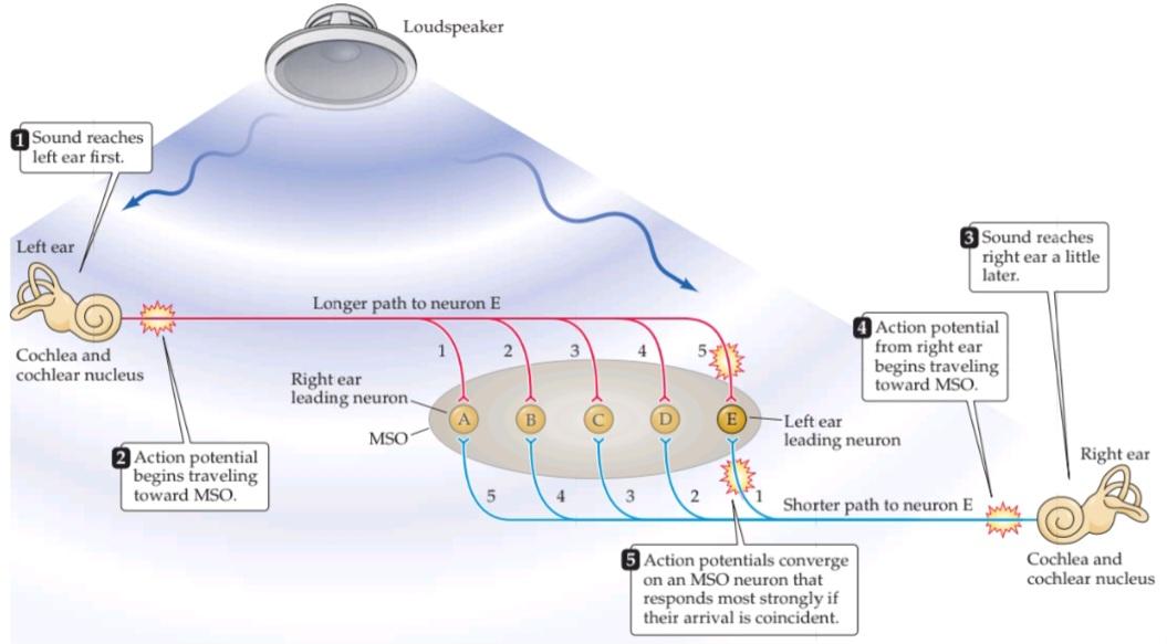
FIGURE 13.14 A model of how the MSO computes the location of a sound by interaural time differences. A given MSO neuron responds most strongly when the two inputs arrive simultaneously, as occurs when the contralateral and ipsilateral inputs precisely compensate (via their different lengths) for differences in the time of arrival of a sound at the two ears. The systematic (and inverse) variation in the delay lengths of the two inputs creates a map of sound location. In this model, neuron E in the MSO would be most sensitive to sounds located to the left, and neuron A to sounds from the right; neuron C would respond best to sounds coming from directly in front of the listener. (After Jeffress, 1948.)
شکل ۱۳.۱۴ مدلی از چگونگی محاسبه مکان صدا توسط MSO با استفاده از اختلاف زمان بین دو گوش. یک نورون MSO مشخص زمانی که دو ورودی به طور همزمان میرسند، قویترین پاسخ را میدهد، همانطور که زمانی اتفاق میافتد که ورودیهای طرف مقابل و همان طرف (از طریق طولهای مختلف خود) تفاوتهای زمان رسیدن صدا به دو گوش را به طور دقیق جبران میکنند. تغییر سیستماتیک (و معکوس) در طول تأخیر دو ورودی، نقشهای از مکان صدا ایجاد میکند. در این مدل، نورون E در MSO بیشترین حساسیت را به صداهای واقع در سمت چپ و نورون A به صداهای سمت راست خواهد داشت. نورون C بهترین پاسخ را به صداهایی که مستقیماً از جلوی شنونده میآیند، میدهد. (به نقل از جفرس، ۱۹۴۸.)
Sound localization perceived on the basis of interaural time differences requires phase-locked information from the periphery, which, as already emphasized, is available to humans only for frequencies below 3 kHz. Therefore, a second mechanism must come into play at higher frequencies. One clue to the solution is that at frequencies higher than about 2 kHz, the human head begins to act as an acoustical obstacle, because the wavelengths of the sounds are too short to bend around it. As a result, when high frequency sounds are directed toward one side of the head, an acoustical “shadow” of lower intensity is created at the far ear. These intensity differences provide a second cue about the location of a sound. The circuits that compute the position of a sound source on this basis are found in the lateral superior olive (LSO) and the medial nucleus of the trapezoid body (MNTB) (Figure 13.15). Excitatory axons project directly from the ipsilateral anteroventral cochlear nucleus to the LSO (as well as to the MSO; see Figure 13.14). Note that the LSO also receives inhibitory input from the contralateral ear via an inhibitory neuron in the MNTB. This excitatory inhibitory interaction results in a net excitation of the LSO on the same side of the head as the sound source. For sounds arising directly lateral to the listener’s head, firing rates will be highest in the LSO on that side; in this circumstance, the excitation via the ipsilateral anteroventral cochlear nucleus will be maximal, and inhibition from the contralateral MNTB minimal. In contrast, sounds arising closer to the listener’s midline will elicit lower firing rates in the ipsilateral LSO because of increased inhibition arising from the contralateral MNTB. For sounds arising at the midline, or from the other side, the increased inhibition arising from the MNTB is powerful enough to completely silence LSO activity. Note that each LSO encodes only sounds arising in the ipsilateral hemifield; therefore, it takes both LSOS to represent the full range of horizontal positions.
مکانیابی صدا که بر اساس اختلاف زمان بین دو گوش درک میشود، نیازمند اطلاعات قفلشده فاز از محیط است که همانطور که قبلاً تأکید شد، فقط برای فرکانسهای کمتر از ۳ کیلوهرتز در دسترس انسان است. بنابراین، مکانیسم دومی باید در فرکانسهای بالاتر وارد عمل شود. یکی از سرنخهای راهحل این است که در فرکانسهای بالاتر از حدود ۲ کیلوهرتز، سر انسان به عنوان یک مانع صوتی عمل میکند، زیرا طول موج صداها برای خم شدن در اطراف آن بسیار کوتاه است. در نتیجه، هنگامی که صداهای فرکانس بالا به سمت یک طرف سر هدایت میشوند، یک “سایه” صوتی با شدت کمتر در گوش دور ایجاد میشود. این اختلاف شدتها، نشانه دومی در مورد مکان صدا ارائه میدهند. مدارهایی که موقعیت منبع صدا را بر این اساس محاسبه میکنند، در زیتون فوقانی جانبی (LSO) و هسته داخلی جسم ذوزنقهای (MNTB) یافت میشوند (شکل ۱۳.۱۵). آکسونهای تحریکی مستقیماً از هسته حلزونی قدامی-شکمی همان طرف به LSO (و همچنین به MSO) ساطع میشوند؛ به شکل ۱۳.۱۴ مراجعه کنید. توجه داشته باشید که LSO همچنین ورودی مهاری را از گوش مقابل از طریق یک نورون مهاری در MNTB دریافت میکند. این تعامل تحریکی-مهاری منجر به تحریک خالص LSO در همان طرف سر به عنوان منبع صدا میشود. برای صداهایی که مستقیماً در سمت جانبی سر شنونده ایجاد میشوند، نرخ شلیک در LSO در آن طرف بالاترین خواهد بود. در این شرایط، تحریک از طریق هسته حلزونی قدامی-شکمی همان طرف حداکثر و مهار از MNTB طرف مقابل حداقل خواهد بود. در مقابل، صداهایی که به خط میانی شنونده نزدیکتر هستند، به دلیل افزایش مهار ناشی از MNTB طرف مقابل، نرخ شلیک کمتری را در LSO همان طرف ایجاد میکنند. برای صداهایی که از خط میانی یا از طرف دیگر برمیخیزند، افزایش مهار ناشی از MNTB به اندازه کافی قدرتمند است تا فعالیت LSO را به طور کامل خاموش کند. توجه داشته باشید که هر LSO فقط صداهایی را که از نیمه میدان همان طرف ناشی میشوند، رمزگذاری میکند. بنابراین، برای نشان دادن طیف کامل موقعیتهای افقی، به هر دو LSOS نیاز است.

FIGURE 13.15 LSO neurons encode sound location through interaural intensity differences. (A) LSO neurons receive direct excitation from the ipsilateral cochlear nucleus; input from the contralateral cochlear nucleus is relayed via inhibitory interneurons in the MNTB. (B) This arrangement of excitation inhibition makes LSO neurons fire most strongly in response to sounds arising directly lateral to the listener on the same side as the LSO, because excitation from the ipsilateral input will be great and inhibition from the contralateral input will be small. In contrast, sounds arising from in front of the listener, or from the opposite side, will silence the LSO output, because excitation from the ipsilateral input will be minimal, but inhibition driven by the contralateral input will be great. Note that LSOS are paired and bilaterally symmetrical; each LSO encodes only the location of sounds arising from the ipsilateral hemifield.
شکل ۱۳.۱۵ نورونهای LSO مکان صدا را از طریق تفاوتهای شدت بین دو گوش کدگذاری میکنند. (الف) نورونهای LSO تحریک مستقیم را از هسته حلزونی همان طرف دریافت میکنند؛ ورودی از هسته حلزونی طرف مقابل از طریق نورونهای رابط مهاری در MNTB رله میشود. (ب) این ترتیب مهار تحریک باعث میشود نورونهای LSO در پاسخ به صداهایی که مستقیماً در کنار شنونده در همان طرف LSO ایجاد میشوند، به شدت شلیک کنند، زیرا تحریک از ورودی همان طرف زیاد و مهار از ورودی طرف مقابل کم خواهد بود. در مقابل، صداهایی که از جلوی شنونده یا از طرف مقابل ناشی میشوند، خروجی LSO را خاموش میکنند، زیرا تحریک از ورودی همان طرف حداقل خواهد بود، اما مهار ناشی از ورودی طرف مقابل زیاد خواهد بود. توجه داشته باشید که LSOS جفت شده و متقارن دو طرفه هستند. هر LSO فقط مکان صداهایی را که از نیمه میدان همان طرف ناشی میشوند، کدگذاری میکند.
In summary, two separate pathways and two separate mechanisms-enable us to localize sound along the azimuth. Humans process interaural time differences in the MSO and interaural intensity differences in the LSO. These two pathways eventually merge in the midbrain auditory centers. The elevation of sound sources is determined by spectral filtering mediated by the external pinnae. Experimental evidence suggests that the spectral “notches” created by the shape of the pinnae are detected by neurons in the dorsal cochlear nucleus. Thus, binaural cues play an important role in localizing the azimuthal position of sound sources, whereas spectral cues are used to localize the elevation of sound sources.
به طور خلاصه، دو مسیر جداگانه و دو مکانیسم جداگانه ما را قادر میسازند تا صدا را در امتداد آزیموت مکانیابی کنیم. انسانها تفاوتهای زمانی بین دو گوش را در MSO و تفاوتهای شدت بین دو گوش را در LSO پردازش میکنند. این دو مسیر در نهایت در مراکز شنوایی مغز میانی با هم ادغام میشوند. ارتفاع منابع صدا توسط فیلتر طیفی با واسطه لاله گوش خارجی تعیین میشود. شواهد تجربی نشان میدهد که “شکافهای” طیفی ایجاد شده توسط شکل لاله گوش توسط نورونهای هسته حلزون پشتی شناسایی میشوند. بنابراین، نشانههای دوگوشی نقش مهمی در مکانیابی موقعیت آزیموت منابع صدا دارند، در حالی که نشانههای طیفی برای مکانیابی ارتفاع منابع صدا استفاده میشوند.
Monaural Pathways from the Cochlear Nucleus to the Nuclei of the Lateral Lemniscus
The binaural pathways for sound localization are only part of the output of the cochlear nucleus. This fact is hardly surprising, given that auditory perception involves much more than locating the position of the sound source. A second major set of pathways from the cochlear nucleus bypasses the superior olive and terminates in the nuclei of the lateral lemniscus on the contralateral side of the brainstem (see Figure 13.13). These particular pathways respond to sound arriving at one ear only and are thus referred to as monaural. Some cells in the lateral lemniscus nuclei signal the onset of sound, regardless of its intensity or frequency. Other cells in the lateral lemniscus nuclei process other temporal aspects of sound, such as duration. The role of these pathways in processing temporal features of sound is not yet known. As with the outputs of the superior olivary nuclei, the pathways from the nuclei of the lateral lemniscus converge at the midbrain.
مسیرهای تکگوشی از هسته حلزونی تا هستههای لمنیسکوس جانبی
مسیرهای دوگوشی برای مکانیابی صدا تنها بخشی از خروجی هسته حلزونی هستند. این واقعیت چندان تعجبآور نیست، با توجه به اینکه ادراک شنوایی چیزی بیش از تعیین موقعیت منبع صدا را در بر میگیرد. دومین مجموعه اصلی از مسیرها از هسته حلزونی، زیتون بالایی را دور میزند و در هستههای لمنیسکوس جانبی در سمت مقابل ساقه مغز خاتمه مییابد (شکل ۱۳.۱۳ را ببینید). این مسیرهای خاص به صدایی که فقط به یک گوش میرسد پاسخ میدهند و بنابراین به عنوان تکگوشی شناخته میشوند. برخی از سلولهای هستههای لمنیسکوس جانبی، صرف نظر از شدت یا فرکانس آن، شروع صدا را اعلام میکنند. سلولهای دیگر در هستههای لمنیسکوس جانبی، جنبههای زمانی دیگری از صدا، مانند مدت زمان را پردازش میکنند. نقش این مسیرها در پردازش ویژگیهای زمانی صدا هنوز مشخص نیست. همانند خروجیهای هستههای زیتونی فوقانی، مسیرهای هستههای لمنیسکوس جانبی در مغز میانی به هم میرسند.
Integration in the Inferior Colliculus
Auditory pathways ascending via the olivary and lemniscal complexes, as well as other projections that arise directly from the cochlear nucleus, project to the midbrain auditory center, also known as the inferior colliculus. In examining how integration occurs in the inferior colliculus, it is again instructive to turn to the most completely analyzed auditory mechanism, the binaural system for localizing sound. As already noted, space is not mapped on the auditory receptor surface; thus, the perception of auditory space must somehow be synthesized by circuitry in the lower brainstem and midbrain. Experiments in the barn owl show that the convergence of binaural inputs in the midbrain produces something entirely new, relative to the periphery namely a computed topographical representation of auditory space. Neurons within this auditory space map in the colliculus respond best to sounds originating in a specific region of space; thus, they have both a preferred elevation and a preferred azimuthal location. Although the circuit mechanisms underlying sound localization in humans are unknown, humans have a clear perception of both the elevational and azimuthal components of a sound’s location, indicating that our brains contain a neural representation of auditory space.
ادغام در کالیکولوس تحتانی
مسیرهای شنوایی که از طریق کمپلکسهای زیتونی و لمنیسکال بالا میروند، و همچنین سایر برآمدگیهایی که مستقیماً از هسته حلزونی ناشی میشوند، به مرکز شنوایی مغز میانی، که به عنوان کالیکولوس تحتانی نیز شناخته میشود، امتداد مییابند. در بررسی چگونگی وقوع ادغام در کالیکولوس تحتانی، باز هم آموزنده است که به کاملترین مکانیسم شنوایی، یعنی سیستم دوگوشی برای مکانیابی صدا، بپردازیم. همانطور که قبلاً اشاره شد، فضا روی سطح گیرنده شنوایی نقشهبرداری نمیشود. بنابراین، درک فضای شنوایی باید به نحوی توسط مدار در ساقه مغز تحتانی و مغز میانی سنتز شود. آزمایشها در جغد انبار نشان میدهد که همگرایی ورودیهای دوگوشی در مغز میانی، چیزی کاملاً جدید، نسبت به حاشیه – یعنی یک نمایش توپوگرافی محاسبهشده از فضای شنوایی – تولید میکند. نورونهای درون این نقشه فضای شنوایی در کالیکولوس، به صداهایی که از یک منطقه خاص از فضا سرچشمه میگیرند، بهترین پاسخ را میدهند. بنابراین، آنها هم ارتفاع ترجیحی و هم مکان آزیموتال ترجیحی دارند. اگرچه مکانیسمهای مداری زیربنایی مکانیابی صدا در انسان ناشناخته است، اما انسانها درک روشنی از مؤلفههای ارتفاعی و سمتی مکان صدا دارند که نشان میدهد مغز ما دارای یک بازنمایی عصبی از فضای شنوایی است.
Another important property of the inferior colliculus is its ability to process sounds with complex temporal patterns. Many neurons in the inferior colliculus respond only frequency modulated sounds, while others respond only to sounds of specific durations or in specific temporal sequences. Such sounds are typical components of biologically relevant sounds, such as those made by predators, or intraspecific communication sounds, which in humans include speech. In summary, the high degree of convergence in the inferior colliculus of inputs conveying information about simpler cues, such as timing, intensity, and frequency, results in more integrative and complex response properties that are likely to be important to the representation of auditory objects.
یکی دیگر از ویژگیهای مهم کالیکولوس تحتانی، توانایی آن در پردازش صداهایی با الگوهای زمانی پیچیده است. بسیاری از نورونهای کالیکولوس تحتانی فقط به صداهای مدوله شده با فرکانس پاسخ میدهند، در حالی که برخی دیگر فقط به صداهایی با مدت زمان خاص یا در توالیهای زمانی خاص پاسخ میدهند. چنین صداهایی اجزای معمول صداهای مرتبط با زیستشناسی هستند، مانند صداهایی که توسط شکارچیان تولید میشوند، یا صداهای ارتباطی درون گونهای، که در انسان شامل گفتار میشود. به طور خلاصه، درجه بالای همگرایی ورودیهایی که اطلاعات مربوط به نشانههای سادهتر، مانند زمانبندی، شدت و فرکانس را منتقل میکنند، در کالیکولوس تحتانی منجر به ویژگیهای پاسخ یکپارچهتر و پیچیدهتری میشود که احتمالاً برای بازنمایی اشیاء شنیداری مهم هستند.
The Auditory Thalamus
Despite the parallel pathways in the auditory stations of the brainstem and midbrain, the medial geniculate complex (MGC) in the thalamus is an obligatory relay for all ascending auditory information destined for the cortex (see Figure 13.13). Most input to the MGC arises from the inferior colliculus, although a few auditory axons from the lower brainstem bypass the inferior colliculus to reach the auditory thalamus directly. The MGC has several divisions, including the ventral division, which projects to the core region of the auditory cortex, and the medial and dorsal divisions, which are organized like a belt around the ventral division and project to the belt regions that surround the core region of the auditory cortex.
تالاموس شنوایی
با وجود مسیرهای موازی در ایستگاههای شنوایی ساقه مغز و مغز میانی، کمپلکس زانویی میانی (MGC) در تالاموس یک رله اجباری برای تمام اطلاعات شنوایی صعودی است که به قشر مغز میرسند (شکل ۱۳.۱۳ را ببینید). بیشتر ورودیهای MGC از کولیکولوس تحتانی ناشی میشود، اگرچه چند آکسون شنوایی از ساقه مغز تحتانی، کولیکولوس تحتانی را دور میزنند تا مستقیماً به تالاموس شنوایی برسند. MGC دارای چندین بخش است، از جمله بخش شکمی که به ناحیه مرکزی قشر شنوایی امتداد دارد، و بخشهای میانی و پشتی که مانند کمربندی در اطراف بخش شکمی سازماندهی شدهاند و به نواحی کمربندی که ناحیه مرکزی قشر شنوایی را احاطه کردهاند، امتداد دارند.
In some mammals, the strictly maintained tonotopy of the lower brainstem areas is exploited by convergence onto MGC neurons, generating specific responses to certain spectral combinations. The original evidence for this statement came from research on the response properties of cells in the MGC of echolocating bats. Some cells in the socalled belt regions of the bat MGC respond only to combinations of widely spaced frequencies that are specific components of the bat’s echolocation signal and of the echoes that are reflected from objects in the bat’s environment. In the mustached bat, where this phenomenon has been most thoroughly studied, the echolocation pulse has a changing frequency (frequency-modulated, or FM) component that includes a fundamental frequency and one or more harmonics. The fundamental frequency (FM1) has low intensity and sweeps from 30 to 20 kHz. The second harmonic (FM) is the most intense component and sweeps from 60 to 40 kHz. Note that these frequencies do not overlap. Most of the echoes are from the intense FM2 sound, and virtually none arise from the weak FM1, even though the emitted FM, is loud enough for the bat to hear. Apparently, the bat assesses the distance to an object by measuring the delay between the FM, emission and the FM, echo. Certain MGC neurons respond when FM, fol- lows FM, by a specific delay, providing a mechanism for sensing such frequency combinations. Because each neuron responds best to a particular delay, the population of MGC neurons encodes a range of distances.
در برخی از پستانداران، تونوتوپیِ بهشدت حفظشدهی نواحی پایینی ساقهی مغز با همگرایی به نورونهای MGC مورد بهرهبرداری قرار میگیرد و پاسخهای خاصی را به ترکیبات طیفی خاص ایجاد میکند. شواهد اولیه برای این گفته از تحقیقات روی خواص پاسخ سلولهای MGC خفاشهای پژواکیاب بهدست آمده است. برخی از سلولها در نواحی کمربندی خفاش MGC فقط به ترکیبی از فرکانسهای با فاصلهی زیاد پاسخ میدهند که اجزای خاصی از سیگنال پژواکیاب خفاش و پژواکهایی هستند که از اشیاء محیط خفاش منعکس میشوند. در خفاش سبیلموزی، که این پدیده بهطور کامل مورد مطالعه قرار گرفته است، پالس پژواکیاب دارای یک مؤلفهی فرکانس متغیر (مدولهشده با فرکانس یا FM) است که شامل یک فرکانس پایه و یک یا چند هارمونیک است. فرکانس پایه (FM1) شدت کمی دارد و از 30 تا 20 کیلوهرتز تغییر میکند. هارمونیک دوم (FM) شدیدترین مؤلفه است و از 60 تا 40 کیلوهرتز تغییر میکند. توجه داشته باشید که این فرکانسها با هم همپوشانی ندارند. بیشتر پژواکها از صدای شدید FM2 هستند و عملاً هیچ کدام از FM1 ضعیف ناشی نمیشوند، حتی با اینکه FM منتشر شده به اندازه کافی بلند است که خفاش بتواند آن را بشنود. ظاهراً خفاش با اندازهگیری تأخیر بین FM (انتشار) و FM (پژواک) فاصله تا یک جسم را ارزیابی میکند. برخی از نورونهای MGC وقتی FM، با تأخیر خاصی پس از FM، پاسخ میدهند، مکانیسمی برای حس کردن چنین ترکیبات فرکانسی فراهم میکنند. از آنجا که هر نورون به یک تأخیر خاص بهترین پاسخ را میدهد، جمعیت نورونهای MGC طیف وسیعی از فواصل را رمزگذاری میکنند.
Bat sonar illustrates two important points about the function of the auditory thalamus. First, the MGC displays pronounced selectivity for frequency combinations. The mechanism responsible for this selectivity is presumably the ultimate convergence of many inputs from cochlear regions with different spectral sensitivities. Second, cells in the MGC are selective not only for frequency combinations, but also for specific time intervals between the two frequencies. The principle is the same as that described for binaural neurons in the medial superior olive, but in this instance, two monaural signals with different frequency sensitivities coincide, and the time difference is in the millisecond rather than the microsecond range. For echolocating bats, time differences in the millisecond range correspond to target distances of catchable prey. Interestingly, speech sounds change continuously over the range of a few milliseconds, suggesting that MGC neurons in the human capable of integrating acoustical information over the millisecond timescale could facilitate speech perception.
سونار خفاش دو نکته مهم در مورد عملکرد تالاموس شنوایی را نشان میدهد. اول، MGC گزینشپذیری قابل توجهی را برای ترکیبات فرکانسی نشان میدهد. مکانیسم مسئول این گزینشپذیری، احتمالاً همگرایی نهایی بسیاری از ورودیها از نواحی حلزونی با حساسیتهای طیفی مختلف است. دوم، سلولهای MGC نه تنها برای ترکیبات فرکانسی، بلکه برای فواصل زمانی خاص بین دو فرکانس نیز گزینشپذیر هستند. اصل کار همان است که برای نورونهای دوگوشی در زیتون فوقانی میانی شرح داده شده است، اما در این مورد، دو سیگنال تکگوشی با حساسیتهای فرکانسی مختلف بر هم منطبق میشوند و اختلاف زمانی در حد میلیثانیه است نه در حد میکروثانیه. برای خفاشهای پژواکیاب، اختلاف زمانی در حد میلیثانیه با فواصل هدف طعمههای قابل گرفتن مطابقت دارد. جالب توجه است که صداهای گفتاری به طور مداوم در محدوده چند میلیثانیه تغییر میکنند، که نشان میدهد نورونهای MGC در انسان که قادر به ادغام اطلاعات صوتی در مقیاس زمانی میلیثانیه هستند، میتوانند درک گفتار را تسهیل کنند.
In summary, neurons in the MGC receive convergent inputs from spectrally and temporally separate pathways. This complex, by virtue of its convergent inputs, mediates the detection of specific spectral and temporal combinations of sounds. In many species, including humans, varying spectral and temporal cues are especially important features of communication sounds. It is not known whether cells in the human MGC are selective to combinations of sounds, but the processing of speech certainly requires both spectral and temporal combination sensitivity.
به طور خلاصه، نورونهای موجود در MGC ورودیهای همگرا را از مسیرهای طیفی و زمانی جداگانه دریافت میکنند. این مجموعه، به دلیل ورودیهای همگرای خود، واسطه تشخیص ترکیبات طیفی و زمانی خاص صداها است. در بسیاری از گونهها، از جمله انسان، نشانههای طیفی و زمانی متغیر، از ویژگیهای مهم صداهای ارتباطی هستند. مشخص نیست که آیا سلولهای MGC انسان نسبت به ترکیبات صداها گزینشی هستند یا خیر، اما پردازش گفتار قطعاً به حساسیت ترکیبی طیفی و زمانی نیاز دارد.
The Auditory Cortex
The auditory cortex is the major target of the ascending fibers from the MGC, and it plays an essential role in our conscious perception of sound, including recognition of speech and music. Indeed, auditory cortical lesions in humans are often accompanied by deficits in speech and music perception. Despite the obvious importance of auditory cortical processing for sound perception, the auditory cortex has been less completely studied than the auditory periphery and is less well understood than visual cortex (although one should hasten to add that the mechanisms of both peripheral auditory processing and visual cortical processing are far from clear).
قشر شنوایی
قشر شنوایی هدف اصلی فیبرهای صعودی از MGC است و نقش اساسی در درک آگاهانه ما از صدا، از جمله تشخیص گفتار و موسیقی، ایفا میکند. در واقع، ضایعات قشر شنوایی در انسان اغلب با نقص در درک گفتار و موسیقی همراه است. علیرغم اهمیت آشکار پردازش قشر شنوایی برای درک صدا، قشر شنوایی کمتر از قشر شنوایی پیرامونی مورد مطالعه قرار گرفته و کمتر از قشر بینایی شناخته شده است (اگرچه باید فوراً اضافه کرد که مکانیسمهای پردازش شنوایی پیرامونی و پردازش قشر بینایی به هیچ وجه مشخص نیست).
Although the auditory cortex has several subdivisions, as in the visual and somatosensory systems a broad distinction can be made between primary (i.e., core) and secondary (i.e., belt and parabelt) regions. The core region in macaque monkeys comprises three divisions, including auditory area 1 (A1), rostral (R), and rostrotemporal (RT), all of which are located on the lower bank of the lateral sulcus in the medial and posterior part of the superior temporal gyrus (STG) in the temporal lobe. Imaging studies in humans indicate that the core region is located in the transverse temporal gyri (Heschl’s gyri, or Brod mann’s areas 41 and 42), buried in the lateral sulcus. The core region receives point to point input from the ventral division of the MGC; thus, the three divisions of the core region contain precise tonotopic maps. The belt and parabelt regions of the auditory cortex receive more diffuse input from the belt division of the MGC, as well as input from the primary auditory cortex, and are less precise in their tonotopic organization. Additionally, these various auditory cortical areas are strongly interconnected, with reciprocal connections between the core and belt regions, between the belt and parabelt regions, and between these latter two regions and auditory related cortical areas in the ST and the STS (superior temporal sulcus), suggestive of a processing hierarchy.
اگرچه قشر شنوایی دارای چندین زیربخش است، همانطور که در سیستمهای بینایی و حسی-تنی میتوان تمایز گستردهای بین مناطق اولیه (یعنی هسته) و ثانویه (یعنی کمربند و پارابلت) قائل شد. منطقه هسته در میمونهای ماکاک شامل سه بخش است، از جمله ناحیه شنوایی ۱ (A1)، ناحیه سری (R) و ناحیه سری-گیجگاهی (RT) که همه آنها در ساحل پایینی شیار جانبی در قسمت میانی و خلفی شکنج گیجگاهی فوقانی (STG) در لوب گیجگاهی قرار دارند. مطالعات تصویربرداری در انسان نشان میدهد که ناحیه هسته در شکنج گیجگاهی عرضی (شکنجهای هشل یا مناطق ۴۱ و ۴۲ برودمن) واقع شده است که در شیار جانبی پنهان شده است. ناحیه هسته ورودی نقطه به نقطه را از بخش شکمی MGC دریافت میکند. بنابراین، سه بخش ناحیه هسته حاوی نقشههای دقیق تونوتوپیک هستند. نواحی کمربند و پارابلت قشر شنوایی، ورودی پراکندهتری از بخش کمربندی MGC و همچنین ورودی از قشر شنوایی اولیه دریافت میکنند و در سازماندهی تونوتوپیک خود دقت کمتری دارند. علاوه بر این، این نواحی مختلف قشر شنوایی به شدت به هم پیوسته هستند، و ارتباطات متقابلی بین نواحی هسته و کمربند، بین نواحی کمربند و پارابلت، و بین این دو ناحیه اخیر و نواحی قشری مرتبط با شنوایی در ST و STS (شیار گیجگاهی فوقانی) وجود دارد که نشاندهنده یک سلسله مراتب پردازش است.
The core divisions of the auditory cortex each contain a topographical map of the cochlea (Figure 13.16), just as the primary visual cortex (V1) and the primary somatosensory cortex (S1) have topographical maps of their respective sensory epithelia. Just as the visual and somatosensory systems represent their peripheral receptor surfaces in central maps, so the organization of the cochlea is laid out in a central map. Since frequencies are arrayed tonotopically along the length of the basilar membrane, this organization as reflected in A1 is said to comprise a tonotopic map, as do most of the ascending auditory structures between the cochlea and the cortex. Orthogonal to the frequency axis of the tonotopic map are irregular patches of neurons that are excited by both ears (and are therefore called EE cells) interspersed with patches of cells that are excited by one ear and inhibited by the other ear (EI cells). The EE and EI stripes alternate, an arrangement that is reminiscent of the ocular dominance columns in V1 (see Chapter 12).
بخشهای اصلی قشر شنوایی هر کدام حاوی یک نقشه توپوگرافی از حلزون گوش هستند (شکل ۱۳.۱۶)، همانطور که قشر بینایی اولیه (V1) و قشر حسی-پیکری اولیه (S1) نقشههای توپوگرافی از اپیتلیوم حسی مربوط به خود دارند. همانطور که سیستمهای بینایی و حسی-پیکری سطوح گیرنده محیطی خود را در نقشههای مرکزی نشان میدهند، سازماندهی حلزون گوش نیز در یک نقشه مرکزی ترسیم شده است. از آنجایی که فرکانسها به صورت تونوتوپیک در امتداد طول غشای پایه قرار گرفتهاند، گفته میشود که این سازماندهی همانطور که در A1 منعکس شده است، یک نقشه تونوتوپیک را تشکیل میدهد، همانطور که بیشتر ساختارهای شنوایی صعودی بین حلزون و قشر گوش نیز چنین هستند. عمود بر محور فرکانس نقشه تونوتوپیک، تکههای نامنظمی از نورونها وجود دارند که توسط هر دو گوش تحریک میشوند (و بنابراین سلولهای EE نامیده میشوند) که با تکههایی از سلولهایی که توسط یک گوش تحریک و توسط گوش دیگر مهار میشوند (سلولهای EI) در هم آمیختهاند. نوارهای EE و EI به طور متناوب قرار میگیرند، ترتیبی که یادآور ستونهای تسلط چشمی در V1 است (به فصل 12 مراجعه کنید).
The auditory cortex obviously does much more than provide a tonotopic map and respond differentially to ipsilateral and contralateral stimulation. The sorts of sensory processing that occur in the auditory cortex are not fully understood, but they are likely to be important to higher order processing of natural sounds, especially those used for communication (see Box 13B and Chapter 33). One clue about such processing comes from work in marmosets, small New World monkeys with a complex vocal repertoire. The A1 and belt areas of these animals are indeed organized tonotopically, but they also contain neurons that are strongly responsive to spectral combinations that characterize certain vocalizations. The responses of these neurons to the tonal stimuli do not accurately predict their responses to the spectral combinations, suggesting that, in accordance with peripheral optimization, cortical processing is, in part, dedicated to detecting particular intraspecific vocalizations. Recent studies in marmosets and humans also implicate secondary regions of the auditory cortex in the perception of pitch. This percept is especially important to our musical sense and to vocal communication, because it enables us to hear two speech sounds as distinct even when they have overlapping spectral content and arise from the same location. A curious feature of pitch perception is that, for the harmonically complex sounds that typify speech and music, pitch corresponds to the fundamental frequency, even when it is absent from the actual stimulus. This ability of pitch processing to “fill in” a missing frequency further underscores the idea that the auditory cortex is doing far more than faithfully representing what the auditory periphery provides as input.
قشر شنوایی آشکارا کاری بسیار فراتر از ارائه یک نقشه تونوتوپیک و پاسخ متفاوت به تحریک همسو و دگرسو انجام میدهد. انواع پردازش حسی که در قشر شنوایی رخ میدهد به طور کامل شناخته نشده است، اما احتمالاً برای پردازش سطح بالاتر صداهای طبیعی، به ویژه آنهایی که برای ارتباط استفاده میشوند، مهم هستند (به کادر 13B و فصل 33 مراجعه کنید). یک سرنخ در مورد چنین پردازشی از کار روی میمونهای کوچک دنیای جدید با مجموعهای از صداهای پیچیده به دست میآید. نواحی A1 و کمربند این حیوانات در واقع به صورت تونوتوپیک سازماندهی شدهاند، اما همچنین حاوی نورونهایی هستند که به شدت به ترکیبات طیفی که مشخصه آواسازیهای خاص هستند، پاسخ میدهند. پاسخهای این نورونها به محرکهای تنال، پاسخهای آنها به ترکیبات طیفی را به طور دقیق پیشبینی نمیکند، که نشان میدهد، مطابق با بهینهسازی محیطی، پردازش قشری تا حدی به تشخیص آواسازیهای درون گونهای خاص اختصاص دارد. مطالعات اخیر در میمونهای کوچک و انسانها نیز مناطق ثانویه قشر شنوایی را در درک زیر و بمی صدا دخیل میدانند. این درک به ویژه برای حس موسیقی و ارتباط صوتی ما مهم است، زیرا ما را قادر میسازد دو صدای گفتاری را حتی زمانی که محتوای طیفی همپوشانی دارند و از یک مکان سرچشمه میگیرند، به صورت مجزا بشنویم. یکی از ویژگیهای عجیب درک زیر و بمی صدا این است که برای صداهای پیچیده هارمونیک که گفتار و موسیقی را مشخص میکنند، زیر و بمی صدا با فرکانس پایه مطابقت دارد، حتی زمانی که در محرک واقعی وجود ندارد. این توانایی پردازش زیر و بمی صدا برای “پر کردن” یک فرکانس از دست رفته، این ایده را بیشتر برجسته میکند که قشر شنوایی کاری بسیار فراتر از نمایش دقیق آنچه که محیط شنوایی به عنوان ورودی ارائه میدهد، انجام میدهد.


FIGURE 13.16 The human auditory cortex. (A) Diagram showing the brain in left lateral view, including the depths of the lateral sulcus, where part of the auditory cortex occupying the superior temporal gyrus normally lies hidden. The core region is shown in blue; the surrounding belt regions of the auditory cortex are in red. (B) Diagram of the brain in left lateral view, showing locations of human auditory cortical regions related to processing speech sounds in the intact hemisphere. Right: An oblique section (plane of dashed line) shows the cortical areas on the superior surface of the temporal lobe. Note that Wernicke’s area, a region important in comprehending speech, is just posterior to the primary auditory cortex.
شکل ۱۳.۱۶ قشر شنوایی انسان. (الف) نموداری که مغز را در نمای جانبی چپ نشان میدهد، شامل اعماق شیار جانبی، جایی که بخشی از قشر شنوایی که شکنج گیجگاهی فوقانی را اشغال میکند، معمولاً پنهان است. ناحیه مرکزی با رنگ آبی نشان داده شده است؛ نواحی کمربندی اطراف قشر شنوایی با رنگ قرمز نشان داده شدهاند. (ب) نمودار مغز در نمای جانبی چپ، مکانهای نواحی قشر شنوایی انسان مربوط به پردازش صداهای گفتاری در نیمکره سالم را نشان میدهد. راست: یک برش مورب (صفحه خط چین) نواحی قشری را در سطح فوقانی لوب گیجگاهی نشان میدهد. توجه داشته باشید که ناحیه ورنیکه، ناحیهای که در درک گفتار مهم است، درست در خلف قشر شنوایی اولیه قرار دارد.
Another clue about the role of the auditory cortex in speech processing comes from electrocorticographic recordings made in human epilepsy patients from parts of the STG that most likely correspond to parabelt regions in non-human primates (ie., Brodmann’s area 22). These recordings reveal that neural responses to speech sounds spread from posterior to anterior regions of the STG, consistent with a processing hierarchy. Moreover, STG population activity strongly correlates with syllable onsets and offsets, which are especially important to speech intelligibility, and is sensitive to acoustical cues, such as voice onset times, that are important to the perceptual categorization of different speech sounds (e.g., distinguishing between ba and pa). Neural activity in the STG also depends strongly on context and attention: When subjects are instructed to attend to only one of two people speaking simultaneously, their STG neurons robustly encode fine spectrotemporal features of the attended voice but display little or no responsiveness to the other voice. Thus, auditory cortical activity is strongly influenced both by linguistic features and cognitive context, consistent with an influence of experience and task demands on auditory cortical processing of speech.
سرنخ دیگری در مورد نقش قشر شنوایی در پردازش گفتار از ثبتهای الکتروکورتیکوگرافی انجام شده در بیماران صرع انسانی از بخشهایی از STG که به احتمال زیاد با نواحی پارابلت در نخستیسانان غیرانسانی مطابقت دارند (یعنی ناحیه ۲۲ برودمن) به دست میآید. این ثبتها نشان میدهند که پاسخهای عصبی به صداهای گفتاری از نواحی خلفی به قدامی STG گسترش مییابند، که با سلسله مراتب پردازش سازگار است. علاوه بر این، فعالیت جمعیت STG به شدت با شروع و پایان هجاها، که به ویژه برای فهم گفتار مهم هستند، مرتبط است و به نشانههای صوتی، مانند زمان شروع صدا، که برای طبقهبندی ادراکی صداهای گفتاری مختلف (مثلاً تمایز بین ba و pa) مهم هستند، حساس است. فعالیت عصبی در STG همچنین به شدت به زمینه و توجه بستگی دارد: هنگامی که به افراد دستور داده میشود که فقط به یکی از دو نفری که همزمان صحبت میکنند توجه کنند، نورونهای STG آنها به طور قوی ویژگیهای طیفی-زمانی دقیق صدای مورد توجه را رمزگذاری میکنند، اما به صدای دیگر پاسخ کمی یا هیچ پاسخی نشان نمیدهند. بنابراین، فعالیت قشر شنوایی به شدت تحت تأثیر ویژگیهای زبانی و زمینه شناختی قرار میگیرد، که با تأثیر تجربه و الزامات تکلیف بر پردازش گفتار در قشر شنوایی سازگار است.
Sounds that are especially important for intraspecific communication often have a highly ordered temporal structure. In humans, the best example of such time varying signals is speech, where different phonetic sequences are perceived as distinct syllables and words (see Clinical Applications). Behavioral studies in cats and monkeys show that the auditory cortex is especially important for processing temporal sequences of sound. If the auditory cortex is ablated in these animals, they lose the ability to discriminate between two complex sounds that have the same frequency components but differ in temporal sequence. Thus, without the auditory cortex, monkeys cannot discriminate one conspecific communication sound from another. The physiological basis of such temporal sensitivity likely requires neurons that are sensitive to time varying cues in communication sounds. Indeed, electrophysiological recordings from the primary auditory cortices of both marmosets and bats show that some neurons that respond to intraspecific communication sounds do not respond as strongly when the sounds are played in reverse, indicating sensitivity to the sounds’ temporal features. Studies of human patients with bilateral damage to the auditory cortex also reveal severe problems in processing the temporal order of sounds. It seems likely, therefore, that specific regions of the human auditory cortex are specialized for processing elementary speech sounds, as well as other temporally complex acoustical signals, such as music. Thus, Wernicke’s area, which is critical to the comprehension of human language, is contiguous with the secondary auditory area (see Figure 13.16 and Chapter 33).
صداهایی که به ویژه برای ارتباط درون گونهای اهمیت دارند، اغلب ساختار زمانی بسیار منظمی دارند. در انسانها، بهترین نمونه از چنین سیگنالهای متغیر با زمان، گفتار است که در آن توالیهای آوایی مختلف به صورت هجاها و کلمات متمایز درک میشوند (به کاربردهای بالینی مراجعه کنید). مطالعات رفتاری در گربهها و میمونها نشان میدهد که قشر شنوایی به ویژه برای پردازش توالیهای زمانی صدا اهمیت دارد. اگر قشر شنوایی در این حیوانات قطع شود، آنها توانایی تمایز بین دو صدای پیچیده که اجزای فرکانس یکسانی دارند اما در توالی زمانی متفاوت هستند را از دست میدهند. بنابراین، بدون قشر شنوایی، میمونها نمیتوانند یک صدای ارتباطی همنوع را از دیگری تشخیص دهند. اساس فیزیولوژیکی چنین حساسیت زمانی احتمالاً به نورونهایی نیاز دارد که به نشانههای متغیر با زمان در صداهای ارتباطی حساس هستند. در واقع، ثبتهای الکتروفیزیولوژیکی از قشر شنوایی اولیه هر دو میمون مارموست و خفاش نشان میدهد که برخی از نورونهایی که به صداهای ارتباطی درون گونهای پاسخ میدهند، وقتی صداها به صورت معکوس پخش میشوند، به شدت پاسخ نمیدهند، که نشاندهنده حساسیت به ویژگیهای زمانی صداها است. مطالعات بیماران انسانی با آسیب دو طرفه به قشر شنوایی نیز مشکلات شدیدی را در پردازش ترتیب زمانی صداها نشان میدهد. بنابراین، به نظر میرسد که مناطق خاصی از قشر شنوایی انسان برای پردازش صداهای گفتاری ابتدایی و همچنین سایر سیگنالهای صوتی پیچیده زمانی، مانند موسیقی، تخصص یافتهاند. بنابراین، ناحیه ورنیکه، که برای درک زبان انسان حیاتی است، در مجاورت ناحیه شنوایی ثانویه قرار دارد (به شکل ۱۳.۱۶ و فصل ۳۳ مراجعه کنید).
BOX 13B Representing Complex Sounds in the Brains of Bats and Humans
Bost natural sounds are complex, meaning that they differ from the quently used in neurophysiological studies of the auditory system. Only a small minority of natural sounds are tonal, but when they occur the stimuli have a fundamental frequency that largely determines the pitch of the sound, and one or more harmonics of different intensities that contribute to the quality or timbre of a sound. The frequency of a harmonic is, by definition, an integer multiple of the fundamental frequency, and both may be modulated over time. Such frequency modulated (FM) sweeps can rise or fall in frequency, or they can change in a sinusoidal (or some other) fashion. Occasionally, multiple nonharmonic frequencies may be simultaneously present in some communication or musical sounds. In some sounds, a level of spectral splatter, or broadband noise, is embedded within tonal or frequency modulated sounds. The variations in the sound spectrum are typically accompanied by a modulation of the amplitude envelope of the complex sound as well. All of these features can be visualized by spectrographic analysis.
کادر ۱۳ب، نمایش صداهای پیچیده در مغز خفاشها و انسانها
بیشتر صداهای طبیعی پیچیده هستند، به این معنی که با صداهایی که معمولاً در مطالعات نوروفیزیولوژیک سیستم شنوایی استفاده میشوند، متفاوتند. تنها تعداد کمی از صداهای طبیعی، تُنال هستند، اما وقتی این صداها تولید میشوند، محرکها دارای یک فرکانس پایه هستند که تا حد زیادی زیر و بمی صدا را تعیین میکند و یک یا چند هارمونیک با شدتهای مختلف دارند که در کیفیت یا طنین صدا نقش دارند. فرکانس یک هارمونیک، طبق تعریف، مضرب صحیحی از فرکانس پایه است و هر دو ممکن است در طول زمان مدوله شوند. چنین جابجاییهای مدوله شده با فرکانس (FM) میتوانند در فرکانس افزایش یا کاهش یابند، یا میتوانند به صورت سینوسی (یا به روش دیگری) تغییر کنند. گاهی اوقات، چندین فرکانس غیر هارمونیک ممکن است به طور همزمان در برخی از صداهای ارتباطی یا موسیقی وجود داشته باشند. در برخی از صداها، سطحی از پراکندگی طیفی یا نویز پهن باند، در صداهای تُنال یا مدوله شده با فرکانس تعبیه شده است. تغییرات در طیف صدا معمولاً با مدولاسیون پوشش دامنه صدای پیچیده نیز همراه است. همه این ویژگیها را میتوان با تجزیه و تحلیل طیفنگاری تجسم کرد.
How does the brain represent such complex natural sounds? Cognitive studies of complex sound perception provide some understanding of how a large but limited number of neurons in the brain can dynamically represent an infinite variety of natural stimuli in the sensory environment of humans and other animals. bats, specializations for processing complex sounds are apparent. Studies in echolocating bats show that both communication and echolocation sounds (Figure A) are processed not only within some of the same areas, but also within the same neurons in the auditory cortex. In humans, multiple modes of processing are also likely, given the large overlap within the superior and middle temporal gyri in the temporal lobe for the representation of different types of complex sounds.
مغز چگونه چنین صداهای طبیعی پیچیدهای را بازنمایی میکند؟ مطالعات شناختی در مورد درک صداهای پیچیده، درک این موضوع را فراهم میکند که چگونه تعداد زیادی اما محدود از نورونها در مغز میتوانند به صورت پویا انواع نامحدودی از محرکهای طبیعی را در محیط حسی انسان و سایر حیوانات بازنمایی کنند. در خفاشها، تخصصهایی برای پردازش صداهای پیچیده آشکار است. مطالعات در خفاشهای پژواکیاب نشان میدهد که هم صداهای ارتباطی و هم صداهای پژواکیاب (شکل الف) نه تنها در برخی از مناطق مشابه، بلکه در نورونهای مشابه در قشر شنوایی پردازش میشوند. در انسانها، با توجه به همپوشانی زیاد در شکنجهای گیجگاهی فوقانی و میانی در لوب گیجگاهی برای بازنمایی انواع مختلف صداهای پیچیده، احتمال وجود حالتهای مختلف پردازش نیز وجود دارد.
(A) Amplitude envelope (above) and spectrogram (below) of a composite syllable emitted by mustached bats for social communication. This composite consists of two simple syllables: a fixed Sinusoidal FM (SFM). and a bent Upward FM (bUFM) that emerges from the fSFM after some overlap. Each syllable has its own fundamental (fa, and fb,) and multiple harmonics. (Courtesy of Jagmeet Kanwal.)
(الف) پوشش دامنه (بالا) و طیفنگار (پایین) یک هجای مرکب که توسط خفاشهای سبیلدار برای ارتباطات اجتماعی منتشر میشود. این ترکیب از دو هجای ساده تشکیل شده است: یک موج سینوسی ثابت (SFM) و یک موج سینوسی خمیده رو به بالا (bUFM) که پس از کمی همپوشانی از موج سینوسی ثابت (fSFM) خارج میشود. هر هجا هارمونیکهای پایه (fa و fb) و چندگانه خود را دارد. (با احترام از جاگمیت کانوال.)

Asymmetrical representation is another common principle of complex sound processing that results in lateralized (though largely overlapping) representations of natural stimuli. Thus, speech sounds that are important for communication are lateralized to the left in the belt regions of the auditory cortex, whereas environmental sounds that are important for recognizing aspects of the auditory environment are represented in each hemisphere (Figure B). The very different types of musical sounds that can motivate us to march in war or to relax when coping with stress are to a considerable degree lateralized to the right in the belt regions of the auditory cortex. The extent of lateralization for speech, and possibly music, may vary with sex, age, and training. In some species of bats, mice, and primates, processing of natural communication sounds appears to be lateralized to the left hemisphere. In summary, natural sounds are complex, and their representation in the sensory cortex tends to be asymmetrical across the two hemispheres.
بازنمایی نامتقارن یکی دیگر از اصول رایج پردازش پیچیده صدا است که منجر به بازنماییهای جانبی (هرچند تا حد زیادی همپوشانی) محرکهای طبیعی میشود. بنابراین، صداهای گفتاری که برای ارتباط مهم هستند، در نواحی کمربندی قشر شنوایی به سمت چپ جانبی میشوند، در حالی که صداهای محیطی که برای تشخیص جنبههای محیط شنوایی مهم هستند، در هر نیمکره نمایش داده میشوند (شکل B). انواع بسیار متفاوتی از صداهای موسیقی که میتوانند ما را برای رژه رفتن در جنگ یا استراحت هنگام مقابله با استرس ترغیب کنند، تا حد قابل توجهی در نواحی کمربندی قشر شنوایی به سمت راست جانبی میشوند. میزان جانبی شدن گفتار و احتمالاً موسیقی ممکن است با توجه به جنس، سن و آموزش متفاوت باشد. در برخی از گونههای خفاشها، موشها و نخستیسانان، به نظر میرسد پردازش صداهای ارتباطی طبیعی به نیمکره چپ جانبی میشود. به طور خلاصه، صداهای طبیعی پیچیده هستند و نمایش آنها در قشر حسی تمایل دارد در دو نیمکره نامتقارن باشد.
(B) Reconstructed fMRI of BOLD (blood oxygen level-dependent) contrast signal change (average for eight subjects) showing significant (p < 0.001) activation elicited by speech, environmental, and musical sounds on surface views of the left versus the right side of the human brain. Bar graphs show the total significant activation to each category of complex sounds in the core and belt areas of the auditory cortex for the left versus the right side. (Courtesy of Jag- meet Kanwal.)
(ب) fMRI بازسازیشده از تغییر سیگنال کنتراست BOLD (وابسته به سطح اکسیژن خون) (میانگین برای هشت آزمودنی) که نشاندهنده فعالسازی معنادار (p < 0.001) ناشی از صداهای گفتاری، محیطی و موسیقی در نماهای سطحی سمت چپ در مقابل سمت راست مغز انسان است. نمودارهای میلهای، کل فعالسازی معنادار را برای هر دسته از صداهای پیچیده در نواحی هسته و کمربند قشر شنوایی برای سمت چپ در مقابل سمت راست نشان میدهند. (با احترام از Jag-meet Kanwal.)

Rather than serving as the end point of a vertical sensory hierarchy, the auditory cortex integrates a wide range of nonauditory information from other cortical and subcortical regions. One especially important type of nonauditory information is motor related. In fact, auditory cortical activity is modulated by a variety of movements, especially vocal and manual gestures, and motor and auditory cortical regions are reciprocally connected, providing a pathway for motor related signals to influence auditory cortical activity. Moreover, electrical or transmagnetic stimulation of the motor cortex can interfere with speech perception, and EEG recordings from humans and single unit recordings in freely vocalizing marmosets reveal that auditory cortical activity decreases immediately before and during vocalization, consistent with a motor related suppressive signal. Such movement related suppression may serve as part of an active filter to dampen responsiveness to the anticipated acoustical consequences of movements, such as those that produce speech, while also heightening sensitivity to unexpected sounds, including errors in vocal performance. Consistent with this idea, experiments in which marmosets listen to the auditory feedback from their vocalizations through headphones show that auditory cortical neurons are strongly excited rather than suppressed when the frequency of feedback is artificially shifted outside the normal range. More broadly, motor based predictions of sensory feedback figure prominently in forward models of speech learning, which posit that differences in predicted and actual feedback lead to the generation of error signals important to speech learning and maintenance.
قشر شنوایی به جای اینکه به عنوان نقطه پایانی یک سلسله مراتب حسی عمودی عمل کند، طیف گستردهای از اطلاعات غیرشنوایی را از سایر مناطق قشری و زیرقشری ادغام میکند. یکی از انواع مهم اطلاعات غیرشنوایی، اطلاعات مرتبط با حرکت است. در واقع، فعالیت قشر شنوایی توسط حرکات متنوعی، به ویژه حرکات صوتی و دستی، تعدیل میشود و مناطق قشر حرکتی و شنوایی به طور متقابل به هم متصل هستند و مسیری را برای سیگنالهای مرتبط با حرکت فراهم میکنند تا بر فعالیت قشر شنوایی تأثیر بگذارند. علاوه بر این، تحریک الکتریکی یا ترانس مغناطیسی قشر حرکتی میتواند در درک گفتار اختلال ایجاد کند و ضبطهای EEG از انسانها و ضبطهای تک واحدی در میمونهای کوچک با صدای آزاد نشان میدهد که فعالیت قشر شنوایی بلافاصله قبل و در حین صداسازی کاهش مییابد، که با یک سیگنال سرکوبگر مرتبط با حرکت سازگار است. چنین سرکوب مرتبط با حرکت ممکن است به عنوان بخشی از یک فیلتر فعال برای کاهش پاسخگویی به پیامدهای صوتی پیشبینیشده حرکات، مانند حرکاتی که گفتار تولید میکنند، عمل کند، در حالی که حساسیت به صداهای غیرمنتظره، از جمله خطاها در عملکرد صوتی را نیز افزایش میدهد. مطابق با این ایده، آزمایشهایی که در آنها میمونهای کوچک (مارموست) از طریق هدفون به بازخورد شنیداری حاصل از آواهایشان گوش میدهند، نشان میدهد که نورونهای قشر شنوایی وقتی فرکانس بازخورد به طور مصنوعی به خارج از محدوده طبیعی تغییر میکند، به جای سرکوب شدن، به شدت تحریک میشوند. به طور گستردهتر، پیشبینیهای مبتنی بر حرکت بازخورد حسی به طور برجسته در مدلهای رو به جلوی یادگیری گفتار نقش دارند، که فرض میکنند تفاوت در بازخورد پیشبینیشده و واقعی منجر به تولید سیگنالهای خطایی میشود که برای یادگیری و حفظ گفتار مهم هستند.
Summary
Sound waves are transmitted via the external and middle ear to the cochlea of the inner ear, which exhibits a traveling wave when stimulated. For high frequency sounds, the amplitude of the traveling wave reaches a maximum at the base of the cochlea; for low frequency sounds, the traveling wave reaches a maximum at the apical end. The associated motions of the basilar membrane are transduced primarily by the inner hair cells, while the basilar membrane motion is itself actively modulated by the outer hair cells. Damage to the outer or middle ear results in conductive hearing loss, while hair cell damage results in a sensorineural hearing deficit. The tonotopic organization of the cochlea is retained at all levels of the central auditory system. Projections from the cochlea travel via the auditory nerve to the three main divisions of the cochlear nucleus. The targets of the cochlear nucleus neurons include the superior olivary complex and nuclei of the lateral lemniscus, where the binaural cues for sound localization are processed. The inferior colliculus, the target of nearly all of the auditory pathways in the lower brainstem, carries out important integrative functions, such as processing of sound frequencies and integration of the cues for localizing sound in space. The primary auditory cortex, which is also organized tonotopically, supports basic auditory functions, such as frequency discrimination and sound localization, and also plays an important role in processing of intraspecific communication sounds. Populations of neurons in belt areas of the auditory cortex, which have a less strict tonotopic organization, display activity patterns that correlate with speech intelligibility and that are strongly modulated by linguistic features and cognitive context. In the human brain, the major speech comprehension areas reside in the zone immediately adjacent to the auditory cortex, and motor-related activity can strongly modulate auditory cortical responses to vocalization-related auditory feedback, suggestive of a predictive sensorimotor mechanism.
خلاصه
امواج صوتی از طریق گوش خارجی و میانی به حلزون گوش داخلی منتقل میشوند که هنگام تحریک، موج سیار را نشان میدهد. برای صداهای فرکانس بالا، دامنه موج سیار در پایه حلزون به حداکثر میرسد؛ برای صداهای فرکانس پایین، موج سیار در انتهای رأسی به حداکثر میرسد. حرکات مرتبط با غشای پایه در درجه اول توسط سلولهای مویی داخلی منتقل میشوند، در حالی که حرکت غشای پایه به طور فعال توسط سلولهای مویی خارجی تعدیل میشود. آسیب به گوش خارجی یا میانی منجر به کم شنوایی انتقالی میشود، در حالی که آسیب به سلولهای مویی منجر به نقص شنوایی حسی-عصبی میشود. سازماندهی تونوتوپیک حلزون در تمام سطوح سیستم شنوایی مرکزی حفظ میشود. امواج از حلزون از طریق عصب شنوایی به سه بخش اصلی هسته حلزون منتقل میشوند. اهداف نورونهای هسته حلزون شامل کمپلکس زیتونی فوقانی و هستههای لمنیسکوس جانبی است، جایی که نشانههای دوگوشی برای مکانیابی صدا پردازش میشوند. کالیکولوس تحتانی، که هدف تقریباً تمام مسیرهای شنوایی در ساقه مغز پایینی است، عملکردهای ادغامی مهمی مانند پردازش فرکانسهای صدا و ادغام نشانههای مکانیابی صدا در فضا را انجام میدهد. قشر شنوایی اولیه، که به صورت تونوتوپی نیز سازماندهی شده است، از عملکردهای شنوایی اساسی مانند تمایز فرکانس و مکانیابی صدا پشتیبانی میکند و همچنین نقش مهمی در پردازش صداهای ارتباطی درون گونهای ایفا میکند. جمعیتهای نورونی در نواحی کمربندی قشر شنوایی، که سازماندهی تونوتوپی کمتری دارند، الگوهای فعالیتی را نشان میدهند که با قابلیت فهم گفتار مرتبط هستند و به شدت توسط ویژگیهای زبانی و زمینه شناختی تعدیل میشوند. در مغز انسان، نواحی اصلی درک گفتار در ناحیهای قرار دارند که بلافاصله در مجاورت قشر شنوایی قرار دارد و فعالیت مرتبط با حرکت میتواند پاسخهای قشر شنوایی به بازخورد شنوایی مرتبط با آواسازی را به شدت تعدیل کند، که نشاندهنده یک مکانیسم حسی-حرکتی پیشبینیکننده است.
ADDITIONAL READING
کلیک کنید «Reviews»
Fettiplace, R. and K. X. Kim (2014) The physiology of mechanoelectrical transduction in hearing. Phys. Rev. 94: 951-986.
Grothe, B., M. Pecka and D. McApline (2010) Mechanisms of sound localization in mammals. Phys. Rev. 90: 983-1012.
Hackett, T. A. (2015) Anatomic organization of the auditory cortex. In Handbook of Clinical Neurology, vol. 129, G. G. Celesia and G. Hickok (eds.). New York: Elsevier, pp. 27-53.
Hudspeth, A. J. (2001-2002) How the ear’s works work: Mechanoelectrical transduction and amplification by hair cells of the internal ear. Harvey Lect. 97: 41-54.
Hudspeth, A. J. (2008) Making an effort to listen: Mechanical amplification in the ear. Neuron 59: 530-545.
King, A. J. and I. Nelken (2009) Unraveling the principles of auditory cortical processing: Can we learn from the visual system? Nature Neurosci. 12: 698-701.
LeMasurier, M. and P. G. Gillespie (2005) Hair cell mechanotransduction and cochlear amplification. Neuron 48: 403-415.
Leonard, M. K. and E. F. Chang (2014) Dynamic speech representations in the human temporal lobe. Trends Cogn. Sci. 18 (9): 472-479.
Mizrahi, A., A. Shalev and I. Nelken (2014) Single neuron and population coding of natural sounds in the auditory cortex. Curr. Opin. Neurobiol. 24: 103-110.
Nelken, I. (2002) Feature detection by the auditory cortex. In Integrative Functions in the Mammalian Auditory Pathway, D. Oertel, R. Fay and A. N. Popper (eds.). Springer Handbook of Auditory Research, vol. 15. New York: Springer-Verlag, pp. 358-416.
Nelken, I. (2008) Processing of complex sounds in the auditory system. Curr. Opin. Neurobiol. 18: 413-417.
Pickles, J. O. (2015) Auditory pathways: anatomy and physiology. In Handbook of Clinical Neurology, vol. 129, G. G. Celesia and G. Hickok (eds.). New York: Elsevier, pp. 3-25.
Vollrath, M. A., K. Y. Kwan and D. P. Corey (2007) The micromachinery of mechanotransduction in hair cells. Annu. Rev. Neurosci. 30: 339-365.
Zhao, B. and U. Müller (2015) The elusive mechanotransduction machinery of hair cells. Curr. Opin. Neurobiol. 34: 172-179.
کلیک کنید «Important original papers»
Barbour, D. L. and X. Wang (2005) The neuronal representation of pitch in primate auditory cortex. Nature 436: 1161-1165. Brand, A., O.
Behrand, T. Marquardt, D. McAlpine and B. Grothe (2002) Precise inhibition is essential for microsecond interaural time difference coding. Nature 417: 543-547.
Chen, X., U. Leischner, N. L. Rochefort, I. Nelken and A. Konnerth (2011) Functional mapping of single spines in cortical neurons in vivo. Nature 475: 501-505.
Corey, D. P. and A. J. Hudspeth (1979) Ionic basis of the recep- tor potential in a vertebrate hair cell. Nature 281: 675-677.
Crawford, A. C. and R. Fettiplace (1981) An electrical tuning mechanism in turtle cochlear hair cells. J. Physiol. 312: 377-413.
Eliades, S. J. and X. Wang (2008) Neural substrates of vocalization feedback monitoring in primate auditory cortex. Nature 453: 1102-1106.
Fitzpatrick, D. C., J. S. Kanwal, J. A. Butman and N. Suga (1993) Combination sensitive neurons in the primary auditory cortex of the mustached bat. J. Neurosci. 13: 931-940.
Jeffress, L. A. (1948) A place theory of sound localization. J. Comp. Physiol. Psychol. 41: 35-39.
Knudsen, E. I. and M. Konishi (1978) A neural map of auditory space in the owl. Science 200: 795-797.
Mesgarani, N. and E. F. Chang (2012) Selective cortical representation of attended speaker in multi-talker speech percep- tion. Nature 485: 233-236.
Mesgarani, N., C. Cheung, K. Johnson and E. F. Chang (2014) Phonetic feature encoding in human superior temporal gyrus. Science 343: 1006-1010.
Middlebrooks, J. C., A. E. Clock, L. Xu and D. M. Green (1994) A panoramic code for sound location by cortical neurons. Sci- ence 264: 842-844.
Rieke, F., D. A. Bodnar and W. Bialek (1995) Naturalistic stimuli increase the rate and efficiency of information transfer by primary auditory afferents. Proc. Biol. Sci. 262 (1365): 259–265.
Rothschild, G., I. Nelken and A. Mizrahi (2010) Functional organization and population dynamics in the mouse primary auditory cortex. Nature Neurosci. 13: 353-360.
Schneider, D. M., A. Nelson and R. Mooney (2014) A synaptic and circuit basis for corollary discharge in the auditory cortex. Nature 513: 189–194.
Suga, N., W. E. O’Neill and T. Manabe (1978) Cortical neurons sensitive to combinations of information-bearing elements of biosonar signals in the mustache bat. Science 200: 778-781.
von Békésy, G. (1960) Experiments in Hearing. New York: McGraw Hill. (A collection of von Békésy’s original papers.)
کلیک کنید «Books»
Moore, B. C. J. (2003) An Introduction to the Psychology of Hearing. London: Academic Press.
Pickles, J. O. (2013) An Introduction to the Physiology of Hearing, 4th Edition. Leiden: Brill.
Schnupp, J., I. Nelken and A. King (2011) Auditory Neuroscience. Cambridge, MA: MIT Press.
Go to the NEUROSCIENCE 6e Companion Website at oup-arc.com/access/purves-6e for Web Topics, Animations, Flashcards, and more. Go to DASHBOARD for additional resources and assessments.