

علوم اعصاب پروس؛ سیستم دهلیزی و تعادل: نیستاگموس و آزمونهای کالریک با مثالهای بالینی


دعای مطالعه [ نمایش ]
بِسْمِ الله الرَّحْمنِ الرَّحیمِ
اَللّهُمَّ اَخْرِجْنى مِنْ ظُلُماتِ الْوَهْمِ
خدايا مرا بيرون آور از تاريكىهاى وهم،
وَ اَكْرِمْنى بِنُورِ الْفَهْمِ
و به نور فهم گرامى ام بدار،
اَللّهُمَّ افْتَحْ عَلَيْنا اَبْوابَ رَحْمَتِكَ
خدايا درهاى رحمتت را به روى ما بگشا،
وَانْشُرْ عَلَيْنا خَزائِنَ عُلُومِكَ بِرَحْمَتِكَ يا اَرْحَمَ الرّاحِمينَ
و خزانههاى علومت را بر ما باز كن به امید رحمتت اى مهربانترين مهربانان.
آیندهنگاران مغز: «ما مغز را میشناسیم، تا آینده را بسازیم.»
کتاب «علوم اعصاب» اثر پروس و همکاران بهعنوان یکی از جامعترین و معتبرترین منابع در حوزه علوم اعصاب (Neuroscience)، همچنان مرجع کلیدی برای درک پیچیدگیهای مغز و سیستم عصبی است. این اثر با بهرهگیری از تازهترین پژوهشها و توضیحات دقیق درباره سازوکارهای عصبی، پلی میان دانش پایه علوم اعصاب و کاربردهای بالینی ایجاد میکند و نقشی بیبدیل در آموزش، پژوهش و ارتقای دانش مغز و اعصاب ایفا مینماید.
ترجمه دقیق و علمی این شاهکار توسط برند علمی «آیندهنگاران مغز» به مدیریت داریوش طاهری، دسترسی فارسیزبانان به مرزهای نوین دانش علوم اعصاب را ممکن ساخته و رسالتی علمی برای ارتقای آموزش، فهم عمیقتر عملکرد مغز و سیستم عصبی و توسعه روشهای نوین در حوزه سلامت عصبی فراهم آورده است.
» کتاب علوم اعصاب پروس
» » فصل ۱۴: سیستم دهلیزی؛ قسمت اول
در حال ویرایش
» Neuroscience; Dale Purves, et al.
»» CHAPTER 14: The Vestibular System

Overview
THE VESTIBULAR SYSTEM PROCESSES sensory information underlying motor responses to (and perceptions of) self-motion, head position, and spatial orientation relative to gravity, helping to stabilize gaze, head, and posture. The peripheral portion of the vestibular system includes inner ear structures that function as an inertial guidance device made up of small linear accelerometers and angular velocity sensors, continuously reporting information about the motions and position of the head to integrative centers in the brainstem, cerebellum, and cerebral cortices. The central portion of the system includes the vestibular nuclei, which make extensive connections with brainstem and cerebellar structures. The vestibular nuclei also innervate motor neurons controlling extraocular, cervical, and postural muscles, thus mediating stabilization of gaze, head orientation, and posture during movement. Interestingly, the vestibular nuclei also receive visual input, resulting in multisensory integration at the earliest point in central vestibular processing. Although we are normally unaware of its functioning, the vestibular system is a key component in postural reflexes and eye movements and, in concert with the visual and proprioceptive systems, plays a central role in distinguishing self-generated “active” movements of the head and body from passive movements resulting from externally applied forces. This multimodal integration is critical to our perception of self-motion, spatial orientation, and body representation. In summary, the vestibular system gives rise to a “sixth sense” that is critical both to automatic behaviors and to perception, with the consequence that balance, gaze stabilization during head movement, and sense of orientation in space are all adversely affected if the system is damaged.
مرور کلی
سیستم دهلیزی اطلاعات حسی زیربنایی پاسخهای حرکتی به (و درک) حرکت خود، موقعیت سر و جهتگیری فضایی نسبت به جاذبه را پردازش میکند و به تثبیت نگاه، سر و وضعیت بدن کمک میکند. بخش محیطی سیستم دهلیزی شامل ساختارهای گوش داخلی است که به عنوان یک دستگاه هدایت اینرسی متشکل از شتابسنجهای خطی کوچک و حسگرهای سرعت زاویهای عمل میکنند و به طور مداوم اطلاعات مربوط به حرکات و موقعیت سر را به مراکز یکپارچهسازی در ساقه مغز، مخچه و قشر مغز گزارش میدهند. بخش مرکزی سیستم شامل هستههای دهلیزی است که ارتباطات گستردهای با ساختارهای ساقه مغز و مخچه برقرار میکنند. هستههای دهلیزی همچنین نورونهای حرکتی کنترلکننده عضلات خارج چشمی، گردنی و وضعیتی را عصبدهی میکنند و در نتیجه واسطه تثبیت نگاه، جهتگیری سر و وضعیت بدن در حین حرکت هستند. جالب توجه است که هستههای دهلیزی نیز ورودی بصری دریافت میکنند که منجر به ادغام چندحسی در اولین نقطه در پردازش دهلیزی مرکزی میشود. اگرچه ما معمولاً از عملکرد آن بیاطلاع هستیم، سیستم دهلیزی یک جزء کلیدی در رفلکسهای وضعیتی و حرکات چشم است و در هماهنگی با سیستمهای بینایی و حس عمقی، نقش محوری در تمایز حرکات “فعال” خودجوش سر و بدن از حرکات غیرفعال ناشی از نیروهای خارجی اعمال شده ایفا میکند. این ادغام چندوجهی برای درک ما از حرکت خود، جهتگیری فضایی و نمایش بدن بسیار مهم است. به طور خلاصه، سیستم دهلیزی باعث ایجاد “حس ششم” میشود که هم برای رفتارهای خودکار و هم برای ادراک حیاتی است، با این نتیجه که تعادل، تثبیت نگاه در حین حرکت سر و حس جهتگیری در فضا در صورت آسیب دیدن این سیستم، همگی تحت تأثیر منفی قرار میگیرند.
The Vestibular Labyrinth
The main peripheral component of the vestibular system is an elaborate set of interconnected chambers the labyrinth that has much in common, and is in fact continuous, with the cochlea (see Chapter 13). Like the cochlea, the labyrinth is derived from the oticplacode of the embryo and uses the same specialized set of sensory cells hair cellsto transduce physical motion into neural impulses. In the cochlea, the motion arises from airborne sound stimuli; in the labyrinth, the pertinent motions arise from the effects of gravity and from translational and rotational movements of the head.
لابیرنت دهلیزی
جزء محیطی اصلی سیستم دهلیزی، مجموعهای پیچیده از محفظههای به هم پیوسته به نام لابیرنت است که اشتراکات زیادی با حلزون گوش دارد و در واقع پیوسته است (به فصل ۱۳ مراجعه کنید). لابیرنت، مانند حلزون گوش، از پلاکود شنوایی جنینی مشتق شده و از همان مجموعه تخصصی سلولهای حسی، سلولهای مویی، برای تبدیل حرکت فیزیکی به تکانههای عصبی استفاده میکند. در حلزون گوش، حرکت از محرکهای صوتی هوابرد ناشی میشود؛ در لابیرنت، حرکات مربوطه از اثرات گرانش و حرکات انتقالی و چرخشی سر ناشی میشوند.
The labyrinth is buried deep in the temporal bone and consists of the two otolith organs the utricle and saccule and three semicircular canals (Figure 14.1). The elaborate architecture of these components explains why this part of the vestibular system is called the labyrinth. The utricle and saccule are specialized primarily to respond to translational movements of the head and static head position relative to the gravitational axis (i.e., head tilts), whereas the semicircular canals, as their shapes suggest, are specialized for responding to rotations of the head.
لابیرنت در اعماق استخوان گیجگاهی قرار دارد و از دو اندام اتولیتی، اوتریکول و ساکول و سه کانال نیمدایرهای، تشکیل شده است (شکل 14.1). معماری پیچیده این اجزا توضیح میدهد که چرا این بخش از سیستم دهلیزی، لابیرنت نامیده میشود. اوتریکول و ساکول در درجه اول برای پاسخ به حرکات انتقالی سر و موقعیت ثابت سر نسبت به محور گرانش (یعنی کج شدن سر) تخصص یافتهاند، در حالی که کانالهای نیمدایرهای، همانطور که شکل آنها نشان میدهد، برای پاسخ به چرخشهای سر تخصص یافتهاند.

FIGURE 14.1 The labyrinth and its innervation. The vestibular and auditory portions of cranial nerve VIII are shown; the small connection from the vestibular nerve to the cochlea contains auditory efferent fibers. General orientation within the head is shown in Figure 13.4; see also Figure 14.8.
شکل ۱۴.۱ لابیرنت و عصبدهی آن. بخشهای دهلیزی و شنوایی عصب جمجمهای VIII نشان داده شده است؛ اتصال کوچک عصب دهلیزی به حلزون گوش حاوی الیاف وابران شنوایی است. جهتگیری کلی در سر در شکل ۱۳.۴ نشان داده شده است؛ همچنین به شکل ۱۴.۸ مراجعه کنید.
The intimate relationship between the cochlea and the labyrinth goes beyond their common embryonic origin. Indeed, the cochlear and vestibular spaces are actually joined (see Figure 14.1), and the specialized ionic environments of the vestibular end organ are much like those of the cochlea. The membranous sacs within the bone are filled with fluid (endolymph) and are collectively called the membranous labyrinth. Between the bony walls (the osseous labyrinth) and the membranous labyrinth is another fluid, the perilymph, which is similar in composition to cerebrospinal fluid (see Chapter 13). The vestibular hair cells are located in the utricle and saccule and in three jug- like swellings called ampullae, located at the base of the semicircular canals next to the utricle. As in the cochlea, tight junctions seal the apical surfaces of the vestibular hair cells, ensuring that endolymph selectively bathes the hair cell bundle while remaining separate from the perilymph surrounding the basal portion of the hair cell.
رابطهی نزدیک بین حلزون گوش و لابیرنت فراتر از منشأ جنینی مشترک آنهاست. در واقع، فضاهای حلزونی و دهلیزی در واقع به هم متصل هستند (شکل 14.1 را ببینید) و محیطهای یونی تخصصی اندام انتهایی دهلیزی بسیار شبیه به محیطهای یونی حلزون گوش هستند. کیسههای غشایی درون استخوان پر از مایع (اندولنف) هستند و در مجموع لابیرنت غشایی نامیده میشوند. بین دیوارههای استخوانی (لابیرنت استخوانی) و لابیرنت غشایی مایع دیگری به نام پریلنف وجود دارد که از نظر ترکیب مشابه مایع مغزی نخاعی است (به فصل 13 مراجعه کنید). سلولهای مویی دهلیزی در اتریکول و ساکول و در سه برآمدگی کوزهمانند به نام آمپول قرار دارند که در پایه مجاری نیمدایرهای کنار اتریکول واقع شدهاند. همانند حلزون گوش، اتصالات محکم، سطوح رأسی سلولهای مویی دهلیزی را میبندند و تضمین میکنند که اندولنف به طور انتخابی دسته سلولهای مویی را در بر میگیرد و در عین حال از پریلنف اطراف قسمت پایه سلول مویی جدا میماند.
Vestibular Hair Cells
The vestibular hair cells, which, like cochlear hair cells, transduce minute displacements into behaviorally relevant receptor potentials, provide the basis for vestibular function. Vestibular and auditory hair cells are quite similar; Chapter 13 gave a detailed description of hair cell structure and function. As in the case of auditory hair cells, movement of the stereocilia toward the kinocilium in the vestibular end organs opens mechanically gated transduction channels located at the tips of the stereocilia, depolarizing the hair cell and causing neurotransmitter release onto (and excitation of) the vestibular nerve fibers (Figure 14.2A,B). Movement of the stereocilia in the direction away from the kinocilium closes the channels, hyperpolarizing the hair cell and thus reducing vestibular nerve activity. The biphasic nature of the receptor potential means that some transduction channels are open in the absence of stimulation, with the result that hair cells tonically release transmitter, thereby generating considerable spontaneous activity in vestibular nerve fibers (see Figure 14.6). One consequence of these spontaneous action potentials is that the firing rates of vestibular fibers can increase or decrease in a manner that faithfully mimics the receptor potentials produced by the hair cells. Adaptation in vestibular hair cells, mediated by calcium entering through mechanoelectrical transduction (MET) and voltage gated calcium channels, is especially important to vestibular function, as it allows hair cells to continue to signal small changes in head position despite much larger tonic forces of gravity.
سلولهای مویی دهلیزی
سلولهای مویی دهلیزی، که مانند سلولهای مویی حلزون، جابجاییهای جزئی را به پتانسیلهای گیرنده مرتبط با رفتار تبدیل میکنند، اساس عملکرد دهلیزی را فراهم میکنند. سلولهای مویی دهلیزی و شنوایی کاملاً مشابه هستند؛ فصل ۱۳ شرح مفصلی از ساختار و عملکرد سلول مویی ارائه داد. همانند سلولهای مویی شنوایی، حرکت استریوسیلیا به سمت کینوسیلیوم در اندامهای انتهایی دهلیزی، کانالهای انتقال مکانیکی دریچهدار واقع در نوک استریوسیلیا را باز میکند، سلول مویی را دپلاریزه میکند و باعث آزادسازی انتقالدهنده عصبی به (و تحریک) فیبرهای عصبی دهلیزی میشود (شکل ۱۴.۲A، B). حرکت استریوسیلیا در جهت دور از کینوسیلیوم، کانالها را میبندد، سلول مویی را هایپرپلاریزه میکند و در نتیجه فعالیت عصب دهلیزی را کاهش میدهد. ماهیت دوفازی پتانسیل گیرنده به این معنی است که برخی از کانالهای انتقال در غیاب تحریک باز هستند، در نتیجه سلولهای مویی به صورت تونیک فرستنده را آزاد میکنند و در نتیجه فعالیت خودبخودی قابل توجهی در فیبرهای عصبی دهلیزی ایجاد میکنند (شکل 14.6 را ببینید). یکی از پیامدهای این پتانسیلهای عمل خودبخودی این است که سرعت شلیک فیبرهای دهلیزی میتواند به گونهای افزایش یا کاهش یابد که پتانسیلهای گیرنده تولید شده توسط سلولهای مویی را به طور دقیق تقلید کند. سازگاری در سلولهای مویی دهلیزی، که با ورود کلسیم از طریق انتقال مکانیکی-الکتریکی (MET) و کانالهای کلسیم وابسته به ولتاژ ایجاد میشود، به ویژه برای عملکرد دهلیزی مهم است، زیرا به سلولهای مویی اجازه میدهد تا علیرغم نیروهای تونیک بسیار بزرگتر گرانش، به ارسال سیگنال تغییرات کوچک در موقعیت سر ادامه دهند.
Importantly, the hair cell bundles in each vestibular organ have specific orientations (Figure 14.2C). As a result, the organ as a whole is responsive to displacements in all directions. In a given semicircular canal, the hair cells in the ampulla are all polarized in the same direction, and the three semicircular canals are oriented to detect rotational motion around the three cardinal (i.e., x, y, and z) axes. In the utricle and saccule, a specialized area called the striola divides the hair cells into two populations having opposing polarities (see Figures 14.2C and 14.4C). The directional polarization of the receptor surfaces is a principle of organization in the vestibular system, as will become apparent in the following descriptions of the individual vestibular organs.
نکته مهم این است که دستههای سلولهای مویی در هر اندام دهلیزی جهتگیریهای خاصی دارند (شکل 14.2C). در نتیجه، کل اندام به جابجاییها در همه جهات واکنش نشان میدهد. در یک مجرای نیمدایرهای مشخص، سلولهای مویی در آمپولا همگی در یک جهت قطبی شدهاند و سه مجرای نیمدایرهای طوری جهتگیری شدهاند که حرکت چرخشی را حول سه محور اصلی (یعنی x، y و z) تشخیص دهند. در اتریکول و ساکول، یک ناحیه تخصصی به نام استریولا، سلولهای مویی را به دو جمعیت با قطبهای متضاد تقسیم میکند (به شکلهای 14.2C و 14.4C مراجعه کنید). قطبش جهتدار سطوح گیرنده، یک اصل سازماندهی در سیستم دهلیزی است، همانطور که در توضیحات بعدی در مورد اندامهای دهلیزی جداگانه آشکار خواهد شد.
Otolith Organs: The Utricle and Saccule
The two otolith organs, the utricle and the saccule, detect tilting and translational (i.e, linear, as opposed to rotational) movements of the head. Both of these organs contain a sensory epithelium, the macula, which consists of hair cells and associated supporting cells. Overlying the hair cells and their hair bundles is a gelatinous layer; above this layer is a fibrous structure, the otolithic membrane, in which are embedded crystals of calcium carbonate called otoconia (Figure 14.3). The crystals give the otolith organs their name (otolith is Greek for “ear stones”). The otoconia make the otolithic membrane heavier than the structures and fluids surrounding it; thus, when the head tilts, gravity causes the membrane to shift relative to the macula (Figure 14.4). The resulting shearing motion between the otolithic membrane and the macula displaces the hair bundles, which are embedded in the lower, gelatinous surface of the membrane. This displacement of the hair bundles generates a receptor potential in the hair cells. A shearing motion between the macula and the otolithic membrane also occurs when the head undergoes translational movements (Figure 14.5); the greater relative mass of the otolithic membrane causes it to lag behind the macula temporarily, leading to transient displacement of the hair bundle.
اندامهای اتولیتی: اوتریکول و ساکول
دو اندام اتولیتی، اوتریکول و ساکول، حرکات کج شدن و انتقالی (یعنی خطی، در مقابل چرخشی) سر را تشخیص میدهند. هر دوی این اندامها حاوی یک اپیتلیوم حسی به نام ماکولا هستند که از سلولهای مویی و سلولهای پشتیبان مرتبط تشکیل شده است. روی سلولهای مویی و دستههای مویی آنها یک لایه ژلاتینی قرار دارد. بالای این لایه یک ساختار فیبری به نام غشای اتولیتی وجود دارد که در آن کریستالهای کربنات کلسیم به نام اتوکونیا تعبیه شدهاند (شکل 14.3). این کریستالها نام اندامهای اتولیتی را به آنها میدهند (اوتولیت در زبان یونانی به معنای “سنگ گوش” است). اتوکونیا غشای اتولیتی را سنگینتر از ساختارها و مایعات اطراف آن میکند. بنابراین، هنگامی که سر کج میشود، گرانش باعث میشود که غشا نسبت به ماکولا جابجا شود (شکل 14.4). حرکت برشی حاصل بین غشای اتولیتیک و ماکولا، دستههای مو را که در سطح ژلاتینی پایینی غشاء قرار دارند، جابجا میکند. این جابجایی دستههای مو، پتانسیل گیرندهای را در سلولهای مو ایجاد میکند. حرکت برشی بین ماکولا و غشای اتولیتیک همچنین زمانی رخ میدهد که سر تحت حرکات انتقالی قرار میگیرد (شکل 14.5)؛ جرم نسبی بیشتر غشای اتولیتیک باعث میشود که به طور موقت از ماکولا عقب بماند و منجر به جابجایی گذرای دسته مو شود.
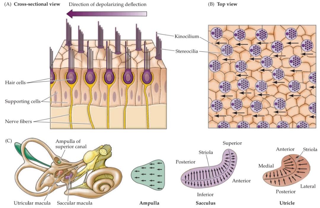
FIGURE 14.2 The morphological polarization of vestibular hair cells and polarization maps of the vestibular organs. (A) A cross section of hair cells shows that the kinocilia of a group of hair cells are all located on the same side of the hair cell. The arrow indicates the direction of deflection that depolarizes the hair cell. (B) View looking down on the hair bundles. (C) In the ampulla located at the base of each semicircular canal, the hair bundles are oriented in the same direction. In the sacculus and utricle, the striola divides the hair cells into populations with opposing hair bundle polarities.
شکل ۱۴.۲ قطبش مورفولوژیکی سلولهای مویی دهلیزی و نقشههای قطبش اندامهای دهلیزی. (الف) برش عرضی سلولهای مویی نشان میدهد که کینوسیلیای یک گروه از سلولهای مویی، همگی در یک طرف سلول مویی قرار دارند. فلش جهت انحرافی را نشان میدهد که سلول مویی را دپلاریزه میکند. (ب) نمای رو به پایین از دستههای مو. (ج) در آمپولا که در پایه هر کانال نیمدایرهای قرار دارد، دستههای مو در یک جهت قرار گرفتهاند. در ساکول و اتریکول، استریولا سلولهای مو را به جمعیتهایی با قطبیتهای دسته مویی مخالف تقسیم میکند.

FIGURE 14.3 Calcium carbonate crystals (otoconia) in the utricular macula of a quail. Each otoconium in this scanning electron micrograph is about 50 μm long. (From Dickman et al., 2004.)
شکل ۱۴.۳ کریستالهای کربنات کلسیم (اتوکونیا) در ماکولای اتریکولار یک بلدرچین. هر اتوکونیوم در این میکروگراف الکترونی روبشی حدود ۵۰ میکرومتر طول دارد. (از دیکمن و همکاران، ۲۰۰۴.)
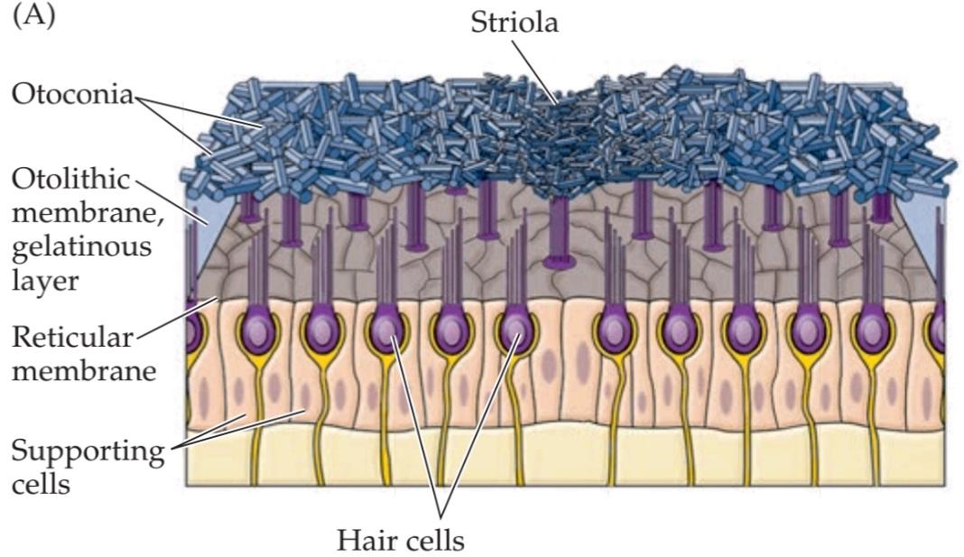

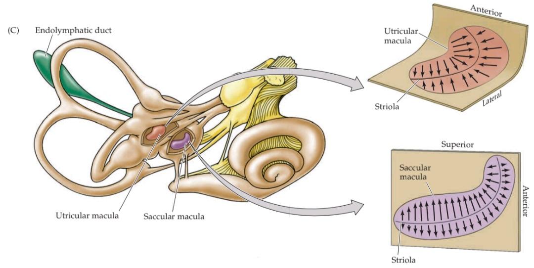
FIGURE 14.4 Morphological polarization of hair cells in the utricular and saccular maculae. (A) Cross section of the utricular macula showing hair bundles projecting into the gelatinous layer when the head is level. (B) Cross section of the utricular macula when the head is tilted. The hair bundles are deflected by the otoconia in the direction of the gravitational force along the macular plane. An equivalent linear acceleration opposite to this force would induce the same deflection of the otoconia and is referred to as the equivalent acceleration. (C) Orientation of the utricular and saccular maculae in the head; arrows show orientation of the kinocilia, as in Figure 14.2. The saccules on either side are oriented more or less vertically, and the utricles more or less horizontally. The striola is a structural landmark consisting of small otoconia arranged in a narrow trench that divides each otolith organ. In the utricular macula, the kinocilia are directed toward the striola. In the saccular macula, the kinocilia point away from the striola. Note that, given the utricle and sacculus on both sides of the body, there is a continuous representation of all directions of head movement.
شکل ۱۴.۴ قطبش مورفولوژیکی سلولهای مویی در ماکولاهای اوتیکولار و ساکولار. (الف) برش عرضی ماکولا اوتیکولار که دستههای مو را نشان میدهد که در حالت سر صاف به داخل لایه ژلاتینی بیرون زدهاند. (ب) برش عرضی ماکولا اوتیکولار هنگام کج شدن سر. دستههای مو توسط اتوکونیا در جهت نیروی گرانش در امتداد صفحه ماکولار منحرف میشوند. یک شتاب خطی معادل در خلاف جهت این نیرو، همان انحراف اتوکونیا را ایجاد میکند و به عنوان شتاب معادل شناخته میشود. (ج) جهتگیری ماکولاهای اوتیکولار و ساکولار در سر؛ فلشها جهتگیری کینوسیلیها را نشان میدهند، همانطور که در شکل 14.2 نشان داده شده است. ساکولها در دو طرف کم و بیش عمودی و اوتیکولها کم و بیش افقی هستند. استرولا یک نقطه عطف ساختاری است که از اتوکونیاهای کوچک تشکیل شده است که در یک شیار باریک قرار گرفتهاند و هر اندام اتولیتی را از هم جدا میکنند. در ماکولای اوتریکولار، کینوسیلیها به سمت استریولا هدایت میشوند. در ماکولای ساکولار، کینوسیلیها از استریولا دور میشوند. توجه داشته باشید که با توجه به اوتریکول و ساکول در دو طرف بدن، نمایش پیوستهای از تمام جهات حرکت سر وجود دارد.
One consequence of the similar effects exerted on otolithic hair cells by certain head tilts and translational movements is that otolith afferents cannot convey information that distinguishes between these two types of stimuli. Consequently, one might expect that these different stimuli would be rendered perceptually equivalent when visual feedback is absent, as occurs in the dark or when the eyes are closed. Nevertheless, blindfolded subjects can discriminate between these two stimulus categories, a feat that depends on the integration of information from the otolith organs and the semicircular canals in the central vestibular system, as described in more detail in the section “Central Pathways for Stabilizing Gaze, Head, and Posture.”
یکی از پیامدهای اثرات مشابهی که بر سلولهای مویی اتولیتی توسط کج کردنهای خاص سر و حرکات انتقالی اعمال میشود، این است که آورانهای اتولیتی نمیتوانند اطلاعاتی را که بین این دو نوع محرک تمایز قائل میشود، منتقل کنند. در نتیجه، میتوان انتظار داشت که این محرکهای مختلف در صورت عدم وجود بازخورد بصری، مانند آنچه در تاریکی یا هنگام بسته بودن چشمها رخ میدهد، از نظر ادراکی معادل باشند. با این وجود، افراد چشمبسته میتوانند بین این دو دسته محرک تمایز قائل شوند، شاهکاری که به ادغام اطلاعات از اندامهای اتولیتی و مجاری نیمدایرهای در سیستم دهلیزی مرکزی بستگی دارد، همانطور که با جزئیات بیشتر در بخش “مسیرهای مرکزی برای تثبیت نگاه، سر و وضعیت بدن” توضیح داده شده است.
As already mentioned, the orientation of the hair cell bundles is organized relative to the striola, which demar- cates the overlying layer of otoconia (see Figure 14.4A). The striola forms an axis of mirror symmetry such that hair cells on opposite sides of the striola have opposing morphological polarizations. Thus, a head tilt along the axis of the striola will excite the hair cells on one side while inhibiting the hair cells on the other side. The saccular macula is oriented vertically and the utricular macula horizontally, with continuous variation in the morphological polarization of the hair cells located in each macula (as shown in Figure 14.4C, where the black arrows indicate the direction of movement that produces excitation). Inspection of the excitatory orientations in the maculae indicates that the utricle responds to translational movements of the head in the horizontal plane and to sideways head tilts, whereas the saccule responds to vertical translational movements of the head and to upward or downward head tilts.
همانطور که قبلاً ذکر شد، جهتگیری دستههای سلولهای مویی نسبت به استرئولا سازماندهی شده است، که لایه پوشاننده اتوکونیا را مشخص میکند (شکل 14.4A را ببینید). استرئولا یک محور تقارن آینهای تشکیل میدهد به طوری که سلولهای مویی در دو طرف مخالف استرئولا، قطبشهای مورفولوژیکی متضادی دارند. بنابراین، کج کردن سر در امتداد محور استرئولا، سلولهای مویی یک طرف را تحریک میکند در حالی که سلولهای مویی طرف دیگر را مهار میکند. ماکولای ساکولار به صورت عمودی و ماکولای اوتریکولار به صورت افقی قرار دارند، با تغییرات مداوم در قطبش مورفولوژیکی سلولهای مویی واقع در هر ماکولا (همانطور که در شکل 14.4C نشان داده شده است، جایی که فلشهای سیاه جهت حرکتی را که باعث تحریک میشود نشان میدهند). بررسی جهتگیریهای تحریکی در ماکولا نشان میدهد که اتریکول به حرکات انتقالی سر در صفحه افقی و کج شدن سر به طرفین پاسخ میدهد، در حالی که ساکول به حرکات انتقالی عمودی سر و کج شدن سر به بالا یا پایین پاسخ میدهد.


FIGURE 14.5 Forces acting on the head displace the otolithic membrane of the utricular macula. For each of the positions and accelerations due to translational movements, some set of hair cells will be maximally excited, while another set will be maximally inhibited. Note that head tilts produce displacements similar to certain accelerations.
شکل ۱۴.۵ نیروهایی که بر سر وارد میشوند، غشای اتولیتیک ماکولای اتریکولار را جابجا میکنند. برای هر یک از موقعیتها و شتابهای ناشی از حرکات انتقالی، مجموعهای از سلولهای مویی به حداکثر تحریک میشوند، در حالی که مجموعهای دیگر به حداکثر مهار میشوند. توجه داشته باشید که کج کردن سر، جابجاییهایی مشابه شتابهای خاص ایجاد میکند.
Note that the saccular and utricular maculae on one side of the head are mirror images of those on the other side. Thus, a tilt of the head to one side has opposite effects on corresponding hair cells of the two utricular maculae. This concept is important in understanding how the central connections of the vestibular periphery mediate the interaction of inputs from the two sides of the head.
توجه داشته باشید که ماکولاهای ساکولار و اتریکولار در یک طرف سر، تصاویر آینهای ماکولاهای طرف دیگر هستند. بنابراین، کج کردن سر به یک طرف، اثرات متضادی بر سلولهای مویی متناظر در دو ماکولا اتریکولار دارد. این مفهوم در درک چگونگی واسطهگری اتصالات مرکزی حاشیه دهلیزی در تعامل ورودیها از دو طرف سر، مهم است.
How Otolith Neurons Sense Head Tilts and Translational Head Movements
🚀 با ما همراه شوید!
تازهترین مطالب و آموزشهای مغز و اعصاب را از دست ندهید. با فالو کردن کانال تلگرام آیندهنگاران مغز، از ما حمایت کنید!