
علوم اعصاب پروس؛ سیستم حسی-پیکری: حس لامسه و حس عمقی


دعای مطالعه [ نمایش ]
بِسْمِ الله الرَّحْمنِ الرَّحیمِ
اَللّهُمَّ اَخْرِجْنى مِنْ ظُلُماتِ الْوَهْمِ
خدايا مرا بيرون آور از تاريكىهاى وهم،
وَ اَكْرِمْنى بِنُورِ الْفَهْمِ
و به نور فهم گرامى ام بدار،
اَللّهُمَّ افْتَحْ عَلَيْنا اَبْوابَ رَحْمَتِكَ
خدايا درهاى رحمتت را به روى ما بگشا،
وَانْشُرْ عَلَيْنا خَزائِنَ عُلُومِكَ بِرَحْمَتِكَ يا اَرْحَمَ الرّاحِمينَ
و خزانههاى علومت را بر ما باز كن به امید رحمتت اى مهربانترين مهربانان.
آیندهنگاران مغز: «ما مغز را میشناسیم، تا آینده را بسازیم.»
کتاب «علوم اعصاب» اثر پروس و همکاران بهعنوان یکی از جامعترین و معتبرترین منابع در حوزه علوم اعصاب، همچنان مرجع کلیدی برای درک پیچیدگیهای مغز و سیستم عصبی است. این اثر با بهرهگیری از تازهترین پژوهشها و توضیحات دقیق درباره سازوکارهای عصبی، پلی میان دانش پایه علوم اعصاب و کاربردهای بالینی ایجاد میکند و نقشی بیبدیل در آموزش، پژوهش و ارتقای دانش مغز و اعصاب ایفا مینماید.
ترجمه دقیق و علمی این شاهکار توسط برند علمی «آیندهنگاران مغز» به مدیریت داریوش طاهری، دسترسی فارسیزبانان به مرزهای نوین دانش علوم اعصاب را ممکن ساخته و رسالتی علمی برای ارتقای آموزش، فهم عمیقتر عملکرد مغز و سیستم عصبی و توسعه روشهای نوین در حوزه سلامت عصبی فراهم آورده است.
» کتاب علوم اعصاب پروس
» » فصل ۹: سیستم حسی-پیکری: حس لامسه و حس عمقی؛ قسمت اول
» Neuroscience; Dale Purves, et al.
»» Chapter 9: The Somatosensory System: Touch and Proprioception
در حال ویرایش
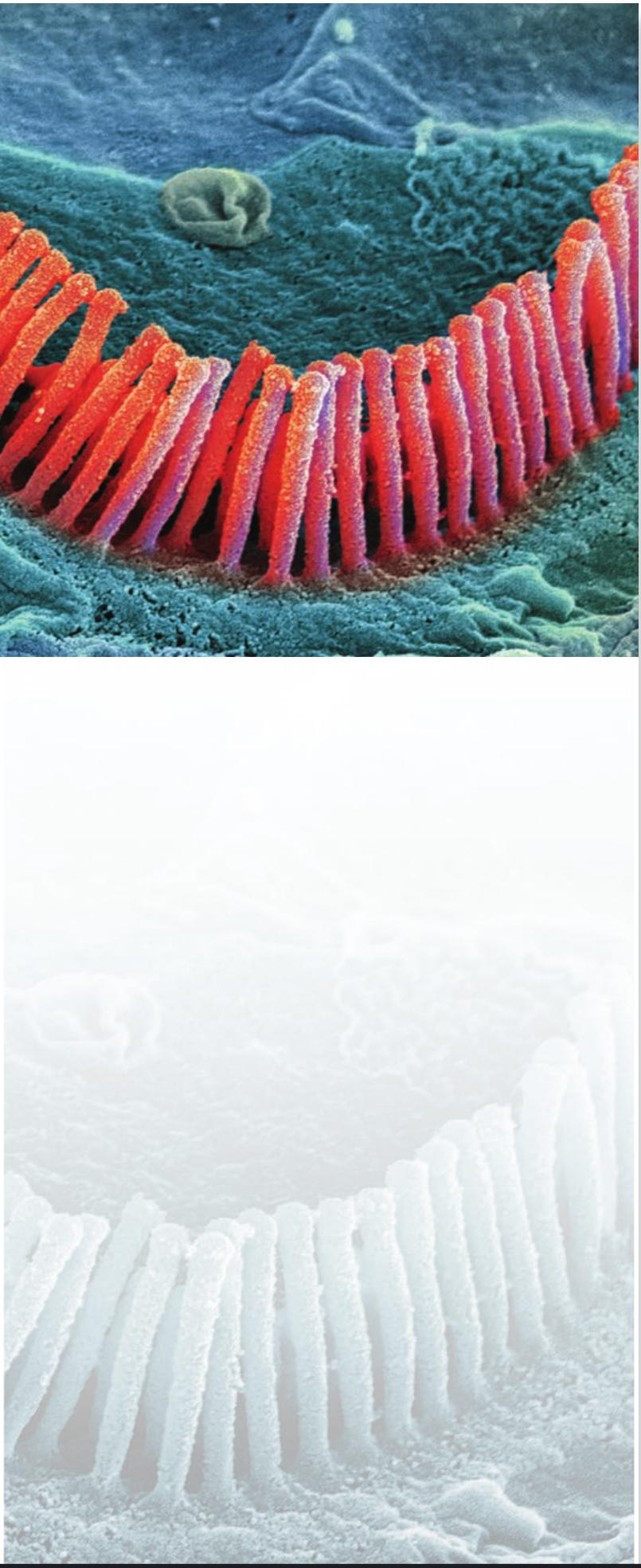
 Scanning electron micrograph of outer hair cells (stereocilia) in the inner ear. Steve Gschmeissner/Science Photo Library.
تصویر میکروسکوپ الکترونی روبشی از سلولهای مویی خارجی (استریوسیلیا) در گوش داخلی. استیو گشمایسنر/کتابخانه عکس علمی.
UNIT II
واحد دوم
Sensation and Sensory Processing
احساس و پردازش حسی
CHAPTER 9 : The Somatosensory System: Touch and Proprioception
CHAPTER 10 : Pain
CHAPTER 11 : Vision: The Eye
CHAPTER 12 : Central Visual Pathways
CHAPTER 13 : The Auditory System
CHAPTER 14 : The Vestibular System
CHAPTER 15 : The Chemical Senses
فصل ۹: سیستم حسی-پیکری: حس لامسه و حس عمقی
فصل ۱۰: درد
فصل ۱۱: بینایی چشم
فصل ۱۲: مسیرهای بینایی مرکزی
فصل ۱۳: سیستم شنوایی
فصل ۱۴: سیستم دهلیزی
فصل ۱۵: حواس شیمیایی
SENSATION ENTAILS THE ABILITY to transduce, encode, and ultimately perceive information generated by stimuli arising from both the external and internal environments. Much of the brain is devoted to these tasks. Although the basic senses somatic sensation, vision, audition, vestibular sensation, and the chemical senses are very different from one another, a few fundamental rules govern the way the nervous system deals with each of these diverse modalities. Highly specialized nerve cells called receptors convert the energy associated with mechanical forces, light, and sound waves and the presence of odorant molecules or ingested chemicals into neural signals that convey Information about stimuli (afferent sensory signals) to the spinal cord and brain. Afferent sensory signals activate central neurons capable of representing both the qualItative and quantitative aspects of the stimulus (what it is and how strong it is) and, in some modalities (somatic sensation, vision, and audition) the location of the stimulus in space (where it is).
احساس مستلزم توانایی انتقال، رمزگذاری و در نهایت درک اطلاعات تولید شده توسط محرکهای ناشی از محیطهای خارجی و داخلی است. بخش زیادی از مغز به این وظایف اختصاص داده شده است. اگرچه حواس پایه – حس پیکری، بینایی، شنوایی، حس دهلیزی و حواس شیمیایی – بسیار متفاوت از یکدیگر هستند، اما چند قانون اساسی نحوه برخورد سیستم عصبی با هر یک از این حالتهای متنوع را کنترل میکند. سلولهای عصبی بسیار تخصصی به نام گیرندهها، انرژی مرتبط با نیروهای مکانیکی، امواج نور و صدا و وجود مولکولهای بو یا مواد شیمیایی بلعیده شده را به سیگنالهای عصبی تبدیل میکنند که اطلاعات مربوط به محرکها (سیگنالهای حسی آوران) را به نخاع و مغز منتقل میکنند. سیگنالهای حسی آوران، نورونهای مرکزی را فعال میکنند که قادر به نمایش جنبههای کیفی و کمی محرک (چه چیزی و چقدر قوی است) و در برخی از حالتها (حس پیکری، بینایی و شنوایی) مکان محرک در فضا (جایی که هست) هستند.
The clinical evaluation of patients routinely requires an assessment of the sensory systems to infer the nature and location of potential neurological problems. Knowledge of where and how the different sensory modalities are transduced, relayed, represented, and further processed to generate appropriate behavioral responses is therefore essential to understanding and treating a wide variety of diseases. Accordingly, these chapters on the neurobiology of sensation also introduce some of the major structure/function relationships in the sensory components of the nervous system.
ارزیابی بالینی بیماران به طور معمول نیازمند ارزیابی سیستمهای حسی برای پی بردن به ماهیت و محل مشکلات عصبی بالقوه است. بنابراین، آگاهی از محل و چگونگی انتقال، انتقال، نمایش و پردازش بیشتر روشهای مختلف حسی برای ایجاد پاسخهای رفتاری مناسب، برای درک و درمان طیف گستردهای از بیماریها ضروری است. بر این اساس، این فصلها در مورد نوروبیولوژی حس، برخی از روابط اصلی ساختار/عملکرد را در اجزای حسی سیستم عصبی نیز معرفی میکنند.
Overview
THE SOMATOSENSORY SYSTEM is arguably the most diverse of the sensory systems, mediating a range of sensations-touch, pressure, vibration, limb position, heat, cold, itch, and pain that are transduced by receptors within the skin, muscles, or joints and conveyed to a variety of CNS targets. Not surprisingly, this complex neurobiological machinery can be divided into functionally distinct subsystems with distinct sets of peripheral receptors and central pathways. One subsystem transmits information from cutaneous mechanoreceptors and mediates the sensations of fine touch, vibration, and pressure. Another originates in specialized receptors that are associated with muscles, tendons, and joints and is responsible for proprioception our ability to sense the position of our own limbs and other body parts in space. A third subsystem arises from receptors that supply information about painful stimuli and changes in temperature as well as nondiscriminative (or sensual) touch. This chapter focuses on the tactile and proprioceptive subsystems; the mechanisms responsible for sensations of pain, temperature, and coarse sensual touch are considered in the following chapter.
مرور کلی
سیستم حسی-پیکری مسلماً متنوعترین سیستم حسی است که طیف وسیعی از احساسات – لمس، فشار، لرزش، موقعیت اندام، گرما، سرما، خارش و درد – را که توسط گیرندههای درون پوست، عضلات یا مفاصل منتقل شده و به اهداف مختلف CNS منتقل میشوند، واسطهگری میکند. جای تعجب نیست که این دستگاه عصبی-زیستی پیچیده را میتوان به زیرسیستمهای عملکردی متمایز با مجموعههای متمایزی از گیرندههای محیطی و مسیرهای مرکزی تقسیم کرد. یک زیرسیستم اطلاعات را از گیرندههای مکانیکی پوستی منتقل میکند و واسطه احساسات لمس ظریف، لرزش و فشار است. دیگری از گیرندههای تخصصی سرچشمه میگیرد که با عضلات، تاندونها و مفاصل مرتبط هستند و مسئول حس عمقی هستند – توانایی ما در حس موقعیت اندامهای خود و سایر قسمتهای بدن در فضا. زیرسیستم سوم از گیرندههایی ناشی میشود که اطلاعات مربوط به محرکهای دردناک و تغییرات دما و همچنین لمس غیرتمایزی (یا حسی) را ارائه میدهند. این فصل بر زیرسیستمهای لمسی و حس عمقی تمرکز دارد. مکانیسمهای مسئول احساس درد، دما و لمس شهوانی خشن در فصل بعد مورد بررسی قرار میگیرند.
Afferent Fibers Convey Somatosensory Information to the Central Nervous System
Somatic sensation originates from the activity of afferent nerve fibers whose peripheral processes ramify within the skin, muscles, or joints (Figure 9.1A). The cell bodies of afferent fibers reside in a series of ganglia that lie alongside the spinal cord and the brainstem and are considered part of the peripheral nervous system. Neurons in the dorsal root ganglia and in the cranial nerve ganglia (for the body and head, respectively) are the critical links supplying CNS circuits with information about sensory events that occur in the periphery.
فیبرهای آوران اطلاعات حسی-پیکری را به سیستم عصبی مرکزی منتقل میکنند
حس پیکری از فعالیت فیبرهای عصبی آوران که فرآیندهای محیطی آنها در پوست، عضلات یا مفاصل منشعب میشود، سرچشمه میگیرد (شکل 9.1A). جسم سلولی فیبرهای آوران در مجموعهای از گانگلیونها قرار دارند که در کنار نخاع و ساقه مغز قرار دارند و بخشی از سیستم عصبی محیطی محسوب میشوند. نورونهای موجود در گانگلیونهای ریشه پشتی و در گانگلیونهای عصب جمجمهای (به ترتیب برای بدن و سر) حلقههای حیاتی هستند که اطلاعات مربوط به رویدادهای حسی که در محیط رخ میدهند را به مدارهای سیستم عصبی مرکزی (CNS) ارائه میدهند.
Action potentials generated in afferent fibers by events that occur in the skin, muscles or joints propagate along the fibers and past the locations of the cell bodies in the ganglia until they reach a variety of targets in the CNS (Figure 9.1B). Peripheral and central components of afferent fibers are continuous, attached to the cell body in the ganglia by a single process. For this reason, neurons in the dorsal root ganglia are often called pseudounipolar. Because of this configuration, conduction of electrical activity through the membrane of the cell body is not an obligatory step in conveying sensory information to central targets. Nevertheless, cell bodies of sensory afferents play a critical role in maintaining the cellular machinery that mediates transduction, conduction, and transmission by sensory afferent fibers.
پتانسیلهای عمل تولید شده در فیبرهای آوران توسط رویدادهایی که در پوست، عضلات یا مفاصل رخ میدهند، در امتداد فیبرها و از محلهای جسم سلولی در گانگلیونها عبور میکنند تا زمانی که به اهداف مختلفی در سیستم عصبی مرکزی برسند (شکل 9.1B). اجزای محیطی و مرکزی فیبرهای آوران پیوسته هستند و توسط یک فرآیند واحد به جسم سلولی در گانگلیونها متصل میشوند. به همین دلیل، نورونهای موجود در گانگلیونهای ریشه پشتی اغلب شبه تک قطبی نامیده میشوند. به دلیل این پیکربندی، هدایت فعالیت الکتریکی از طریق غشای جسم سلولی گامی اجباری در انتقال اطلاعات حسی به اهداف مرکزی نیست. با این وجود، اجسام سلولی آورانهای حسی نقش مهمی در حفظ ماشینآلات سلولی دارند که واسطه انتقال، هدایت و انتقال توسط فیبرهای آوران حسی هستند.
The fundamental mechanism of sensory transduction the process of converting the energy of a stimulus into an electrical signal is similar in all somatosensory afferents: A stimulus alters the permeability of cation channels in the afferent nerve endings, generating a depolarizing current known as a receptor (or generator) potential (Figure 9.2). If sufficient in magnitude, the receptor potential reaches threshold for the generation of action potentials in the afferent fiber; the resulting rate of action potential firing is roughly proportional to the magnitude of the depolarization, as described in Chapters 2 and 3. Recently, the first family of mammalian mechanotransduction channels was identified. It consists of two members: Piezol and Piezo2 (Greek piesi, “pressure”; see Chapter 4). Piezo channels are predicted to have more than 30 transmembrane domains. Purified Piezo proteins reconstituted in artificial lipid bilayers form ion channels that transduce tension in the surrounding membrane. Importantly, Piezo2 is expressed in subsets of sensory afferent neurons as well as in other cells.
مکانیسم اساسی انتقال حسی فرآیند تبدیل انرژی یک محرک به یک سیگنال الکتریکی در تمام آورانهای حسی-پیکری مشابه است: یک محرک، نفوذپذیری کانالهای کاتیونی را در انتهای عصب آوران تغییر میدهد و یک جریان دپلاریزه کننده به نام پتانسیل گیرنده (یا مولد) ایجاد میکند (شکل 9.2). اگر مقدار آن کافی باشد، پتانسیل گیرنده به آستانه تولید پتانسیلهای عمل در فیبر آوران میرسد. سرعت شلیک پتانسیل عمل حاصل تقریباً متناسب با مقدار دپلاریزاسیون است، همانطور که در فصلهای 2 و 3 توضیح داده شده است. اخیراً، اولین خانواده از کانالهای انتقال مکانیکی پستانداران شناسایی شده است. این خانواده از دو عضو تشکیل شده است: پیزول و پیزو2 (به یونانی piesi، “فشار”؛ به فصل 4 مراجعه کنید). پیشبینی میشود کانالهای پیزو بیش از 30 دامنه غشایی داشته باشند. پروتئینهای پیزو خالص شده که در دولایههای لیپیدی مصنوعی بازسازی شدهاند، کانالهای یونی را تشکیل میدهند که تنش را در غشای اطراف منتقل میکنند. نکته مهم این است که Piezo2 در زیرمجموعههایی از نورونهای آوران حسی و همچنین در سایر سلولها بیان میشود.

FIGURE 9.1 Somatosensory afferents convey information from the skin surface to central circuits. (A) The cell bodies of somatosensory afferent fibers conveying information about the body reside in a series of dorsal root ganglia that lie along the spinal cord; those conveying information about the head are found primarily in the trigeminal ganglia. (B) Pseudounipolar neurons in the dorsal root ganglia give rise to peripheral processes that ramify within the skin (or muscles or joints) and central processes that synapse with neurons located in the spinal cord and at higher levels of the nervous system. The peripheral processes of mechanore- ceptor afferents are encapsulated by specialized receptor cells; afferents carrying pain and temperature information terminate in the periphery as free endings.
شکل ۹.۱ آورانهای حسی-پیکری اطلاعات را از سطح پوست به مدارهای مرکزی منتقل میکنند. (الف) جسم سلولی فیبرهای آوران حسی-پیکری که اطلاعات مربوط به بدن را منتقل میکنند، در مجموعهای از گانگلیونهای ریشه پشتی قرار دارند که در امتداد نخاع قرار دارند. آنهایی که اطلاعات مربوط به سر را منتقل میکنند، عمدتاً در گانگلیونهای سه قلو یافت میشوند. (ب) نورونهای شبه تک قطبی در گانگلیونهای ریشه پشتی باعث ایجاد فرآیندهای محیطی میشوند که در پوست (یا عضلات یا مفاصل) منشعب میشوند و فرآیندهای مرکزی که با نورونهای واقع در نخاع و در سطوح بالاتر سیستم عصبی سیناپس برقرار میکنند. فرآیندهای محیطی آورانهای گیرنده مکانیکی توسط سلولهای گیرنده تخصصی محصور شدهاند. آورانهای حامل اطلاعات درد و دما در محیط به صورت انتهای آزاد خاتمه مییابند.
Afferent fiber terminals that detect and transmit touch sensory stimuli (mechanoreceptors) are often encapsulated by specialized receptor cells that help tune the afferent fiber to particular features of somatic stimulation. Afferent fibers that lack specialized receptor cells are referred to as free nerve endings and are especially important in the sensation of pain (see Chapter 10). Afferents that have encapsulated endings generally have lower thresholds for action potential generation and are thus are more sensitive to sensory stimulation than are free nerve endings.
پایانههای فیبر آوران که محرکهای حسی لمسی (گیرندههای مکانیکی) را تشخیص داده و منتقل میکنند، اغلب توسط سلولهای گیرنده تخصصی محصور شدهاند که به تنظیم فیبر آوران برای ویژگیهای خاص تحریک سوماتیک کمک میکنند. فیبرهای آوران که فاقد سلولهای گیرنده تخصصی هستند، به عنوان پایانههای عصبی آزاد شناخته میشوند و به ویژه در احساس درد اهمیت دارند (به فصل 10 مراجعه کنید). آورانهایی که پایانههای کپسولدار دارند، عموماً آستانه پایینتری برای تولید پتانسیل عمل دارند و بنابراین نسبت به پایانههای عصبی آزاد، به تحریک حسی حساستر هستند.
Somatosensory Afferents Convey Different Functional Information
Somatosensory afferents differ significantly in their response properties. These differences, taken together, define distinct classes of afferents, each of which makes unique contributions to somatic sensation. Axon diameter is one factor that differentiates classes of somatosensory afferents (Table 9.1). The largest diameter sensory afferents (designated Ia) are those that supply the sensory receptors in the muscles. Most of the information subserving touch is conveyed by slightly smaller diameter fibers (Aẞ afferents), and information about pain and temperature is conveyed by even smaller diameter fibers (AS and C). The diameter of the axon determines the action potential conduction speed and is well matched to the properties of the central circuits and the various behavior demands for which each type of sensory afferent is employed (see Chapter 16).
آورانهای حسی-پیکری اطلاعات عملکردی متفاوتی را منتقل میکنند
آورانهای حسی-پیکری در ویژگیهای پاسخ خود تفاوت قابل توجهی دارند. این تفاوتها، در کنار هم، دستههای متمایزی از آورانها را تعریف میکنند که هر کدام سهم منحصر به فردی در حس پیکری دارند. قطر آکسون یکی از عواملی است که دستههای آوران حسی-پیکری را متمایز میکند (جدول 9.1). آورانهای حسی با بزرگترین قطر (که با Ia مشخص شدهاند) آنهایی هستند که گیرندههای حسی را در عضلات تغذیه میکنند. بیشتر اطلاعات مربوط به لمس توسط فیبرهای با قطر کمی کوچکتر (آورانهای Aẞ) منتقل میشوند و اطلاعات مربوط به درد و دما توسط فیبرهای با قطر حتی کوچکتر (AS و C) منتقل میشوند. قطر آکسون سرعت هدایت پتانسیل عمل را تعیین میکند و به خوبی با خواص مدارهای مرکزی و خواستههای رفتاری مختلفی که هر نوع آوران حسی برای آنها به کار میرود، مطابقت دارد (به فصل 16 مراجعه کنید).
Another distinguishing feature of sensory afferents is the size of the receptive field for cutaneous afferents, the area of the skin surface over which stimulation results in a significant change in the rate of action potentials (Figure 9.3A). A given region of the body surface is served by sensory afferents that vary significantly in the size of their receptive fields. The size of the receptive field is largely a function of the branching characteristics of the afferent within the skin; smaller arborizations result in smaller receptive fields. Moreover, there are systematic regional variations in the average size of afferent receptive fields that reflect the density of afferent fibers supplying the area. The receptive fields in regions with dense innervation (fingers, lips, toes) are relatively small compared with those in the forearm or back that are innervated by a smaller number of afferent fibers (Figure 9.3B).
یکی دیگر از ویژگیهای متمایز آورانهای حسی، اندازه میدان گیرنده برای آورانهای پوستی است، ناحیهای از سطح پوست که تحریک روی آن منجر به تغییر قابل توجهی در سرعت پتانسیلهای عمل میشود (شکل 9.3A). یک ناحیه مشخص از سطح بدن توسط آورانهای حسی که اندازه میدانهای پذیرنده آنها به طور قابل توجهی متفاوت است، پوشش داده میشود. اندازه میدان گیرنده تا حد زیادی تابع ویژگیهای شاخهبندی آوران در پوست است؛ شاخهبندیهای کوچکتر منجر به میدانهای پذیرنده کوچکتر میشوند. علاوه بر این، تغییرات منطقهای سیستماتیکی در اندازه متوسط میدانهای پذیرنده آوران وجود دارد که نشان دهنده تراکم فیبرهای آوران تغذیه کننده آن ناحیه است. میدانهای پذیرنده در مناطقی با عصبدهی متراکم (انگشتان دست، لبها، انگشتان پا) در مقایسه با میدانهای پذیرنده در ساعد یا پشت که توسط تعداد کمتری از فیبرهای آوران عصبدهی میشوند، نسبتاً کوچک هستند (شکل 9.3B).
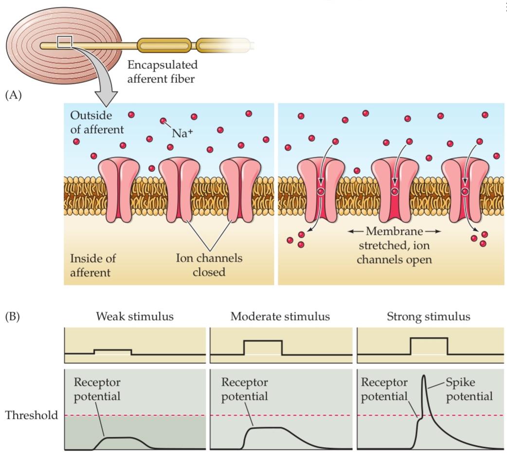
FIGURE 9.2 Transduction in a mechanosensory afferent. The process is illustrated here for a Pacinian corpuscle. (A) Deformation of the capsule leads to a stretching of the membrane of the afferent fiber, increasing the probability of opening mechanotransduction channels in the membrane. (B) Opening of these cation channels leads to depolarization of the afferent fiber (receptor potential). If the afferent is sufficiently depolarized, an action potential is generated and propagates to central targets.
شکل ۹.۲ انتقال در یک آوران حسگرهای مکانیکی. این فرآیند در اینجا برای یک جسمک پاچینی نشان داده شده است. (الف) تغییر شکل کپسول منجر به کشش غشای فیبر آوران میشود و احتمال باز شدن کانالهای انتقال مکانیکی در غشاء را افزایش میدهد. (ب) باز شدن این کانالهای کاتیونی منجر به دپلاریزاسیون فیبر آوران (پتانسیل گیرنده) میشود. اگر آوران به اندازه کافی دپلاریزه شود، یک پتانسیل عمل تولید میشود و به اهداف مرکزی منتشر میشود.
Regional differences in receptive field size and innervation density are the major factors that limit the spatial accuracy with which tactile stimuli can be sensed. Thus, measures of two point discrimination the minimum interstimulus distance required to perceive two simultaneously applied stimuli as distinct vary dramatically across the skin surface (Figure 9.3C). On the fingertips, stimuli (the indentation points produced by the tips of a caliper, for example) are perceived as distinct if they are separated by roughly 2 mm, but the same stimuli applied to the upper arm are not perceived as distinct until they are at least 40 mm apart.
تفاوتهای منطقهای در اندازه میدان گیرنده و تراکم عصبدهی، عوامل اصلی محدودکننده دقت مکانی هستند که با آن میتوان محرکهای لمسی را حس کرد. بنابراین، معیارهای تمایز دو نقطه – حداقل فاصله بین محرکهای مورد نیاز برای درک دو محرک اعمالشده همزمان به صورت مجزا – در سطح پوست به طور چشمگیری متفاوت است (شکل 9.3C). در نوک انگشتان، محرکها (به عنوان مثال، نقاط فرورفتگی ایجاد شده توسط نوک یک کولیس) اگر تقریباً 2 میلیمتر از هم فاصله داشته باشند، متمایز درک میشوند، اما همان محرکهای اعمالشده روی بازو تا زمانی که حداقل 40 میلیمتر از هم فاصله نداشته باشند، متمایز درک نمیشوند.
Sensory afferents are further differentiated by the temporal dynamics of their response to sensory stimulation. Some afferents fire rapidly when a stimulus is first presented, then fall silent in the presence of continued stimulation; others generate a sustained discharge in the presence of an ongoing stimulus (Figure 9.4). Rapidly adapting afferents (those that become quiescent in the face of continued stimulation) are thought to be particularly effective in conveying information about changes in ongoing stimulation such as those produced by stimulus movement. In contrast, slowly adapting afferents are better suited to provide information about the spatial attributes of the stimulus, such as size and shape. At least for some classes of afferent fibers, the adaptation characteristics are attributable to the properties of the receptor cells that encapsulate them. Rapidly adapting afferents that are associated with Pacinian corpuscles (see the following section) become slowly adapting when the corpuscle is removed.
آورانهای حسی با توجه به پویایی زمانی پاسخشان به تحریک حسی، بیشتر متمایز میشوند. برخی از آورانها هنگامی که یک محرک برای اولین بار ارائه میشود، به سرعت فعال میشوند، سپس در حضور تحریک مداوم خاموش میشوند؛ برخی دیگر در حضور یک محرک مداوم، تخلیه مداوم ایجاد میکنند (شکل 9.4). تصور میشود آورانهای با سازگاری سریع (آنهایی که در مواجهه با تحریک مداوم خاموش میشوند) به ویژه در انتقال اطلاعات در مورد تغییرات در تحریک مداوم، مانند تغییرات ایجاد شده توسط حرکت محرک، مؤثر هستند. در مقابل، آورانهایی که به آرامی سازگار میشوند، برای ارائه اطلاعات در مورد ویژگیهای مکانی محرک، مانند اندازه و شکل، مناسبتر هستند. حداقل برای برخی از دستههای فیبرهای آوران، ویژگیهای سازگاری به خواص سلولهای گیرندهای که آنها را در بر میگیرند، نسبت داده میشود. آورانهای با سازگاری سریع که با جسمکهای پاچینی مرتبط هستند (به بخش زیر مراجعه کنید) هنگامی که جسمک برداشته میشود، به آرامی سازگار میشوند.
TABLE 9.1 Somatosensory Afferents That Link Receptors to the Central Nervous System
جدول ۹.۱ آورانهای حسی-پیکری که گیرندهها را به سیستم عصبی مرکزی متصل میکنند
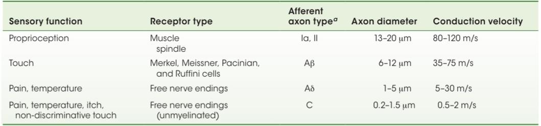
“During the 1920s and 1930s, there was a virtual cottage industry classifying axons according to their conduction velocity. Three main categories were discerned, called A, B, and C. A comprises the largest and fastest axons, C the smallest and slowest. Mechanoreceptor axons generally fall into category A. The A group is further broken down into subgroups designated α (the fastest), β, and δ (the slowest). To make matters even more confusing, muscle afferent axons are usually classified into four additional groups-I (the fastest), II, III, and IV (the slowest) with subgroups designated by lowercase roman letters! (After Rosenzweig et al., 2005.)
“در طول دهههای ۱۹۲۰ و ۱۹۳۰، یک صنعت خانگی مجازی وجود داشت که آکسونها را بر اساس سرعت هدایت آنها طبقهبندی میکرد. سه دسته اصلی به نامهای A، B و C تشخیص داده شدند. A شامل بزرگترین و سریعترین آکسونها و C کوچکترین و کندترین آنها است. آکسونهای گیرنده مکانیکی عموماً در دسته A قرار میگیرند. گروه A به زیرگروههای α (سریعترین)، β و δ (کندترین) تقسیم میشود. برای اینکه موضوع گیجکنندهتر شود، آکسونهای آوران عضلانی معمولاً به چهار گروه اضافی – I (سریعترین)، II، III و IV (کندترین) – طبقهبندی میشوند که زیرگروهها با حروف کوچک رومی مشخص میشوند! (به نقل از روزنزویگ و همکاران، ۲۰۰۵.)
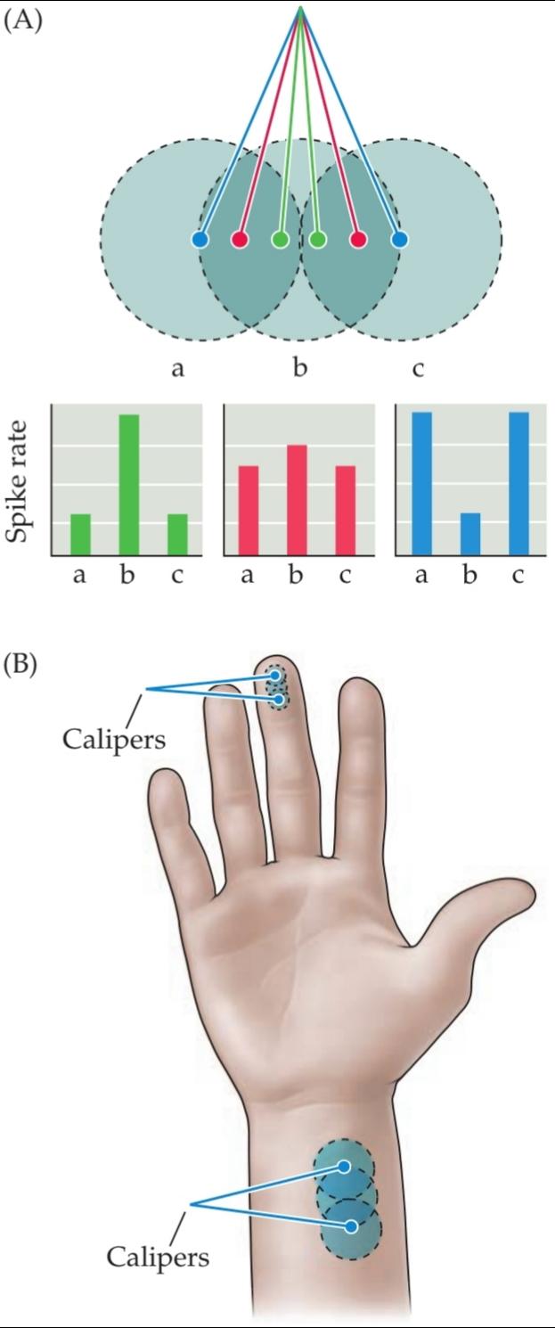

FIGURE 9.3 Receptive fields and the two point discrimination threshold. (A) Patterns of activity in three mechanosensory afferent fibers with overlapping receptive fields a, b, and c on the skin surface. When two point discrimination stimuli are closely spaced (green dots and histogram), there is a single focus of neural activity, with afferent b firing most actively. As the stimuli are moved farther apart (red dots and histogram), the activity in afferents a and c increases and the activity in b decreases. At some separation distance (blue dots and histogram), the activity in a and c exceeds that in b to such an extent that two discrete foci of stimulation can be identified. This differential pattern of activity forms the basis for the two point discrimination threshold. Stimulation applied to the center of the receptive field tends to evoke stronger responses than stimuli applied at more eccentric locations within the receptive field (see Figure 1.14). (B) The two point discrimination threshold in the fingers is much finer than that in the wrist because of differences in the sizes of afferent receptive fields that is, the separation distance necessary to produce two distinct foci of neural activity in the population of afferents innervating the lower arm is much greater than that for the afferents innervating the fingertips. (C) Differences in the two-point discrimination threshold across the surface of the body. Somatic acuity is much higher in the fingers, toes, and face than in the arms, legs, or torso. (C after Weinstein, 1968.)
شکل ۹.۳ میدانهای پذیرنده و آستانه تمایز دو نقطهای. (الف) الگوهای فعالیت در سه فیبر آوران حسگرهای مکانیکی با میدانهای پذیرنده همپوشانی a، b و c در سطح پوست. هنگامی که محرکهای تمایز دو نقطهای از هم فاصله کمی دارند (نقاط سبز و هیستوگرام)، یک کانون فعالیت عصبی وجود دارد که در آن آوران b بیشترین فعالیت را از خود نشان میدهد. با دورتر شدن محرکها از یکدیگر (نقاط قرمز و هیستوگرام)، فعالیت در آورانهای a و c افزایش و فعالیت در b کاهش مییابد. در فاصلهای از هم جدا (نقاط آبی و هیستوگرام)، فعالیت در a و c از فعالیت در b فراتر میرود تا حدی که میتوان دو کانون مجزای تحریک را شناسایی کرد. این الگوی افتراقی فعالیت، اساس آستانه تمایز دو نقطهای را تشکیل میدهد. تحریک اعمال شده به مرکز میدان گیرنده، پاسخهای قویتری نسبت به محرکهای اعمال شده در مکانهای غیرمتعارفتر در میدان گیرنده ایجاد میکند (شکل ۱.۱۴ را ببینید). (ب) آستانه تشخیص دو نقطه در انگشتان دست بسیار کمتر از مچ دست است، به دلیل تفاوت در اندازه میدانهای پذیرنده آوران، یعنی فاصله جدایی لازم برای ایجاد دو کانون مجزا از فعالیت عصبی در جمعیت آورانهایی که ساعد را عصبدهی میکنند، بسیار بیشتر از آورانهایی است که نوک انگشتان را عصبدهی میکنند. (ج) تفاوت در آستانه تشخیص دو نقطه در سطح بدن. تیزبینی سوماتیک در انگشتان دست، پا و صورت بسیار بیشتر از بازوها، پاها یا تنه است. (ج پس از واینشتاین، ۱۹۶۸.)
Finally, sensory afferents respond differently to the qualities of somatosensory stimulation. Due to differences in the properties of the channels expressed in sensory afferents, or to the filter properties of the specialized receptor cells that encapsulate many sensory afferents, generator potentials are produced only by a restricted set of stimuli that impinge on a given afferent fiber. For example, the afferents encapsulated within specialized receptor cells in the skin respond vigorously to mechanical deformation of the skin surface, but not to changes in temperature or to the presence of mechanical forces or chemicals that are known to elicit painful sensations. The latter stimuli are especially effective in driving the responses of sensory afferents known as nociceptors (see Chapter 10) that terminate in the skin as free nerve endings. Further subtypes of mechanoreceptors and nociceptors are identified on the basis of their distinct responses to somatic stimulation.
در نهایت، آورانهای حسی به کیفیت تحریک حسی-پیکری پاسخ متفاوتی میدهند. به دلیل تفاوت در خواص کانالهای بیانشده در آورانهای حسی، یا به دلیل خواص فیلتر سلولهای گیرنده تخصصی که بسیاری از آورانهای حسی را در بر میگیرند، پتانسیلهای مولد فقط توسط مجموعهای محدود از محرکها که به یک فیبر آوران معین برخورد میکنند، تولید میشوند. به عنوان مثال، آورانهای محصور شده در سلولهای گیرنده تخصصی در پوست به شدت به تغییر شکل مکانیکی سطح پوست پاسخ میدهند، اما به تغییرات دما یا وجود نیروهای مکانیکی یا مواد شیمیایی که به عنوان ایجادکننده احساسات دردناک شناخته میشوند، پاسخ نمیدهند. محرکهای اخیر به ویژه در هدایت پاسخهای آورانهای حسی معروف به گیرندههای درد (به فصل 10 مراجعه کنید) که به عنوان پایانههای عصبی آزاد در پوست خاتمه مییابند، مؤثر هستند. زیرگروههای بیشتری از گیرندههای مکانیکی و گیرندههای درد بر اساس پاسخهای متمایز آنها به تحریک سوماتیک شناسایی میشوند.
While a given sensory afferent can give rise to multiple peripheral branches, the transduction properties of all the branches of a single fiber are identical. As a result, somatosensory afferents constitute parallel pathways that differ in conduction velocity, receptive field size, dynamics, and effective stimulus features. As will become apparent, these different pathways remain segregated through several stages of central processing, and their activity contributes in unique ways to the extraction of somatosensory information that is necessary for the appropriate control of both goaloriented and reflexive movements.
در حالی که یک آوران حسی معین میتواند شاخههای محیطی متعددی ایجاد کند، خواص انتقال تمام شاخههای یک فیبر واحد یکسان است. در نتیجه، آورانهای حسی-پیکری مسیرهای موازی تشکیل میدهند که در سرعت هدایت، اندازه میدان گیرنده، دینامیک و ویژگیهای محرک مؤثر متفاوت هستند. همانطور که آشکار خواهد شد، این مسیرهای مختلف در طول چندین مرحله از پردازش مرکزی از هم جدا میمانند و فعالیت آنها به روشهای منحصر به فردی در استخراج اطلاعات حسی-پیکری که برای کنترل مناسب حرکات هدفمند و رفلکسی ضروری است، نقش دارد.
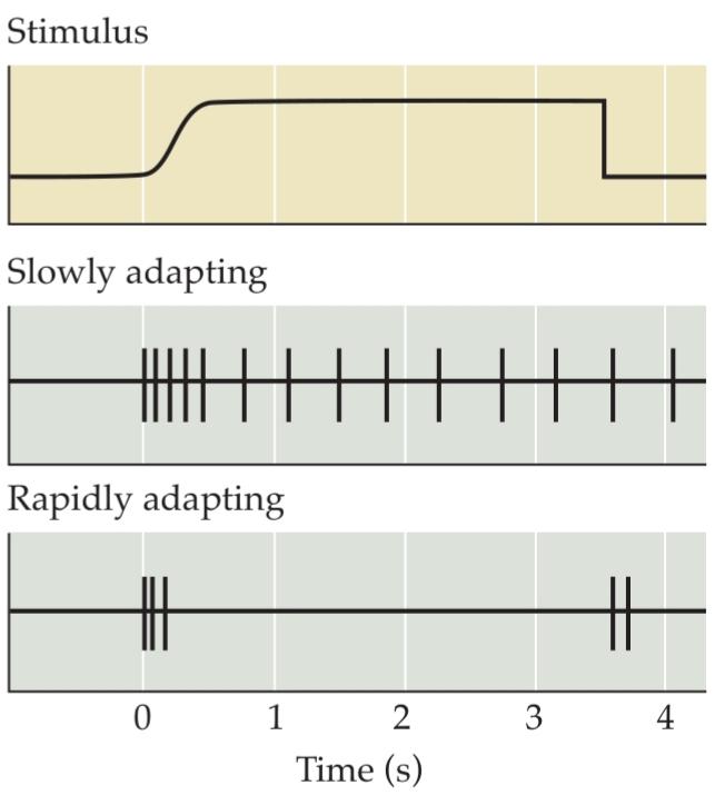
FIGURE 9.4 Slowly and rapidly adapting mechanoreceptors provide different information. Slowly adapting receptors continue responding to a stimulus, whereas rapidly adapting receptors respond only at the onset (and often the offset) of stimulation. These functional differences allow mechanoreceptors to provide information about both the static (via slowly adapting receptors) and dynamic (via rapidly adapting receptors) qualities of a stimulus.
شکل ۹.۴ گیرندههای مکانیکی با سازگاری آهسته و سریع، اطلاعات متفاوتی ارائه میدهند. گیرندههای با سازگاری آهسته به پاسخ دادن به یک محرک ادامه میدهند، در حالی که گیرندههای با سازگاری سریع فقط در شروع (و اغلب در زمان جبران) تحریک پاسخ میدهند. این تفاوتهای عملکردی به گیرندههای مکانیکی اجازه میدهد تا اطلاعاتی در مورد ویژگیهای استاتیک (از طریق گیرندههای با سازگاری آهسته) و دینامیک (از طریق گیرندههای با سازگاری سریع) یک محرک ارائه دهند.
Mechanoreceptors Specialized to Receive Tactile Information
«فصل نهم کتاب علوم اعصاب پروس به زبان انگلیسی»
🚀 با ما همراه شوید!
تازهترین مطالب و آموزشهای مغز و اعصاب را از دست ندهید. با فالو کردن کانال تلگرام آیندهنگاران مغز، از ما حمایت کنید!




بسیار عالی و جذاب