مبانی علوم اعصاب اریک کندل؛ مغز سالخورده؛ تشخیص و عوامل خطر برای آلزایمر


دعای مطالعه [ نمایش ]
بِسْمِ الله الرَّحْمنِ الرَّحیمِ
اَللّهُمَّ اَخْرِجْنى مِنْ ظُلُماتِ الْوَهْمِ
خدايا مرا بيرون آور از تاريكىهاى وهم،
وَ اَكْرِمْنى بِنُورِ الْفَهْمِ
و به نور فهم گرامى ام بدار،
اَللّهُمَّ افْتَحْ عَلَيْنا اَبْوابَ رَحْمَتِكَ
خدايا درهاى رحمتت را به روى ما بگشا،
وَانْشُرْ عَلَيْنا خَزائِنَ عُلُومِكَ بِرَحْمَتِكَ يا اَرْحَمَ الرّاحِمينَ
و خزانههاى علومت را بر ما باز كن به امید رحمتت اى مهربانترين مهربانان.
«مبانی علوم اعصاب» اثر دکتر اریک کندل و همکاران، یکی از جامعترین و معتبرترین منابع در حوزه علوم اعصاب محسوب میشود.
ترجمه دقیق این کتاب، با مدیریت داریوش طاهری و تحت نظارت برند علمی آیندهنگاران مغز، با الهام از تلاشهای علمی و تدریس اساتیدی همچون دکتر محمدتقی جغتایی و دکتر ثریا مهرابی، به فارسی برگردانده شده است.
این ترجمه، دسترسی به دانش عمیق علوم اعصاب را برای دانشجویان و پژوهشگران رشتههای پزشکی، علوم اعصاب، روانشناسی، علوم شناختی، هوش مصنوعی، گفتاردرمانی، مهندسی پزشکی و تمام علاقهمندان به درک عملکرد ذهن و مغز فراهم کرده و گامی در راستای ارتقای دانش و بهبود کیفیت زندگی است.
» کتاب مبانی علوم اعصاب اریک کندل
» » فصل شصت و چهارم: مغز سالخورده؛ قسمت سوم
در حال ویرایش
Principles of Neural Science; Eric R. Kandel
»» The Aging Brain
Risk Factors for Alzheimer Disease Have Been Identified
عوامل خطر برای بیماری آلزایمر شناسایی شده است
Alzheimer Disease Can Now Be Diagnosed well but Available Treatments Are Unsatisfactory
اکنون میتوان بیماری آلزایمر را به خوبی تشخیص داد اما درمانهای موجود رضایت بخش نیست
Highlights
نکات برجسته
Risk Factors for Alzheimer Disease Have Been Identified
عوامل خطر برای بیماری آلزایمر شناسایی شده است
Very few individuals develop AD because they bear autosomal dominant mutant alleles of the APP or presenilin genes, and these are generally of the early-onset variety. Hardly any cases of late-onset AD are due to mutations in APP or presenilin genes. Can we, then, predict AD in such individuals?
تعداد بسیار کمیاز افراد به AD مبتلا میشوند زیرا آنها دارای آللهای جهش یافته اتوزومال غالب ژنهای APP یا presenilin هستند و اینها معمولاً از انواع زودرس هستند. تقریباً هیچ موردی از AD دیررس ناشی از جهش در ژنهای APP یا presenilin است. پس آیا ما میتوانیم AD را در چنین افرادی پیش بینی کنیم؟
The major risk factor is age. The disease is pre- sent in a vanishingly small fraction of people younger than age 60 (many of those being autosomal dominant cases), 1% to 3% of those between ages 60 and 70, 3% to 12% of those between ages 70 and 80, and 25% to 40% of those older than age 85. Knowing that elderly people are prime candidates for AD is of little therapeutic use, however, because modern medicine can do nothing to slow the passage of time. Therefore, there has been intense interest in other factors that affect the incidence of AD.
عامل خطر اصلی سن است. این بیماری در بخش بسیار کوچکی از افراد جوانتر از 60 سال ناپدید میشود (بسیاری از آنهایی که موارد اتوزوم غالب هستند)، 1 تا 3 درصد از افراد بین 60 تا 70 سال، 3 تا 12 درصد از افراد بین 70 تا 80 سال، و 25 تا 40 درصد از افراد 85 سالخورده میدانند که سن بالای 85 سال دارند. با این حال، استفاده درمانی کمیدارد، زیرا پزشکی مدرن هیچ کاری برای کند کردن گذر زمان انجام نمیدهد. بنابراین، علاقه شدیدی به سایر عوامل موثر بر بروز AD وجود داشته است.
To date, the most significant genetic risk factors discovered for late-onset AD are the alleles of the gene APOE. The ApoE protein is an apolipoprotein. In the blood, it plays an important role in plasma cholesterol metabolism. It is also expressed at high levels in the brain, most prominently by astrocytes and to some extent by microglia. In the brain, where its normal function has not been clarified, it is secreted as a component of high density-like lipoproteins. In humans, there are three alleles of the APOE gene, APOE2, APOE3, and APOE4, which differ from each other by at most two amino acids. People with the APOE4 allele are at risk for AD, whereas those with the APOE2 allele are protected against AD relative to people who have the most common APOE3/APOE3 genotype. The APOE4 allele is present in about 25% of the general population but present in as many as 60% of those with AD. One copy of the APOE4 allele increases the risk of AD by about 3.7-fold, and two copies by about 12-fold, relative to someone who is ApoE3/E3 (Figure 64-14). One copy of the APOE2 allele decreases the risk for AD by about 40% relative to being APOE3/APOE3.
تا به امروز، مهم ترین عوامل خطر ژنتیکی کشف شده برای AD دیررس، آللهای ژن APOE هستند. پروتئین ApoE یک آپولیپوپروتئین است. در خون، نقش مهمیدر متابولیسم کلسترول پلاسما دارد. همچنین در سطوح بالایی در مغز، به طور برجسته توسط آستروسیتها و تا حدی توسط میکروگلیا بیان میشود. در مغز، جایی که عملکرد طبیعی آن مشخص نشده است، به عنوان جزئی از لیپوپروتئینهای شبه چگالی بالا ترشح میشود. در انسان سه آلل ژن APOE به نامهای APOE2، APOE3 و APOE4 وجود دارد که حداکثر دو اسید آمینه با یکدیگر تفاوت دارند. افراد دارای آلل APOE4 در معرض خطر ابتلا به AD هستند، در حالی که افرادی که دارای آلل APOE2 هستند در برابر AD نسبت به افرادی که دارای رایج ترین ژنوتیپ APOE3/APOE3 هستند محافظت میشوند. آلل APOE4 در حدود 25٪ از جمعیت عمومیوجود دارد، اما در 60٪ از افراد مبتلا به AD وجود دارد. یک کپی از آلل APOE4 خطر ابتلا به AD را حدود 3.7 برابر و دو کپی حدود 12 برابر نسبت به فردی که ApoE3/E3 است افزایش میدهد (شکل 14-64). یک کپی از آلل APOE2 نسبت به APOE3/APOE3 بودن حدود 40 درصد خطر ابتلا به AD را کاهش میدهد.
The mechanism by which APOE4 predisposes to AD and APOE2 protects against AD is uncertain, but ApoE4 clearly promotes Aβ aggregation by diminishing Aβ clearance and promoting fibrillization (ApoE4 > ApoE3> ApoE2). It may also act through additional mechanisms such as influencing tau, the innate immune system, cholesterol metabolism, or synaptic plasticity, although these pathways remain to be worked out.
مکانیسمیکه توسط آن APOE4 مستعد ابتلا به AD میشود و APOE2 در برابر AD محافظت میکند نامشخص است، اما ApoE4 به وضوح با کاهش کلیرانس Aβ و ترویج فیبریلیزاسیون باعث افزایش تجمع Aβ میشود (ApoE4 > ApoE3 > ApoE2). همچنین ممکن است از طریق مکانیسمهای اضافی مانند تأثیرگذاری بر تاو، سیستم ایمنی ذاتی، متابولیسم کلسترول یا شکلپذیری سیناپسی عمل کند، اگرچه این مسیرها هنوز باید بررسی شوند.
A number of other genes and genetic loci influence risk for late-onset AD. Some are common variants that alter risk only slightly, whereas other rarer variants increase risk to a greater extent (Figure 64-14). For example, relatively rare mutations in the gene TREM2 double or triple the risk for AD, similar to having one copy of the APOE4 allele. This is interesting because TREM2 as well as another gene associated with risk for AD, CD33, are expressed only in microglia. Along with other emerging cellular and animal model data, this finding suggests that the innate immune system is involved in AD pathogenesis. A number of other rare variants that increase risk to varying degrees are under investigation. It seems likely that these developments will ultimately result in a more personalized clinical approach to determining risk for AD, especially as treatments for the disease emerge.
تعدادی از ژنها و جایگاههای ژنتیکی دیگر بر خطر ابتلا به AD با شروع دیررس تأثیر میگذارند. برخی از انواع رایج هستند که ریسک را فقط اندکی تغییر میدهند، در حالی که سایر انواع نادرتر خطر را به میزان بیشتری افزایش میدهند (شکل 64-14). برای مثال، جهشهای نسبتاً نادر در ژن TREM2 خطر ابتلا به AD را دو یا سه برابر میکند، مشابه داشتن یک کپی از آلل APOE4. این جالب است زیرا TREM2 و همچنین ژن دیگر مرتبط با خطر AD، CD33، تنها در میکروگلیا بیان میشوند. همراه با سایر دادههای مدل سلولی و حیوانی در حال ظهور، این یافته نشان میدهد که سیستم ایمنی ذاتی در پاتوژنز AD نقش دارد. تعدادی از انواع نادر دیگر که خطر را به درجات مختلف افزایش میدهند در دست بررسی هستند. به نظر میرسد که این پیشرفتها در نهایت منجر به یک رویکرد بالینی شخصیتر برای تعیین خطر ابتلا به AD، به ویژه با ظهور درمانهای این بیماری میشود.
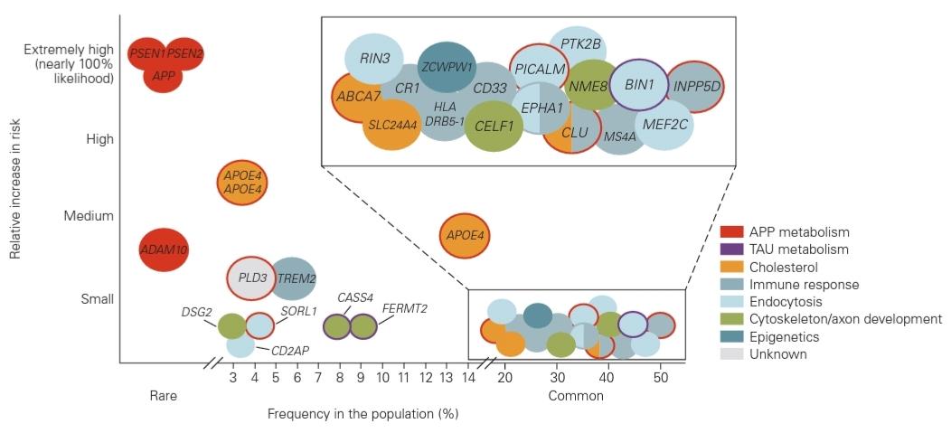
Figure 64-14 The risk for Alzheimer disease due to rare and common genetic variants. Data are from genome-wide association studies (GWAS). Mutations in the three genes that cause early-onset familial Alzheimer disease (PSEN1, PSEN2, and APP) are rare but result in Alzheimer disease in virtually 100% of people with these mutations if they live to middle age. There have been a number of common genetic changes found located in regions around or in genes that are relatively frequent in the population (eg, ABCA7, CLU, BIN1) that affect risk for Alzheimer disease but to a very small degree. The one common and strong genetic risk factor for Alzheimer disease that is present in about 20% to 25% of the population (allele frequency ~15%) is APOE4. One copy of APOE4 increases risk approximately 3.7-fold and two copies increase risk approximately 12-fold relative to people who are homozygous for APOE3. (Adapted, with permission, from Karch and Goate 2015.)
شکل 64-14 خطر ابتلا به بیماری آلزایمر به دلیل انواع ژنتیکی نادر و رایج. دادهها از مطالعات انجمن گسترده ژنوم (GWAS) است. جهش در سه ژن که باعث شروع زودهنگام بیماری آلزایمر خانوادگی میشوند (PSEN1، PSEN2 و APP) نادر است، اما اگر تا میانسالی زندگی کنند، تقریباً در 100٪ افراد مبتلا به این جهشها منجر به بیماری آلزایمر میشود. تعدادی از تغییرات ژنتیکی رایج در نواحی اطراف یا در ژنهایی که در جمعیت نسبتاً متداول هستند (مانند ABCA7، CLU، BIN1) یافت شدهاند که بر خطر ابتلا به بیماری آلزایمر تأثیر میگذارند، اما به میزان بسیار کمی. یکی از عوامل خطر ژنتیکی رایج و قوی برای بیماری آلزایمر که در حدود 20 تا 25 درصد از جمعیت (فرکانس آللی ~ 15 درصد) وجود دارد، APOE4 است. یک کپی از APOE4 خطر را تقریباً 3.7 برابر و دو نسخه در مقایسه با افرادی که هموزیگوت برای APOE3 هستند تقریباً 12 برابر خطر را افزایش میدهد. (اقتباس شده، با اجازه، از Karch and Goate 2015.)
Alzheimer Disease Can Now Be Diagnosed Well but Available Treatments Are Unsatisfactory
اکنون میتوان بیماری آلزایمر را به خوبی تشخیص داد، اما درمانهای موجود رضایت بخش نیست
Diagnosing AD at its earliest stages in the absence of biomarkers can be challenging, as its initial symptoms can be similar to those of normal age-related cognitive decline or of other related diseases. Nevertheless, diagnosis of mild to moderate dementia due to AD is usually fairly accurate. In fact, during the past few decades, the ability to accurately diagnose the disease has improved, largely because of three factors.
تشخیص AD در مراحل اولیه آن در غیاب نشانگرهای زیستی میتواند چالش برانگیز باشد، زیرا علائم اولیه آن میتواند مشابه علائم زوال شناختی طبیعی مرتبط با سن یا سایر بیماریهای مرتبط باشد. با این وجود، تشخیص زوال عقل خفیف تا متوسط ناشی از AD معمولاً نسبتاً دقیق است. در واقع، طی چند دهه گذشته، توانایی تشخیص دقیق این بیماری عمدتاً به دلیل سه عامل بهبود یافته است.
First, protocols for physical, neurological, and neuropsychological examination have become more sophisticated and standardized. Second, increased knowledge of the structural changes revealed by magnetic resonance imaging (MRI) have helped in diagnosing AD at early stages. For example, it is now possible to predict, with approximately 80% accuracy, which patients with MCI will develop AD based on the cortical thinning and ventricular enlargement visible by MRI. These imaging and diagnostic methods also assist in distinguishing dementia syndromes from each other and relating structural to functional defects. For example, patients with the disease known as behavioral variant of frontotemporal dementia experience personality changes early on, and MRI at that stage reveals atrophy of the frontal and/or temporal lobes. Likewise, initial difficulties in AD usually center on memory and attention, and MRI reveals initial alterations in the medial temporal cortex and hippocampus.
اول، پروتکلهای معاینه فیزیکی، عصبی و عصبروانشناختی پیچیدهتر و استاندارد شدهاند. دوم، افزایش دانش از تغییرات ساختاری آشکار شده توسط تصویربرداری رزونانس مغناطیسی (MRI) به تشخیص AD در مراحل اولیه کمک کرده است. به عنوان مثال، اکنون میتوان با دقت تقریباً 80 درصد پیش بینی کرد که کدام بیماران مبتلا به MCI بر اساس نازک شدن قشر مغز و بزرگ شدن بطن قابل مشاهده توسط MRI به AD مبتلا میشوند. این روشهای تصویربرداری و تشخیصی همچنین به تشخیص سندرمهای زوال عقل از یکدیگر و مرتبط ساختن نقصهای ساختاری به عملکرد کمک میکنند. به عنوان مثال، بیماران مبتلا به این بیماری که به عنوان نوع رفتاری زوال عقل فرونتومپورال شناخته میشود، تغییرات شخصیتی را در مراحل اولیه تجربه میکنند و MRI در آن مرحله آتروفی لوبهای پیشانی و/یا گیجگاهی را نشان میدهد. به همین ترتیب، مشکلات اولیه در AD معمولاً بر حافظه و توجه متمرکز است و MRI تغییرات اولیه را در قشر گیجگاهی داخلی و هیپوکامپ نشان میدهد.
Third, and perhaps most promising, amyloid plaques and neurofibrillary tangles can be visualized by positron emission tomography (PET) using compounds that avidly bind fibrillar forms of Aβ or aggregated forms of tau. The first of these, Pittsburgh compound B (PIB), binds with high affinity to fibrillar Aβ; its radioactive form, labeled with short-lived isotopes of carbon or fluorine, is readily detected by PET (Figure 64-15). The US Food and Drug Administration (FDA) has approved three amyloid imaging agents: florbetapir (Amyvid), flutemetamol (Vizamyl), and florbetaben (Neuraceq).
سومین، و شاید امیدوارکنندهترین، پلاکهای آمیلوئید و درهمتنیدگیهای نوروفیبریلاری را میتوان با توموگرافی انتشار پوزیترون (PET) با استفاده از ترکیباتی که مشتاقانه به اشکال فیبریل Aβ یا اشکال تجمع یافته تاو متصل میکنند، مشاهده کرد. اولین مورد، ترکیب پیتسبورگ B (PIB)، با میل ترکیبی بالا به Aβ فیبریلار متصل میشود. شکل رادیواکتیو آن که با ایزوتوپهای کربن یا فلوئور کوتاهمدت برچسبگذاری شده است، به آسانی توسط PET شناسایی میشود (شکل 64-15). سازمان غذا و داروی ایالات متحده (FDA) سه عامل تصویربرداری آمیلوئیدی را تأیید کرده است: فلوربتاپیر (Amyvid)، فلوتمتامول (Vizamyl) و فلوربتابن (Neuraceq).
The availability of safe molecular markers of AD allows early stages of the disease to be identified before clinical symptoms are present. Of equal importance, it allows for improved selection of patients for clinical trials and keener selection of subjects for detailed analyses of normal aging. It is important to note that these changes can also be detected in the cerebrospinal fluid, where the level of Aβ42 drops when amyloid deposition is present and total tau and phosphorylated forms of tau increase with neurodegeneration and tau aggregation.
در دسترس بودن نشانگرهای مولکولی ایمن AD اجازه میدهد تا مراحل اولیه بیماری قبل از ظهور علائم بالینی شناسایی شوند. از اهمیت یکسانی برخوردار است، این امکان را برای انتخاب بهتر بیماران برای کارآزماییهای بالینی و انتخاب دقیقتر افراد برای تجزیه و تحلیل دقیق پیری طبیعی فراهم میکند. توجه به این نکته مهم است که این تغییرات را میتوان در مایع مغزی نخاعی نیز شناسایی کرد، جایی که سطح Aβ42 با وجود رسوب آمیلوئید کاهش مییابد و تاو تاو و اشکال فسفریله تاو با تخریب عصبی و تجمع تاو افزایش مییابد.
Of course, improved diagnosis of AD is most useful if treatments are available that can halt or slow its progression at an early stage. While we still do not have a treatment that delays the onset or slows the progression of AD, there is hope that we are not too far off from being able to mitigate symptoms. Although there is no definitive proof, there is good evidence that a variety of lifestyle factors decrease risk for AD. These include high levels of education, cognitive stimulation, staying socially engaged, regular exercise, not being overweight, and getting appropriate amounts of sleep. Present-day therapies focus on treating associated symptoms such as depression, agitation, sleep disorders, hallucinations, and delusions.
البته، تشخیص بهبود یافته AD در صورتی مفیدتر است که درمانی در دسترس باشد که بتواند پیشرفت آن را در مراحل اولیه متوقف یا کند کند. در حالی که ما هنوز درمانی نداریم که شروع را به تأخیر بیندازد یا پیشرفت AD را کند کند، اما این امیدواری وجود دارد که بتوانیم علائم را کاهش دهیم. اگرچه هیچ مدرک قطعی وجود ندارد، شواهد خوبی وجود دارد که نشان میدهد انواع عوامل سبک زندگی خطر ابتلا به AD را کاهش میدهد. اینها شامل سطوح بالای تحصیلات، تحریک شناختی، درگیر ماندن در اجتماع، ورزش منظم، نداشتن اضافه وزن و خوابیدن به مقدار مناسب است. درمانهای امروزی بر درمان علائم مرتبط مانند افسردگی، بی قراری، اختلالات خواب، توهمات و هذیان تمرکز دارند.

Figure 64-15 Positron emission tomography scans can visualize amyloid plaques in the living brain. The density of Aβ plaques is indicated by the red regions in these images made after administration of Pittsburgh compound B (PIB), a fluorescent analog of thioflavin T. (Images reproduced, with permission, from R. Buckner.)
شکل 64-15 اسکن توموگرافی گسیل پوزیترون میتواند پلاکهای آمیلوئید را در مغز زنده تجسم کند. چگالی پلاکهای A با نواحی قرمز رنگ در این تصاویر نشان داده میشود که پس از تجویز ترکیب B Pittsburgh (PIB)، یک آنالوگ فلورسنت تیوفلاوین T. (تصاویر با کسب اجازه از R. Buckner، تکثیر شدهاند.)
One of the principal therapeutic targets to date has been the cholinergic system in the basal forebrain, a region of the brain that is damaged in AD and that contributes to attention. Acetylcholinesterase inhibitors increase levels of acetylcholine by inhibiting its breakdown and represent one of the few drug classes approved by the FDA for treatment of AD. Another drug, the N-methyl-D-aspartate (NMDA) receptor antagonist memantine, also improves symptoms in individuals with mild to moderate dementia due to AD. It is believed that memantine’s action modulates glutamate-mediated neurotransmission. Nevertheless, these drugs exert only a modest effect on cognitive functions and the activities of daily living.
یکی از اهداف درمانی اصلی تا به امروز، سیستم کولینرژیک در پیشمغز قاعدهای بوده است، ناحیهای از مغز که در AD آسیب دیده و به توجه کمک میکند. مهارکنندههای استیل کولین استراز با مهار تجزیه استیل کولین، سطح استیل کولین را افزایش میدهند و یکی از معدود کلاسهای دارویی تایید شده توسط FDA برای درمان AD هستند. داروی دیگر، ممانتین آنتاگونیست گیرنده N-methyl-D-aspartate (NMDA) نیز علائم را در افراد مبتلا به زوال عقل خفیف تا متوسط ناشی از AD بهبود میبخشد. اعتقاد بر این است که اثر ممانتین انتقال عصبی با واسطه گلوتامات را تعدیل میکند. با این وجود، این داروها تنها تأثیر متوسطی بر عملکردهای شناختی و فعالیتهای زندگی روزمره دارند.
Recent advances in our understanding of the cell-biological basis of AD have produced several promising new therapeutic targets, all of which are being explored intensively. One approach is to develop drugs that reduce or modulate the activity of the β- and y-secretases that cleave APP to generate Aβ peptides and the associated soluble extracellular and intracellular fragments. In fact, decreasing either β- or y-secretase levels in transgenic mice that overexpress mutant APP decreases Aβ deposition and, in some cases, functional abnormalities.
پیشرفتهای اخیر در درک ما از اساس سلولی-بیولوژیکی AD چندین هدف درمانی جدید امیدوارکننده ایجاد کرده است که همه آنها به شدت در حال بررسی هستند. یک رویکرد توسعه داروهایی است که فعالیت β- و y-secretases را کاهش میدهند یا تعدیل میکنند که APP را برای تولید پپتیدهای Aβ و قطعات محلول خارج سلولی و داخل سلولی مرتبط میشکنند. در واقع، کاهش سطح β- یا y-secretase در موشهای تراریخته که APP جهش یافته را بیش از حد بیان میکنند، رسوب Aβ و در برخی موارد، ناهنجاریهای عملکردی را کاهش میدهد.
Accordingly, pharmaceutical companies have developed drugs that decrease or modulate levels of B- and y-secretases in humans. An obstacle to this approach is that the secretases also act on substrates other than APP, so decreasing their levels can have deleterious side effects. This is especially true for y-secretase, whose inhibition has led to toxicity in human trials for AD. There are now several ẞ-secretase inhibitors in clinical trials for AD, and it is likely such drugs will also move into trials for what is called preclinical AD, when AD pathology is accumulating but there is no sign yet of cognitive decline (Figure 64-16). The goal of this therapy would be to delay or prevent the onset of cognitive decline and dementia.
بر این اساس، شرکتهای داروسازی داروهایی تولید کرده اند که سطوح B- و y-secretases را در انسان کاهش یا تعدیل میکند. یک مانع برای این رویکرد این است که سکرتازها روی سوبستراهای دیگری غیر از APP نیز عمل میکنند، بنابراین کاهش سطح آنها میتواند عوارض جانبی مضری داشته باشد. این امر به ویژه در مورد y-secretase که مهار آن منجر به سمیت در آزمایشات انسانی برای AD شده است، صادق است. در حال حاضر چندین مهارکننده ẞ-secretase در کارآزماییهای بالینی برای AD وجود دارد، و احتمالاً چنین داروهایی برای آنچه که AD پیش بالینی نامیده میشود، زمانی که آسیبشناسی AD در حال انباشته شدن است اما هنوز نشانهای از زوال شناختی وجود ندارد، وارد آزمایشهایی شوند (شکل 64-16). هدف این درمان به تاخیر انداختن یا جلوگیری از شروع زوال شناختی و زوال عقل خواهد بود.
Another approach is to decrease levels of Aβ through immunological means. Both immunization with Aβ, which leads to generation of antibodies to Aβ, and passive transfer of Aβ antibodies have been tested in transgenic mouse models of AD. Both treatments have been shown to reduce levels of Aβ, Aβ toxicity, and plaques (Figure 64-17). The mechanisms of enhanced Aβ clearance are not completely clear. Serum antibodies likely serve as a “sink,” resulting in Aβ peptides with low molecular weight being cleared more extensively from the brain into the circulation, thus changing the equilibrium of Aβ in different compartments and promoting removal of Aβ from the brain.
رویکرد دیگر کاهش سطح Aβ از طریق روشهای ایمونولوژیک است. هم ایمن سازی با Aβ که منجر به تولید آنتی بادیهای Aβ میشود و هم انتقال غیر فعال آنتی بادیهای Aβ در مدلهای تراریخته موش AD آزمایش شده اند. هر دو درمان نشان داده شده است که سطوح Aβ، سمیت Aβ و پلاکها را کاهش میدهند (شکل 64-17). مکانیسمهای افزایش ترخیص کالا از گمرک Aβ کاملاً مشخص نیست. آنتیبادیهای سرم احتمالاً بهعنوان یک «سینک» عمل میکنند و در نتیجه پپتیدهای Aβ با وزن مولکولی پایین به طور گستردهتری از مغز به گردش خون پاک میشوند، بنابراین تعادل Aβ در بخشهای مختلف تغییر میکند و باعث حذف Aβ از مغز میشود.

Figure 64-16 Relationship of biomarker changes to cognitive and clinical changes in Alzheimer disease (AD). In cognitively normal people who are going to develop AD dementia, one of the first physical signs is the initiation of Aβ aggregation in the brain in the form of amyloid plaques. While people are still cognitively normal, amyloid plaques continue to accumulate. At some point, about 5 years before any clear-cut cognitive decline, tau accumulation begins to increase in the neocortex, inflammation and oxidative stress increase, and brain network connections and metabolism begin to decline. Neuronal and synaptic loss and brain atrophy also begin. This period-when the patient remains cognitively normal but AD-type pathology is building up-is termed preclinical AD. Once there is enough neuronal and synaptic dysfunction as well as cell loss, very mild dementia and mild cognitive impairment become detectable. At that time, amyloid deposition has almost reached its peak. As dementia worsens to mild, moderate, and severe stages, neurofibrillary tangles form, and neuronal and synaptic dysfunction, inflammation, cell death, and brain atrophy worsen. (Adapted, with permission, from Perrin, Fagan, and Holtzman 2009.)
شکل 64-16 رابطه تغییرات نشانگرهای زیستی با تغییرات شناختی و بالینی در بیماری آلزایمر (AD). در افراد از نظر شناختی عادی که قرار است به زوال عقل AD مبتلا شوند، یکی از اولین علائم فیزیکی شروع تجمع Aβ در مغز به شکل پلاکهای آمیلوئید است. در حالی که افراد هنوز از نظر شناختی طبیعی هستند، پلاکهای آمیلوئید همچنان به تجمع میرسند. در برخی موارد، حدود 5 سال قبل از هر زوال شناختی واضح، تجمع تاو در نئوکورتکس شروع به افزایش میکند، التهاب و استرس اکسیداتیو افزایش مییابد و ارتباطات شبکه مغز و متابولیسم شروع به کاهش میکند. از دست دادن عصبی و سیناپسی و آتروفی مغز نیز شروع میشود. این دوره – زمانی که بیمار از نظر شناختی نرمال میماند اما آسیب شناسی نوع AD در حال ایجاد است – AD پیش بالینی نامیده میشود. هنگامیکه اختلال عملکرد عصبی و سیناپسی به اندازه کافی و همچنین از دست دادن سلول وجود داشته باشد، زوال عقل بسیار خفیف و اختلال شناختی خفیف قابل تشخیص است. در آن زمان رسوب آمیلوئید تقریباً به اوج خود رسیده است. با بدتر شدن زوال عقل به مراحل خفیف، متوسط و شدید، گرههای نوروفیبریلاری شکل میگیرد و اختلال عملکرد عصبی و سیناپسی، التهاب، مرگ سلولی و آتروفی مغز بدتر میشود. (اقتباس شده، با اجازه، از Perrin، Fagan، و Holtzman 2009.)

Figure 64-17 Immunization with antibodies to the Aβ peptide clears amyloid plaques and preserves cognitive performance in mice expressing the peptide. Mice that develop Aβ deposition in the form of amyloid plaques were immunized with the Aβ peptide. This led to production of anti- bodies against Aβ.
شکل 64-17 ایمن سازی با آنتی بادیهای پپتید Aβ، پلاکهای آمیلوئید را پاک میکند و عملکرد شناختی را در موشهای بیان کننده پپتید حفظ میکند. موشهایی که رسوب Aβ را به شکل پلاکهای آمیلوئید ایجاد میکنند با پپتید Aβ ایمن سازی شدند. این منجر به تولید آنتی بادی علیه Aβ شد.
A. Comparison of amyloid plaque deposition in the cerebral cortex of mice overexpressing a mutant APP transgene (APP transgenic mice) that develop amyloid plaques. The mice that were immunized with the Aβ peptide have substantially reduced amyloid plaque deposition. (Adapted from Brody and Holtzman 2008.)
الف. مقایسه رسوب پلاک آمیلوئید در قشر مغز موشهایی که بیش از حد یک ترانس ژن APP جهش یافته (موش تراریخته APP) را بیان میکنند که پلاکهای آمیلوئید ایجاد میکند. موشهایی که با پپتید Aβ واکسینه شده بودند رسوب پلاک آمیلوئید را به میزان قابل توجهی کاهش دادند. (برگرفته از برودی و هولتزمن 2008.)
B. Cognitive performance (a memory test) in two groups of APP transgenic mice. One group was immunized with an irrelevant protein, the other with the Aβ peptide. The mice vaccinated with Aβ performed at levels close to normal animals, whereas mice immunized with the irrelevant protein showed severe impairment in memory. (Adapted, with permission, from Janus et al. 2000.)
ب. عملکرد شناختی (آزمون حافظه) در دو گروه موش تراریخته APP. یک گروه با یک پروتئین نامربوط و گروه دیگر با پپتید Aβ واکسینه شدند. موشهای واکسینهشده با Aβ در سطوحی نزدیک به حیوانات عادی عمل کردند، در حالی که موشهایی که با پروتئین نامربوط واکسینه شده بودند، اختلال شدید در حافظه نشان دادند. (اقتباس شده، با اجازه، از Janus و همکاران 2000.)
It is also clear that in the brain several anti-Aβ antibodies bind either soluble or fibrillar Aβ, or both.
همچنین واضح است که در مغز چندین آنتی بادی ضد Aβ به Aβ محلول یا فیبریلار یا هر دو متصل میشود.
Those that bind to aggregated forms of Aβ can stimulate microglia-mediated phagocytosis to remove Aβ, although there is also plaque removal that is not dependent on microglial-mediated phagocytosis. Anti- bodies to soluble Aβ that enter the brain may decrease soluble Aβ toxicity. These findings suggest that immunotherapeutic strategies may be successful in AD patients, especially if they are given early enough in the disease course, prior to significant neuronal damage and loss. There are multiple human trials underway using active and passive immunotherapies against Aβ both in preclinical and mild AD.
آنهایی که به اشکال تجمع یافته Aβ متصل میشوند میتوانند فاگوسیتوز با واسطه میکروگلیا را برای حذف Aβ تحریک کنند، اگرچه حذف پلاک نیز وجود دارد که به فاگوسیتوز با واسطه میکروگلیال وابسته نیست. آنتیبادیهایی که به Aβ محلول وارد مغز میشوند، ممکن است سمیت Aβ محلول را کاهش دهند. این یافتهها نشان میدهد که استراتژیهای ایمونوتراپی ممکن است در بیماران مبتلا به AD موفقیتآمیز باشد، بهویژه اگر در اوایل دوره بیماری، قبل از آسیب و از دست دادن نورون قابلتوجه به آنها داده شود. آزمایشهای انسانی متعددی با استفاده از ایمونوتراپیهای فعال و غیرفعال علیه Aβ در هر دو بیماری پیشبالینی و خفیف در حال انجام است.
In addition to targeting Aβ, clinical trials have also begun targeting tau. This is being done with active and passive immunization against tau as well as with small molecules that, in cell culture and animal models, can decrease tau aggregation. A number of studies in animal models have shown that certain anti-tau antibodies can decrease the amount of aggregated, hyperphosphorylated tau in the central nervous system and in some cases improve function. Although tau is predominantly a cytoplasmic protein, one of the reasons that anti-tau antibodies may be having an effect is that, as discussed above, tau aggregates may spread from cell to cell in the extracellular space in a prion-like fashion. It is in this space that an antibody may be able to interact with tau and block this process.
علاوه بر هدف قرار دادن Aβ، آزمایشات بالینی نیز هدف قرار دادن تاو را آغاز کرده اند. این کار با ایمن سازی فعال و غیرفعال در برابر تاو و همچنین با مولکولهای کوچکی انجام میشود که در کشت سلولی و مدلهای حیوانی میتوانند تجمع تاو را کاهش دهند. تعدادی از مطالعات در مدلهای حیوانی نشان دادهاند که برخی از آنتیبادیهای ضد تاو میتوانند میزان تاو تجمع یافته و هیپرفسفریله شده در سیستم عصبی مرکزی را کاهش دهند و در برخی موارد عملکرد را بهبود بخشند. اگرچه تاو عمدتاً یک پروتئین سیتوپلاسمیاست، یکی از دلایلی که ممکن است آنتیبادیهای ضد تاو تأثیر بگذارند، این است که، همانطور که در بالا مورد بحث قرار گرفت، تجمعات تاو ممکن است از سلولی به سلول دیگر در فضای خارج سلولی به شکل پریونی پخش شوند. در این فضا است که یک آنتی بادی ممکن است بتواند با تاو تعامل داشته باشد و این فرآیند را مسدود کند.
Highlights
نکات برجسته
1. It is only in the past 50 years that a large percentage of the population has lived into the eighth to tenth decades of life. With this increase, neuroscientists have been able to study changes in the brain that occur with normal aging as well as in individuals who develop age-related brain disorders.
1. تنها در 50 سال گذشته است که درصد زیادی از جمعیت در دهههای هشتم تا دهم زندگی میکنند. با این افزایش، دانشمندان علوم اعصاب توانستهاند تغییراتی را در مغز که با پیری طبیعی و همچنین در افرادی که به اختلالات مغزی مرتبط با سن مبتلا میشوند، روی میدهد، مطالعه کنند.
2. Subtle changes in a variety of brain functions occur with age, including declines in speed of processing and memory storage and changes in sleep. The underlying basis for these changes is likely brain atrophy and loss of white matter integrity. In general, however, there is not a significant decrease in neuronal number that accounts for changes in brain function that occur with normal aging.
2. تغییرات ظریف در انواع عملکردهای مغز با افزایش سن رخ میدهد، از جمله کاهش سرعت پردازش و ذخیره حافظه و تغییر در خواب. اساس اساسی این تغییرات احتمالاً آتروفی مغز و از دست دادن یکپارچگی ماده سفید است. با این حال، به طور کلی، کاهش قابل توجهی در تعداد نورونها وجود ندارد که علت تغییرات در عملکرد مغز باشد که با افزایش سن طبیعی رخ میدهد.
3. The changes in cognition that occur in normal aging are not disabling. When memory and often other areas of cognitive function decline more than is expected with age such that it is noticeable to others and mildly affects one’s day-to- day life, this syndrome is called mild cognitive impairment (MCI).
3. تغییرات شناختی که در پیری طبیعی رخ میدهد ناتوان کننده نیست. هنگامیکه حافظه و غالباً سایر بخشهای عملکرد شناختی با افزایش سن بیش از حد انتظار کاهش مییابد به طوری که برای دیگران قابل توجه است و به طور ملایمیبر زندگی روزمره فرد تأثیر میگذارد، به این سندرم اختلال شناختی خفیف (MCI) میگویند.
4. MCI is not a disease, it is a syndrome. About 50% of individuals with MCI have Alzheimer disease (AD) as the underlying cause of the MCI. Other conditions that can cause MCI include depression, cerebrovascular disease, Lewy body dis- ease, metabolic disorders, and drugs, prescribed for other diseases, that cause central nervous system side effects.
4. MCI یک بیماری نیست، یک سندرم است. حدود 50 درصد از افراد مبتلا به MCI بیماری آلزایمر (AD) را به عنوان علت اصلی MCI دارند. سایر شرایطی که میتوانند باعث MCI شوند عبارتند از افسردگی، بیماری عروق مغزی، بیماری بدن لوی، اختلالات متابولیک، و داروهای تجویز شده برای سایر بیماریها که باعث عوارض جانبی سیستم عصبی مرکزی میشوند.
5. AD is the most common cause of dementia and manifests as loss of memory and other cognitive abilities sufficient to impair social and occupational functions. AD accounts for about 70% of cases of dementia in the United States, with the remainder caused primarily by cerebrovascular disease, Parkinson and Lewy body dementia, and frontotemporal dementia.
5. AD شایع ترین علت زوال عقل است و به صورت از دست دادن حافظه و سایر تواناییهای شناختی کافی برای مختل کردن عملکردهای اجتماعی و شغلی ظاهر میشود. AD حدود 70 درصد از موارد زوال عقل در ایالات متحده را تشکیل میدهد و بقیه عمدتاً ناشی از بیماری عروق مغزی، دمانس بدن پارکینسون و لوی و زوال عقل فرونتومپورال است.
6. The pathology of AD is characterized by the accumulation of aggregated forms of two proteins in the brain, the Aβ peptide and tau. Aβ accumulates in a fibrillar form in extracellular structures called amyloid plaques in the brain parenchyma as well as in the walls of arterioles (where it is called cerebral amyloid angiopathy). Tau accumulates in neurofibrillary tangles in cell bodies and dendrites.
6. پاتولوژی AD با تجمع اشکال تجمع یافته دو پروتئین در مغز، پپتید Aββ و tau مشخص میشود. Aβ به شکل فیبریلار در ساختارهای خارج سلولی به نام پلاکهای آمیلوئید در پارانشیم مغز و همچنین در دیواره شریانها (که آنژیوپاتی آمیلوئید مغزی نامیده میشود) تجمع مییابد. تاو در پیچ و تابهای عصبی فیبریلاری در بدن سلولی و دندریتها تجمع مییابد.
7. In addition to the accumulation of protein aggregates in the Alzheimer brain, marked brain atrophy as well as synaptic and neuronal loss occurs as the disease progresses. There is also a strong neuroinflammatory response, especially around amyloid plaques, which involves microglia and astrocytes.
7. علاوه بر تجمع تودههای پروتئینی در مغز آلزایمر، آتروفی مشخص مغز و همچنین از دست دادن سیناپسی و عصبی با پیشرفت بیماری رخ میدهد. همچنین یک پاسخ عصبی التهابی قوی، به ویژه در اطراف پلاکهای آمیلوئید، که میکروگلیا و آستروسیتها را درگیر میکند، وجود دارد.
8. The pathology of AD begins about 15 years prior to the onset of cognitive decline or the MCI phase of the disease. Aβ accumulation in the neocortex appears to initiate the disease with markedly abnormal levels, followed by the spread of tau aggregates from the medial temporal lobe to other regions of the neocortex. This phase of Alzheimer pathology prior to symptom onset is known as preclinical AD.
8. آسیب شناسی AD حدود 15 سال قبل از شروع زوال شناختی یا مرحله MCI بیماری شروع میشود. به نظر میرسد تجمع Aβ در نئوکورتکس باعث شروع بیماری با سطوح غیرطبیعی قابل توجهی میشود، و به دنبال آن تودههای تاو از لوب گیجگاهی داخلی به سایر مناطق نئوکورتکس گسترش مییابد. این مرحله از آسیب شناسی آلزایمر قبل از شروع علائم به عنوان AD پیش بالینی شناخته میشود.
9. Significant data suggest that certain aggregated forms of the Aβ peptide lead to synaptic and neuronal damage in the Alzheimer brain, but a much better correlate of the cognitive decline is the presence and accumulation of aggregated forms of the tau protein.
9. دادههای قابل توجهی نشان میدهد که اشکال تجمع یافته خاصی از پپتید Aβ منجر به آسیب سیناپسی و عصبی در مغز آلزایمر میشود، اما ارتباط بسیار بهتری با زوال شناختی وجود و تجمع اشکال تجمعی پروتئین تاو است.
10. There are two major forms of AD. The first is a dominantly inherited AD, which accounts for less than 1% of Alzheimer patients and is caused by mutations in one of three genes encoding the proteins APP, PS1, and PS2; this form leads to clinical disease onset between the ages of 30 and 50. Genetic, biochemical, and other studies have shown that the genes that cause autosomal dominant AD do so through early accumulation of the Aβ peptide in the brain. The second form, late- onset AD, with an age of onset of 65 years or later, accounts for more than 99% of cases. Although age is the greatest risk factor for late-onset AD, genetics also contribute. The APOE gene is by far the biggest genetic contributor to AD, with the APOE4 variant increasing risk and the APOE2 variant decreasing risk. There are a number of other common genetic variants in other genes that influence risk. There are also rare variants in other genes such as TREM2 that increase risk to a level similar to that associated with one copy of APOE4. Nonetheless, there is general agreement that major features of pathogenesis are similar in sporadic and familial AD.
10. دو شکل عمده از AD وجود دارد. اولی یک بیماری آلزایمر غالباً ارثی است که کمتر از 1٪ از بیماران آلزایمر را تشکیل میدهد و به دلیل جهش در یکی از سه ژن کد کننده پروتئینهای APP، PS1 و PS2 ایجاد میشود. این شکل منجر به شروع بیماری بالینی در سنین 30 تا 50 سالگی میشود. مطالعات ژنتیکی، بیوشیمیایی و سایر مطالعات نشان داده اند که ژنهایی که باعث AD غالب اتوزومیمیشوند این کار را از طریق تجمع اولیه پپتید Aβ در مغز انجام میدهند. شکل دوم، AD دیر شروع، با سن شروع 65 سال یا بالاتر، بیش از 99٪ موارد را تشکیل میدهد. اگرچه سن بزرگترین عامل خطر برای دیررس بیماری AD است، ژنتیک نیز در این امر نقش دارد. ژن APOE تا حد زیادی بزرگترین عامل ژنتیکی در ابتلا به بیماری آلزایمر است، با افزایش خطر ابتلا به نوع APOE4 و کاهش خطر نوع APOE2. تعدادی از انواع ژنتیکی رایج دیگر در ژنهای دیگر وجود دارد که بر خطر تأثیر میگذارد. همچنین انواع نادری در ژنهای دیگر مانند TREM2 وجود دارد که خطر را تا سطحی مشابه با یک کپی از APOE4 افزایش میدهد. با این وجود، توافق کلی وجود دارد که ویژگیهای اصلی پاتوژنز در AD پراکنده و خانوادگی مشابه است.
11. Besides clinical symptoms and signs of AD, amyloid and tau imaging and cerebrospinal fluid markers can determine that Alzheimer pathology is present in a living person with or without cognitive decline.
11. علاوه بر علائم بالینی و نشانههای AD، تصویربرداری آمیلوئید و تاو و نشانگرهای مایع مغزی نخاعی میتوانند مشخص کنند که پاتولوژی آلزایمر در یک فرد زنده با یا بدون زوال شناختی وجود دارد.
12. There are currently only symptomatic therapies for AD that have modest benefit at best. A number of potential disease-modifying therapies that influence the production, clearance, and aggregation of either Aβ or tau are being tested in humans. Although none of these therapies is yet approved, there is hope that over the next several years one or more of these therapies will begin to show a clear benefit.
12. در حال حاضر فقط درمانهای علامتی برای بیماری AD وجود دارد که در بهترین حالت دارای فایده اندکی هستند. تعدادی از درمانهای بالقوه اصلاحکننده بیماری که بر تولید، پاکسازی و تجمع Aβ یا tau تأثیر میگذارند، روی انسانها آزمایش میشوند. اگرچه هیچ یک از این درمانها هنوز تایید نشده است، اما این امید وجود دارد که طی چندین سال آینده، یک یا چند مورد از این درمانها شروع به نشان دادن سود آشکار کنند.
کلیک کنید تا Selected Reading نمایش داده شود
Joshua R. Sanes David M. Holtzman
Brody DL, Holtzman DM. 2008. Active and passive immunotherapy for neurodegenerative disorders. Annu Rev Neurosci 31:175–193.
Buckner RL. 2004. Memory and executive function in aging and AD: multiple factors that cause decline and reserve factors that compensate. Neuron 44:195–208.
Goedert M, Eisenberg DS, Crowther RA. 2017. Propagation of tau aggregates and neurodegeneration. Annu Rev Neurosci 40:189–210.
Haass C, Selkoe DJ. 2007. Soluble protein oligomers in neurodegeneration: lessons from the Alzheimer’s amyloid beta- peptide. Nat Rev Mol Cell Biol 8:101-112.
Holtzman DM, Herz J, Bu G. 2012. Apolipoprotein E and apolipoprotein E receptors: normal biology and roles in Alzheimer disease. Cold Spring Harb Perspect Med 2:a006312.
Holtzman DM, Morris JC, Goate AM. 2011. Alzheimer’s dis- ease: the challenge of the second century. Sci Transl Med 3:77sr1.
Kenyon C. 2005. The plasticity of aging: insights from long- lived mutants. Cell 120:449–460.
Musiek ES, Holtzman DM. 2015. Three dimensions of the amyloid hypothesis: time, space and “wingmen.” Nat Neurosci 18:800-806.
Sanders DW, Kaufman SK, Holmes BB, Diamond MI. 2016. Prions and protein assemblies that convey biological information in health and disease. Neuron 89:433–448.
کلیک کنید تا References نمایش داده شود
Andrews-Hanna JR, Snyder AZ, Vincent JL, et al. 2007. Disruption of large-scale brain systems in advanced aging. Neuron 56:924-935.
Arias E. 2004. United States Life Tables, 2001. National Vital Statistics Reports, Vol. 52, No. 14. Hyattsville, MD: National Center for Health Statistics.
Arnold SE, Hyman BT, Flory J, Damasio AR, Van Hoesen GW. 1991. The topographical and neuroanatomical distribution of neurofibrillary tangles and neuritic plaques in the cerebral cortex of patients with Alzheimer’s disease. Cereb Cortex 1:103–116.
Bard F, Cannon C, Barbour R, et al. 2000. Peripherally administered antibodies against amyloid beta-peptide enter the central nervous system and reduce pathology in a mouse model of Alzheimer disease. Nat Med 6:916-919.
Bateman RJ, Xiong C, Benzinger TL, et al. 2012. Clinical and biomarker changes in dominantly inherited Alzheimer’s disease. N Engl J Med 367:795–804.
Bishop NA, Lu T, Yankner BA. 2010. Neural mechanisms of ageing and cognitive decline. Nature 464:529–535.
Brown-Borg H, Borg K, Meliska C, Bartke A. 1996. Dwarf mice and the ageing process. Nature 384:33.
Cai H, Wang Y, McCarthy D, et al. 2001. BACE1 is the major beta-secretase for generation of A-beta peptides by neurons. Nat Neurosci 4:233-234.
Choi SH, Kim YH, Hebisch M, et al. 2014. A three-dimensional human neural cell culture model of Alzheimer’s disease. Nature 515:274-278.
Cleary JP, Walsh DM, Hofmeister JJ, et al. 2005. Natural oligomers of the amyloid-beta protein specifically disrupt cognitive function. Nat Neurosci 8:79-84.
Cohen E, Dillin A. 2008. The insulin paradox: aging, proteotoxicity and neurodegeneration. Nat Rev Neurosci 9:759-767.
Corder EH, Saunders AM, Strittmatter WJ, et al. 1993. Gene dose of apolipoprotein E type 4 allele and the risk of Alzheimer disease in late onset families. Science 261:921-923.
De Strooper B, Saftig P, Craessaerts K, et al. 1998. Deficiency of presenilin-1 inhibits the normal cleavage of amyloid precursor protein. Nature 391:387–390.
Dickstein DL, Kabaso D, Rocher AB, Luebke JI, Wearne SL, Hof PR. 2007. Changes in the structural complexity of the aged brain. Aging Cell 6:275-284.
Fitzpatrick AWP, Falcon B, He S, et al. 2017. Cryo-EM structures of tau filaments from Alzheimer’s disease. Nature 547:185-190.
Glenner GG, Wong CW. 1984. Alzheimer’s disease: initial report of the purification and characterization of a novel cerebrovascular amyloid protein. Biochem Biophys Res Commun 120:885-890.
Goate A, Chartier-Harlin MC, Mullan M, et al. 1991. Segregation of a missense mutation in the amyloid precursor protein gene with familial Alzheimer’s disease. Nature 349:704-706.
Guerreiro R, Wojtas A, Bras J, et al. 2013. TREM2 variants in Alzheimer’s disease. N Engl J Med 368:117-127.
Hansson O, Zetterberg H, Buchhave P, Londos E, Blennow K, Minthon L. 2006. Association between CSF biomarkers and incipient Alzheimer’s disease in patients with mild cognitive impairment: a follow-up study. Lancet Neurol 5:228-234.
Hebert LE, Scherr PA, Bienias JL, Bennett DA, Evans DA. 2003. Alzheimer disease in the US population: prevalence estimates using the 2000 census. Arch Neurobiol 60: 1119-1122.
Hekimi S, Guarente L. 2003. Genetics and the specificity of the aging process. Science 299:1351-1354.
Hsiao K, Chapman P, Nilsen S, et al. 1996. Correlative memory deficits, Aβ elevation, and amyloid plaques in trans- genic mice. Science 274:99-102.
Huttenlocher PR. 2002. Neural Plasticity: The Effects of Environment on the Development of the Cerebral Cortex. Cambridge, MA: Harvard Univ. Press.
Janus C, Pearson J, McLaurin J, et al. 2000. A beta peptide immunization reduces behavioural impairment and plaques in a model of Alzheimer’s disease. Nature 408:979-982.
Johnson KA, Schultz A, Betensky RA, et al. 2016. Tau positron emission tomographic imaging in aging and early Alzheimer disease. Ann Neurol 79:110-119.
Jonsson T, Atwal JK, Steinberg S, et al. 2007. A mutation in APP protects against Alzheimer’s disease and age-related cognitive decline. Nature 488:96-99.
Kang J, Lemaire HG, Unterbeck A, et al. 1987. The precursor of Alzheimer’s disease amyloid A4 protein resembles a cell-surface receptor. Nature 325:733–736.
Kang JE, Lim MM, Bateman RJ, et al. 2009. Amyloid-beta dynamics are regulated by orexin and the sleep-wake cycle. Science 326:1005-1007.
Karch CM, Goate AM. 2015. Alzheimer’s disease risk genes and mechanisms of disease pathogenesis. Biol Psychiatry 77:43-51.
Klunk WE, Engler H, Nordberg A, et al. 2004. Imaging brain amyloid in Alzheimer’s disease with Pittsburgh Compound-B. Ann Neurol 55:306–319.
Lesne S, Koh MT, Kotilinek L, et al. 2006. A specific amyloid-beta protein assembly in the brain impairs memory. Nature 440:352–357.
Levy-Lahad E, Wasco W, Poorkaj P, et al. 1995. Candidate gene for the chromosome 1 familial Alzheimer’s disease locus. Science 269:973-977.
Morgan D, Diamond DM, Gottschall PE, et al. 2000. A beta peptide vaccination prevents memory loss in an animal model of Alzheimer’s disease. Nature 408:982-985.
Morris JC, McKeel DW Jr, Storandt M, et al. 1991. Very mild Alzheimer’s disease: informant-based clinical, psychometric, and pathologic distinction from normal aging. Neurology 41:469–478.
Oddo S, Caccamo A, Shepherd JD, et al. 1996. Mediators of long-term memory performance across the life span. Psychol Aging 11:621–637.
Park DC, Smith AD, Lautenschlager G, et al. 1996. Mediators of long-term memory performance across the life span. Psychol Aging. 11:621–37.
Perrin RJ, Fagan AM, Holtzman DM. 2009. Multimodal techniques for diagnosis and prognosis of Alzheimer’s dis- ease. Nature 461:916-922.
Price JL, Davis PB, Morris JC, White DL. 1991. The distribution of tangles, plaques and related immunohistochemical markers in healthy aging and Alzheimer’s disease. Neurobiol Aging 12:295–312.
Rubin EH, Storandt M, Miller JP, et al. 1998. A prospective study of cognitive function and onset of dementia in cognitively healthy elders. Arch Neurol 55:395–401.
Sanders DW, Kaufman SK, DeVos SL, et al. 2014. Distinct tau prion strains propagate in cells and mice and define different tauopathies. Neuron 82:1271-1278.
Schenk D, Barbour R, Dunn W, et al. 1999. Immunization with amyloid attenuates Alzheimer-disease-like pathology in the PDAPP mouse. Nature 400:173-177.
Sevigny J, Chiao P, Bussière T, et al. 2016 The antibody aducanumab reduces Aβ plaques in Alzheimer’s disease. Nature 537:50-56.
Shi Y, Yamada K, Liddelow SA, et al. 2017. ApoE4 markedly exacerbates tau-mediated neurodegeneration in a mouse model of tauopathy. Nature 549:523-527.
Sperling RA, Aisen PS, Beckett LA, et al. 2011. Toward defining the preclinical stages of Alzheimer’s disease: recommendations from the National Institute on Aging-Alzheimer’s Association workgroups on diagnostic guidelines for Alzheimer’s disease. Alzheimers Dement 7:280–292.
Strehler BL. 1975. Implications of aging research for society. Fed Proc 34:5-8.
Valdez G, Tapia JC, Kang H, et al. 2010. Attenuation of age- related changes in mouse neuromuscular synapses by caloric restriction and exercise. Proc Natl Acad Sci USA 107:14863-14868.
Van Broeckhoven C, Haan J, Bakker E, et al. 1990. Amyloid beta protein precursor gene and hereditary cerebral hemorrhage with amyloidosis (Dutch). Science 248:1120-1122.
Yanamandra K, Kfoury N, Jiang H, et al. 2013. Anti-tau antibodies that block tau aggregate seeding in vitro markedly decrease pathology and improve cognition in vivo. Neuron 80:402–414.
Yi L, Seroude L, Benzer S. 1998. Extended life-span and stress resistance in the Drosophila mutant Methuselah. Science 282:943-946.
»» ابتدای فصل مغز سالمند
»» فصل اول: فصل مغز و رفتار
»» تمامی کتاب