مبانی علوم اعصاب اریک کندل؛ مغز سالخورده؛ بیماری آلزایمر شایع ترین علت زوال عقل


دعای مطالعه [ نمایش ]
بِسْمِ الله الرَّحْمنِ الرَّحیمِ
اَللّهُمَّ اَخْرِجْنى مِنْ ظُلُماتِ الْوَهْمِ
خدايا مرا بيرون آور از تاريكىهاى وهم،
وَ اَكْرِمْنى بِنُورِ الْفَهْمِ
و به نور فهم گرامى ام بدار،
اَللّهُمَّ افْتَحْ عَلَيْنا اَبْوابَ رَحْمَتِكَ
خدايا درهاى رحمتت را به روى ما بگشا،
وَانْشُرْ عَلَيْنا خَزائِنَ عُلُومِكَ بِرَحْمَتِكَ يا اَرْحَمَ الرّاحِمينَ
و خزانههاى علومت را بر ما باز كن به امید رحمتت اى مهربانترين مهربانان.
«مبانی علوم اعصاب» اثر دکتر اریک کندل و همکاران، یکی از جامعترین و معتبرترین منابع در حوزه علوم اعصاب محسوب میشود.
ترجمه دقیق این کتاب، با مدیریت داریوش طاهری و تحت نظارت برند علمی آیندهنگاران مغز، با الهام از تلاشهای علمی و تدریس اساتیدی همچون دکتر محمدتقی جغتایی و دکتر ثریا مهرابی، به فارسی برگردانده شده است.
این ترجمه، دسترسی به دانش عمیق علوم اعصاب را برای دانشجویان و پژوهشگران رشتههای پزشکی، علوم اعصاب، روانشناسی، علوم شناختی، هوش مصنوعی، گفتاردرمانی، مهندسی پزشکی و تمام علاقهمندان به درک عملکرد ذهن و مغز فراهم کرده و گامی در راستای ارتقای دانش و بهبود کیفیت زندگی است.
» کتاب مبانی علوم اعصاب اریک کندل
» » فصل شصت و چهارم: مغز سالخورده؛ قسمت دوم
در حال ویرایش
Principles of Neural Science; Eric R. Kandel
»» The Aging Brain
Alzheimer Disease Is the Most Common Cause of Dementia
بیماری آلزایمر شایع ترین علت زوال عقل است
The Brain in Alzheimer Disease Is Altered by Atrophy, Amyloid Plaques, and Neurofibrillary Tangles
مغز در بیماری آلزایمر توسط آتروفی، پلاکهای آمیلوئید و گرههای نوروفیبریلاری تغییر میکند.
Amyloid Plaques Contain Toxic Peptides That Contribute to Alzheimer Pathology
پلاکهای آمیلوئید حاوی پپتیدهای سمیهستند که به آسیب شناسی آلزایمر کمک میکنند
Neurofibrillary Tangles Contain
درهم پیچیده عصبی فیبریلاری
Microtubule-Associated Proteins
پروتئینهای مرتبط با میکروتوبول
Alzheimer Disease Is the Most Common Cause of Dementia
بیماری آلزایمر شایع ترین علت زوال عقل است
In 1901, Alois Alzheimer examined a middle-aged woman who had developed a progressive loss of cognitive abilities. Her memory became increasingly impaired. She could no longer orient herself, even in her own home, and she hid objects in her apartment. At times, she believed that people intended to murder her.
در سال 1901، آلویس آلزایمر یک زن میانسال را که به تدریج تواناییهای شناختی خود را از دست داده بود، معاینه کرد. حافظه او به طور فزاینده ای ضعیف شد. او دیگر نمیتوانست حتی در خانه خودش جهت گیری کند و اشیاء را در آپارتمانش پنهان میکرد. گاهی اوقات او معتقد بود که مردم قصد قتل او را دارند.
She was institutionalized in a psychiatric hospital and died approximately 5 years after she was first seen by Dr. Alzheimer. After death, Alzheimer performed an autopsy that revealed specific alterations in the cerebral cortex, described below. The constellation of behavioral symptoms and physical alterations was subsequently given the name Alzheimer disease (AD).
او در یک بیمارستان روانی بستری شد و تقریباً 5 سال پس از اولین ملاقات توسط دکتر آلزایمر درگذشت. پس از مرگ، آلزایمر کالبد شکافی انجام داد که تغییرات خاصی را در قشر مغز نشان داد که در زیر توضیح داده شده است. مجموعه علائم رفتاری و تغییرات فیزیکی متعاقباً بیماری آلزایمر (AD) نام گرفت.
This case caught Alzheimer’s attention because it occurred in middle age; the initial clinical manifestations of AD (usually memory loss and decreased executive function) most commonly appear after age 65. The prevalence of AD at age 70 is about 2%, whereas after age 80, it is greater than 20%. Early-onset cases before age 65 are often familial (autosomal dominant AD), and gene mutations have been discovered in many of these patients, as we shall discuss below. In fact, new genetic tests on preserved brain samples from Alzheimer’s first case recently showed that her disease resulted from a mutation of a gene called presenilin-1, the most common cause of familial or dominantly inherited AD. Late-onset AD (onset at age 65 or greater) is more often sporadic, implying that there is no single causative gene as occurs in dominantly inherited AD. Nonetheless, it is clear that genetics contribute greatly to risk for even late-onset AD more likely through variants that affect susceptibility, along with environ- mental and other contributing factors that are just now being uncovered.
این مورد توجه آلزایمر را به خود جلب کرد زیرا در میانسالی رخ داد. تظاهرات بالینی اولیه AD (معمولاً از دست دادن حافظه و کاهش عملکرد اجرایی) معمولاً بعد از سن 65 سالگی ظاهر میشود. شیوع AD در 70 سالگی حدود 2٪ است در حالی که بعد از 80 سالگی بیشتر از 20٪ است. موارد شروع زودرس قبل از 65 سالگی اغلب خانوادگی هستند (AD غالب اتوزومی)، و جهشهای ژنی در بسیاری از این بیماران کشف شده است، همانطور که در ادامه به آن خواهیم پرداخت. در واقع، آزمایشهای ژنتیکی جدید بر روی نمونههای مغزی حفظشده از اولین مورد آلزایمر اخیراً نشان داد که بیماری او ناشی از جهش ژنی به نام پرسنیلین-1 است که شایعترین علت بیماری آلزایمر خانوادگی یا غالباً ارثی است. AD دیررس (شروع در سن 65 سالگی یا بیشتر) اغلب پراکنده است، به این معنی که هیچ ژن مسبب منفردی وجود ندارد که در AD غالباً ارثی رخ میدهد. با این وجود، واضح است که ژنتیک به احتمال زیاد از طریق انواعی که حساسیت را تحت تأثیر قرار میدهند، همراه با عوامل محیطی و سایر عوامل مؤثر که به تازگی کشف شدهاند، به میزان زیادی در خطر ابتلا به بیماری AD با شروع دیررس نقش دارد.
Both early-onset and late-onset varieties of AD usually present with a selective defect in episodic memory and executive function. At first, language, strength, reflexes, and sensory abilities and motor skills are nearly normal. Gradually, however, memory and attention are lost, along with cognitive abilities such as problem solving, language, calculation, and visuospatial perception. Unsurprisingly, these cognitive losses lead to behavioral alterations, and some patients develop psychotic symptoms such as hallucinations and delusions. All patients suffer progressive impairment of mental functions and activities of daily living; in the late stages, they become mute, incontinent, and bedridden.
هر دو نوع شروع زودرس و دیررس AD معمولاً با یک نقص انتخابی در حافظه اپیزودیک و عملکرد اجرایی ظاهر میشوند. در ابتدا، زبان، قدرت، رفلکسها، و تواناییهای حسی و مهارتهای حرکتی تقریبا طبیعی هستند. اما به تدریج حافظه و توجه همراه با تواناییهای شناختی مانند حل مسئله، زبان، محاسبه و ادراک فضایی از بین میرود. جای تعجب نیست که این ضایعات شناختی منجر به تغییرات رفتاری میشود و برخی از بیماران دچار علائم روان پریشی مانند توهم و هذیان میشوند. همه بیماران از اختلال پیشرونده در عملکردهای ذهنی و فعالیتهای روزمره زندگی رنج میبرند. در مراحل پایانی، لال، بی اختیاری و بستری میشوند.
Alzheimer disease affects approximately one-eighth of people older than 65 years. More than 5 million people in the United States now suffer from dementia due to AD. Because the elderly population is increasing rapidly, the population at risk for AD is growing rapidly. During the next 25 years, the number of people with AD in the United States is expected to triple, as will the cost of caring for patients no longer able to care for themselves. Thus, AD is one of society’s major public health problems.
بیماری آلزایمر تقریباً یک هشتم افراد بالای 65 سال را تحت تأثیر قرار میدهد. در حال حاضر بیش از 5 میلیون نفر در ایالات متحده از زوال عقل ناشی از AD رنج میبرند. از آنجا که جمعیت سالمندان به سرعت در حال افزایش است، جمعیت در معرض خطر ابتلا به AD به سرعت در حال رشد است. در طول 25 سال آینده، انتظار میرود تعداد افراد مبتلا به AD در ایالات متحده سه برابر شود، و همچنین هزینه مراقبت از بیمارانی که دیگر قادر به مراقبت از خود نیستند، افزایش مییابد. بنابراین، AD یکی از مشکلات عمده بهداشت عمومیجامعه است.
The Brain in Alzheimer Disease Is Altered by Atrophy, Amyloid Plaques, and Neurofibrillary Tangles
مغز در بیماری آلزایمر توسط آتروفی، پلاکهای آمیلوئید و گرههای نوروفیبریلاری تغییر میکند.
Three categories of brain abnormalities are found in AD. First, because of neuronal and synaptic loss, the brain is atrophied, with narrowed gyri, widened sulci, reduced brain weight, and enlarged ventricles (Figure 64-8). These changes are also seen in milder forms in cognitively intact elderly people who die from other causes. Thus, AD is a neurodegenerative disease.
سه دسته از ناهنجاریهای مغزی در AD یافت میشوند. اول، به دلیل از دست دادن نورونها و سیناپسیها، مغز آتروفی میشود، با شکنج باریک، شیارهای باز، کاهش وزن مغز، و بطنهای بزرگ شده (شکل 64-8). این تغییرات همچنین به اشکال خفیفتر در سالمندانی که از نظر شناختی سالم هستند و به دلایل دیگر میمیرند، دیده میشود. بنابراین، AD یک بیماری نورودژنراتیو است.

Figure 64-8 Overt pathological changes in the brain of individuals with Alzheimer disease. When compared to age- matched normal brains, the brain of an Alzheimer patient displays marked shrinkage and ventricular enlargement. (See also Figure 64-3.) (Whole brain photos reproduced, with permission, from University of Alabama at Birmingham Department of Pathology PEIR Digital Library [http://peir.net]; brain slice photos reproduced, with permission, from A.C. McKee.)
شکل 64-8 تغییرات پاتولوژیک آشکار در مغز افراد مبتلا به بیماری آلزایمر. در مقایسه با مغزهای نرمال منطبق بر سن، مغز یک بیمار آلزایمری کوچک شدگی و بزرگ شدن بطن مشخصی را نشان میدهد. (همچنین به شکل 64-3 مراجعه کنید.) (عکسهای کل مغز با مجوز از دانشگاه آلاباما در بخش آسیبشناسی بیرمنگام کتابخانه دیجیتال PEIR [http://peir.net] بازتولید شده است؛ عکسهای برش مغز تکثیر شده، با اجازه از A.C. McKee.)

Figure 64-9 Plaques and tangles in the Alzheimer brain. A section of cerebral cortex from the brain of an individual with severe Alzheimer disease shows characteristic plaques and neurofibrillary tangles. (Images reproduced, with permission, from James Goldman.)
شکل 64-9 پلاکها و گرهها در مغز آلزایمر. بخشی از قشر مغز از مغز یک فرد مبتلا به بیماری آلزایمر شدید، پلاکهای مشخصه و گرههای نوروفیبریلاری را نشان میدهد. (تصاویر با اجازه جیمز گلدمن تکثیر شده است.)
Left: The diagram shows a neuron containing neurofibrillary tangles in the cell body and axon. Amyloid plaques are shown in the neuropil; one of them surrounds a dendrite, which displays an altered, swollen shape. Tangles, composed of bundles of paired helical filaments, are comprised of abnormal polymers of hyperphosphorylated tau protein, and amyloid plaques are extracellular deposits of polymers of the amyloid-β (Aβ) peptide.
سمت چپ: نمودار یک نورون حاوی گرههای نوروفیبریلاری در جسم سلولی و آکسون را نشان میدهد. پلاکهای آمیلوئید در نوروپیل نشان داده شده است. یکی از آنها یک دندریت را احاطه کرده است که شکلی متورم و تغییر یافته را نشان میدهد. درهم، متشکل از دستهای از رشتههای مارپیچ جفتی، از پلیمرهای غیرعادی پروتئین تاو هیپرفسفریله شده تشکیل شدهاند و پلاکهای آمیلوئید رسوبات خارج سلولی پلیمرهای پپتید آمیلوئید-بتا (Aβ) هستند.
Middle: A section of neocortex from a patient with Alzheimer disease treated with a silver stain shows neuronal cell bodies containing neurofibrillary tangles and neuropil containing amyloid plaques.
وسط: بخشی از نئوکورتکس از یک بیمار مبتلا به بیماری آلزایمر که تحت درمان با رنگ نقره ای قرار گرفته است، بدن سلولهای عصبی حاوی رگههای نوروفیبریلار و نوروپیل حاوی پلاکهای آمیلوئید را نشان میدهد.
Right: A higher magnification of the cortex shows neurofibrillary tangles in neuronal cell bodies and a healthy neuron with- out a tangle. Many thin silver-positive cell processes are seen in the neuropil.
راست: بزرگنمایی بیشتر قشر، گرههای نوروفیبریلاری را در بدن سلولهای عصبی و یک نورون سالم بدون درهم تنیدگی نشان میدهد. بسیاری از فرآیندهای سلولی نازک نقره ای مثبت در نوروپیل دیده میشود.
Second, the brains of AD patients contain extracellular plaques composed predominantly of an aggregated form of a peptide called amyloid-ẞ, or Aβ, which is cleaved from a normally produced protein. Aggregates of Aβ are called amyloid plaques. Much of the Aβ in plaques is fibrillar; aggregates of Aβ appear in a B-pleated sheet conformation along with other proteins that co-aggregate with Aβ (Figure 64-9). Amyloid can be detected when stained with dyes such as Congo red, and is refractive when viewed in polarized light or when stained with thioflavin S and viewed with fluorescence optics. The extracellular deposits of amyloid are surrounded by swollen axons and dendrites (neuritic dystrophy). These neuronal processes in turn are surrounded by the cell processes of activated astrocytes and microglia (inflammatory cells). Aβ can also form amyloid deposits in the walls of arterioles in the brain, producing what is known as cerebral amyloid angiopathy. This occurs to varying extents in up to 90% of patients who develop AD, but it can also occur independently of AD. Cerebral amyloid angiopathy can lead to ischemic stroke, and it is a common cause of hemorrhagic stroke in the elderly.
دوم، مغز بیماران مبتلا به AD حاوی پلاکهای خارج سلولی است که عمدتاً از شکل تجمعی یک پپتید به نام آمیلوئید-ẞ یا Aβ تشکیل شده است که از یک پروتئین طبیعی تولید میشود. به تودههای Aβ پلاکهای آمیلوئید میگویند. بیشتر Aβ در پلاکها فیبریل است. دانههای Aβ در یک ترکیب ورقه چین دار B به همراه سایر پروتئینهایی که با Aβ هم انباشته میشوند ظاهر میشوند (شکل 64-9). آمیلوئید را میتوان با رنگ آمیزی با رنگهایی مانند قرمز کنگو تشخیص داد و هنگامیکه در نور قطبی دیده میشود یا زمانی که با تیوفلاوین S رنگ آمیزی میشود و با اپتیک فلورسانس مشاهده میشود منکسر است. رسوبات خارج سلولی آمیلوئید توسط آکسونها و دندریتهای متورم احاطه شده است (دیستروفی عصبی). این فرآیندهای عصبی به نوبه خود توسط فرآیندهای سلولی آستروسیتهای فعال و میکروگلیا (سلولهای التهابی) احاطه شده اند. Aβ همچنین میتواند رسوبات آمیلوئیدی را در دیواره شریانهای مغز ایجاد کند و آنچه را که به عنوان آنژیوپاتی آمیلوئید مغزی شناخته میشود ایجاد کند. این امر در 90 درصد بیماران مبتلا به AD به میزانهای متفاوتی رخ میدهد، اما میتواند مستقل از AD نیز رخ دهد. آنژیوپاتی آمیلوئید مغزی میتواند منجر به سکته مغزی ایسکمیک شود و یکی از علل شایع سکته مغزی هموراژیک در افراد مسن است.
Third, many neurons that are affected by Alzheimer pathology but still alive have cytoskeletal abnormalities, the most dramatic of which is the accumulation of neurofibrillary tangles and neuropil threads (Figure 64-9). The tangles are filamentous inclusions in the cell bodies and dendrites that contain paired helical filaments and 15-nm straight filaments. These filaments are made up of an aggregated form of the normal microtubule-associated protein tau.
سوم، بسیاری از نورونهایی که تحت تأثیر آسیب شناسی آلزایمر قرار دارند اما هنوز زنده هستند، دارای ناهنجاریهای اسکلت سلولی هستند که چشمگیرترین آنها انباشته شدن پیچهای عصبی فیبریلاری و نخهای نوروپیل است (شکل 64-9). درهمتنهها آخالهای رشتهای در جسمهای سلولی و دندریتهایی هستند که حاوی رشتههای مارپیچ زوجی و رشتههای مستقیم ۱۵ نانومتری هستند. این رشتهها از شکل تجمعی پروتئین تاو مرتبط با میکروتوبول طبیعی تشکیل شده اند.
In AD, tangles do not occur uniformly through- out the brain, but rather affect specific regions. The entorhinal cortex, the hippocampus, parts of the neo- cortex, and the nucleus basalis are especially vulnerable (Figure 64-10). Alterations in the entorhinal cortex and hippocampus likely underlie the problems with episodic memory that are among the first symptoms of AD. Abnormalities in the basal forebrain cholinergic systems may contribute to cognitive difficulties and attention deficits. These cholinergic abnormalities contrast with those in frontostriatal circuits that correlate with age-related cognitive decline in normal subjects. The combination of anatomical differences, pathological changes, widespread neuronal death, and genetic mutations (see below) argue against the idea, once prevalent, that AD is an aberrant form of normal aging processes.
در پس از میلاد، گره خوردن به طور یکنواخت در سراسر مغز رخ نمیدهد، بلکه بر مناطق خاصی تأثیر میگذارد. قشر آنتورینال، هیپوکامپ، بخشهایی از نئوکورتکس و هسته بازالیس به ویژه آسیب پذیر هستند (شکل 64-10). تغییرات در قشر انتورینال و هیپوکامپ احتمالا زمینه ساز مشکلات حافظه اپیزودیک است که جزو اولین علائم بیماری AD هستند. ناهنجاریهای موجود در سیستمهای کولینرژیک پیشمغز پایه ممکن است به مشکلات شناختی و نقص توجه کمک کند. این ناهنجاریهای کولینرژیک با مواردی که در مدارهای پیشانی استریتال هستند که با کاهش شناختی مرتبط با سن در افراد عادی مرتبط است، در تضاد هستند. ترکیبی از تفاوتهای آناتومیکی، تغییرات پاتولوژیک، مرگ گسترده عصبی و جهشهای ژنتیکی (به پایین مراجعه کنید) مخالف این ایده است که زمانی رایج شد که AD یک شکل ناهنجار از فرآیندهای طبیعی پیری است.

Figure 64-10 Neurofibrillary tangles and senile plaques are concentrated in different regions of the Alzheimer brain. (Adapted, with permission, from Arnold et al. 1991. Copyright © 1991, Oxford University Press.)
شکل 64-10 گرههای نوروفیبریلاری و پلاکهای پیری در نواحی مختلف مغز آلزایمر متمرکز شده اند. (اقتباس شده، با اجازه، از آرنولد و همکاران 1991. حق چاپ © 1991، انتشارات دانشگاه آکسفورد.)
Amyloid Plaques Contain Toxic Peptides That Contribute to Alzheimer Pathology
پلاکهای آمیلوئید حاوی پپتیدهای سمیهستند که به آسیب شناسی آلزایمر کمک میکنند
The main constituent of amyloid plaques, aggregates of Aβ peptides, were first isolated in the early 1980s by centrifugation, based on their low solubility. The predominant peptides were 40 and 42 amino acids in length (the 40 residues plus two additional amino acids at the carboxy terminal end). Biochemical studies showed that the Aβ42 peptide nucleates more rapidly than Aβ40 into amyloid fibrils.
ماده اصلی تشکیل دهنده پلاکهای آمیلوئید، تودههای پپتیدهای Aβ، برای اولین بار در اوایل دهه 1980 توسط سانتریفیوژ، بر اساس حلالیت کم آنها جدا شد. پپتیدهای غالب دارای طول 40 و 42 اسید آمینه بودند (40 باقیمانده به اضافه دو اسید آمینه اضافی در انتهای کربوکسی). مطالعات بیوشیمیایی نشان داد که پپتید Aβ42 سریعتر از Aβ40 به فیبرهای آمیلوئید هسته میشود.
Considerable experimental evidence indicates that Aβ42 drives the initial aggregation, although Aβ40 also accumulates to a significant extent, especially in cerebral amyloid angiopathy. For neurons in culture, the forms of the Aβ42 peptide that are larger than a monomer are generally more toxic than aggregated forms of Aβ40. These results implicate Aβ42 as a key driver of amyloid formation as well as Aβ toxicity.
شواهد تجربی قابل توجهی نشان میدهد که Aβ42 باعث تجمع اولیه میشود، اگرچه Aβ40 نیز به میزان قابل توجهی تجمع مییابد، به خصوص در آنژیوپاتی آمیلوئید مغزی. برای نورونهای موجود در فرهنگ، اشکال پپتید Aβ42 که بزرگتر از یک مونومر هستند، عموماً سمیتر از اشکال تجمعی Aβ40 هستند. این نتایج Aβ42 را به عنوان یک محرک اصلی تشکیل آمیلوئید و همچنین سمیت Aβ دخیل میکند.
Once it was discovered that Aβ peptides 38 to 43 amino acids in length are formed by cleavage of a precursor protein, researchers set out to isolate the precursor. The precursor was found in the mid-1980s, molecularly cloned, and named the amyloid precursor protein (APP). It is a large transmembrane glycoprotein that is present in all types of cells but is expressed at its highest levels in neurons. The normal functions of APP in the brain are not understood.
هنگامیکه کشف شد که پپتیدهای Aβ به طول 38 تا 43 اسید آمینه با جدا کردن یک پروتئین پیش ساز تشکیل میشوند، محققان شروع به جداسازی پیش ساز کردند. این پیش ساز در اواسط دهه 1980 یافت شد، به صورت مولکولی شبیه سازی شد و پروتئین پیش ساز آمیلوئید (APP) نام گرفت. این یک گلیکوپروتئین گذرنده بزرگ است که در همه انواع سلولها وجود دارد اما در نورونها در بالاترین سطح خود بیان میشود. عملکرد طبیعی APP در مغز درک نشده است.
How is APP processed to form Aβ peptides? The answer has turned out to be complex. Three enzymes, α-, β-, and γ-secretase, cut APP into pieces. The β- and γ-secretases cleave APP to generate soluble extracellular fragments that are released into the interstitial fluid. These are the Aβ peptides, which include part of the transmembrane segment of APP (Figure 64-11). The cleavage by γ-secretase is unusual in that it occurs in a membrane-spanning portion of APP, a region long thought to be immune from hydrolysis because it is surrounded by lipids rather than water. Cleavage by α-secretase in the middle of the Aβ sequence prevents the formation of Aβ peptides.
چگونه APP برای تشکیل پپتیدهای Aβ پردازش میشود؟ پاسخ پیچیده است. سه آنزیم α-، β- و γ-secretase، APP را به قطعات تقسیم میکنند. β- و γ-secretases APP را برای تولید قطعات محلول خارج سلولی که در مایع بینابینی آزاد میشوند، میشکافند. اینها پپتیدهای Aβ هستند که شامل بخشی از بخش گذرنده APP هستند (شکل 64-11). جدا شدن توسط γ-secretase از این جهت غیر معمول است که در بخش پوشش غشایی APP رخ میدهد، منطقه ای که مدتها تصور میشد از هیدرولیز مصون است زیرا به جای آب توسط لیپیدها احاطه شده است. برش توسط α-secretase در وسط توالی Aβ از تشکیل پپتیدهای Aβ جلوگیری میکند.
The enzymes that account for α-, β-, and γ-secretases have been isolated and characterized. The enzyme α-secretase is a member of a large family of extracellular proteases called ADAM (a disintegrin and metalloproteinase) that are responsible for degrading many components of the extracellular matrix. β-secretase, called BACE1 (B-site APP cleaving enzyme 1), is a transmembrane protein in central neurons that is concentrated in synapses. Brain cells derived from mutant mice lacking BACE1 do not produce Aβ peptides, proving that BACE1 is indeed the neuronal β-secretase. 7-Secretase, the most complicated of the three, is actually a multiprotein complex that cleaves several different transmembrane proteins. As would be expected, given its peculiar ability to act within the membrane, γ-secretase itself includes several transmembrane proteins. Two of these are called presenilin-1 and presenilin-2, reflecting their association with AD. Other components of the complex include the transmembrane proteins nicastrin, Aph-1, and Pen-2.
آنزیمهایی که α-، β- و γ-secretases را تشکیل میدهند، جدا شده و مشخص شده اند. آنزیم α-سکرتاز عضوی از خانواده بزرگی از پروتئازهای خارج سلولی به نام ADAM (یک دی اینتگرین و متالوپروتئیناز) است که مسئول تجزیه بسیاری از اجزای ماتریکس خارج سلولی است. β-secretase، به نام BACE1 (آنزیم جداکننده APP B-site 1)، یک پروتئین غشایی در نورونهای مرکزی است که در سیناپسها متمرکز شده است. سلولهای مغزی مشتق شده از موشهای جهش یافته فاقد BACE1 پپتیدهای Aβ تولید نمیکنند و ثابت میکند که BACE1 در واقع β-secretase عصبی است. 7-Secretase، پیچیده ترین از این سه، در واقع یک کمپلکس چند پروتئینی است که چندین پروتئین غشایی مختلف را میشکافد. همانطور که انتظار میرود، با توجه به توانایی عجیب آن در عمل در غشاء، γ-secretase خود شامل چندین پروتئین گذرنده است. دو مورد از اینها پرسنیلین-1 و پرسنیلین-2 نامیده میشوند که نشان دهنده ارتباط آنها با AD است. سایر اجزای این مجموعه شامل پروتئینهای گذرنده نیکاسترین، Aph-1 و Pen-2 است.

Figure 64-11 Processing of the amyloid precursor protein, generation of the Aβ peptide, and downstream effects on tau aggregation. The Aβ peptide is produced from the amyloid precursor protein (APP), a transmembrane protein, via cleavage by two enzymes, ẞ-secretase and γ-secretase. (Cleavage by α-secretase prevents Aβ production.) Presenilin is the active enzymatic component of the γ-secretase complex and cleaves APP at several sites within the membrane to produce Aβ peptides of different lengths such as Aβ38, AB40, and Aβ42. Several mutations in APP that are just outside of the Aβ region or within the coding sequence of Aβ cause forms of autosomal dominant Alzheimer disease (AD). The amino acids (blue) in the APP/Aβ amino acid sequence represent the normal amino acids in APP; amino acids in green (below the normal sequence) are those that cause familial AD or cerebral amyloid angiopathy (CAA). Aβ is predominantly produced from APP within endosomes. A variety of molecules and synaptic activity regulate Aβ levels. There is evidence that Aβ aggregation is influenced by the Aβ-binding molecules ApoE and clusterin, which likely interact in the extracellular space of the brain. A variety of molecules and processes affect Aβ clearance from the interstitial fluid (ISF) that is present in the extracellular space of the brain, including neprilysin and insulin-degrading enzyme (IDE), as well as cerebral spinal fluid and interstitial fluid bulk flow. LRP1 and RAGE (receptor for advanced glycation end products) appear to influence Aβ transport across the blood-brain barrier. The concentration and type of Aβ influence aggregation (Aβ42 is more fibrillogenic). Once it aggregates into oligomers and fibrils, it can be directly toxic to cells, induce inflammation, and exacerbate the conversion of soluble tau to aggregated tau through mechanisms that remain unclear. In addition to Aβ, a variety of factors influence tau aggregation and toxicity, including tau levels, sequence, and phosphorylation state. (Abbreviation: AICD, APP intracellular domain.)
شکل 64-11 پردازش پروتئین پیش ساز آمیلوئید، تولید پپتید A و اثرات پایین دستی بر تجمع تاو. پپتید Aβ از پروتئین پیش ساز آمیلوئید (APP)، یک پروتئین غشایی، از طریق برش توسط دو آنزیم ẞ-secretase و γ-secretase تولید میشود. (شکاف توسط α-secretase از تولید Aβ جلوگیری میکند.) Presenilin جزء آنزیمیفعال کمپلکس γ-secretase است و APP را در چندین مکان درون غشاء میشکافد تا پپتیدهای Aβ با طولهای مختلف مانند Aβ38، AB40 و Aβ42 تولید کند. چندین جهش در APP که درست خارج از ناحیه Aβ یا در توالی کدگذاری Aβ هستند، باعث ایجاد اشکال بیماری آلزایمر اتوزومال غالب (AD) میشوند. اسیدهای آمینه (آبی) در توالی اسید آمینه APP / Aβ اسیدهای آمینه طبیعی در APP را نشان میدهد. اسیدهای آمینه سبز (زیر توالی طبیعی) آنهایی هستند که باعث AD خانوادگی یا آنژیوپاتی آمیلوئید مغزی (CAA) میشوند. Aβ عمدتاً از APP درون اندوزومها تولید میشود. انواع مولکولها و فعالیت سیناپسی سطوح Aβ را تنظیم میکنند. شواهدی وجود دارد که تجمع Aβ تحت تأثیر مولکولهای Aβ-binding ApoE و clusterin است که احتمالاً در فضای خارج سلولی مغز تعامل دارند. انواع مولکولها و فرآیندها بر پاکسازی Aβ از مایع بینابینی (ISF) که در فضای خارج سلولی مغز وجود دارد، از جمله نپریلیسین و آنزیم تجزیه کننده انسولین (IDE) و همچنین مایع نخاعی مغزی و جریان حجمیمایع بین بافتی تأثیر میگذارد. به نظر میرسد LRP1 و RAGE (گیرنده برای محصولات نهایی گلیکوزیشن پیشرفته) بر انتقال Aβ در سراسر سد خونی مغزی تأثیر میگذارند. غلظت و نوع Aβ بر تجمع تأثیر میگذارد (Aβ42 فیبریلوژنیک تر است). هنگامیکه به الیگومرها و فیبریلها تبدیل میشود، میتواند مستقیماً برای سلولها سمیباشد، التهاب ایجاد کند و تبدیل تاو محلول به تاو تجمعی را از طریق مکانیسمهایی که نامشخص باقی میمانند تشدید کند. علاوه بر Aβ، عوامل مختلفی بر تجمع و سمیت تاو تأثیر میگذارند، از جمله سطوح تاو، توالی و حالت فسفوریلاسیون. (مخفف: AICD، APP دامنه درون سلولی.)
Although the biochemical properties of Aβ and APP are interesting, the critical question is whether they have a part in the debilitating symptoms of AD. The disease might be caused by Aβ accumulation, but Aβ might itself be a result of another pathological process or even be an innocuous correlate. Genetic evidence in humans and experimental animals has been critical in demonstrating that APP and, specifically, Aβ play a central role in AD.
اگرچه خواص بیوشیمیایی Aβ و APP جالب است، اما سوال مهم این است که آیا آنها در علائم ناتوان کننده AD نقشی دارند یا خیر. این بیماری ممکن است ناشی از تجمع Aβ باشد، اما Aβ ممکن است خود نتیجه یک فرآیند پاتولوژیک دیگر یا حتی یک ارتباط بی ضرر باشد. شواهد ژنتیکی در انسان و حیوانات آزمایشی برای نشان دادن اینکه APP و به طور خاص، Aβ نقش مرکزی در AD ایفا میکنند بسیار مهم بوده است.
The first clue came from the observation that the APP gene lies on chromosome 21, which is present in three copies rather than the normal two in people with Down syndrome (also known as trisomy 21). All people with Down syndrome who live to middle age develop AD pathology and dementia, with onset around 50 years. This association is consistent with the idea that APP predisposes to AD by overproducing APP and Aβ by 50% throughout life. Nevertheless, copies of many genes are present in three copies in individuals with trisomy 21, and initially, it was not clear that triplication of APP in Down syndrome was responsible for AD in this population. Subsequently, rare families were found in which both AD and cerebral amyloid angiopathy developed in the absence of Down syndrome due to duplication of just the APP locus on human chromosome 21. This is strong evidence that overexpression of APP alone is enough to lead to AD and cerebral amyloid angiopathy.
اولین سرنخ از مشاهدات حاصل شد که ژن APP روی کروموزوم 21 قرار دارد، که در سه نسخه به جای دو نسخه طبیعی در افراد مبتلا به سندرم داون (همچنین به عنوان تریزومی21 شناخته میشود) وجود دارد. همه افراد مبتلا به سندرم داون که تا میانسالی زندگی میکنند، دچار آسیب شناسی AD و زوال عقل میشوند که حدود 50 سال شروع میشود. این ارتباط با این ایده سازگار است که APP با تولید بیش از حد APP و Aβ به میزان 50٪ در طول زندگی، مستعد ابتلا به AD است. با این وجود، کپیهایی از بسیاری از ژنها در سه نسخه در افراد مبتلا به تریزومی21 وجود دارد و در ابتدا مشخص نبود که سه برابر شدن APP در سندرم داون مسئول AD در این جمعیت باشد. متعاقباً، خانوادههای نادری یافت شدند که در آنها هم AD و هم آنژیوپاتی آمیلوئید مغزی در غیاب سندرم داون به دلیل تکثیر تنها جایگاه APP در کروموزوم 21 انسانی ایجاد شد. این شواهد قوی است که بیان بیش از حد APP به تنهایی برای منجر به AD و آنژیوپاتی آمیلوئید مغزی کافی است.
More direct genetic evidence came from analysis of the rare patients with dominantly inherited AD, in whom the onset of symptoms is usually between 30 and 50 years of age. In the late 1980s, several research groups began using methods of molecular cloning to identify the genes mutated in dominantly inherited AD. Remarkably, the first three genes identified were those encoding the proteins APP, presenilin-1, and presenilin-2 (Figure 64-12). Many different mutations in these three genes have been found, and the majority influence cleavage of APP, increasing the production of Aβ peptides or specifically the proportion of the more aggregation-prone Aβ42 species. Interestingly, some APP mutations occur within the Aβ sequence itself and do not affect Aβ production but do affect Aβ aggregation and clearance from the brain.
شواهد ژنتیکی مستقیم تری از تجزیه و تحلیل بیماران نادر مبتلا به بیماری آلزایمر ارثی غالب بدست آمد که در آنها شروع علائم معمولاً بین 30 تا 50 سالگی است. در اواخر دهه 1980، چندین گروه تحقیقاتی شروع به استفاده از روشهای شبیهسازی مولکولی برای شناسایی ژنهای جهشیافته در AD بهطور غالب ارثی کردند. قابل توجه است که سه ژن اول شناسایی شده آنهایی بودند که پروتئینهای APP، presenilin-1 و presenilin-2 را کد میکنند (شکل 64-12). بسیاری از جهشهای مختلف در این سه ژن یافت شدهاند، و اکثریت آنها بر برش APP تأثیر میگذارند، تولید پپتیدهای Aβ یا به طور خاص نسبت گونههای Aβ42 مستعد تجمع بیشتر را افزایش میدهند. جالب توجه است، برخی از جهشهای APP در خود توالی Aβ رخ میدهند و بر تولید Aβ تأثیر نمیگذارند، اما بر تجمع و پاکسازی Aβ از مغز تأثیر میگذارند.
Some APP mutations are amino acid substitutions flanking the Aβ region. Cells that express a double mutation at the β-secretase cleavage site (the so-called Swedish mutation), which is required for Aβ formation, secrete several-fold more Aβ peptide than cells expressing wild type APP. Interestingly, another mutation in APP adjacent to the β-secretase site was recently discovered. This mutation appears to protect against AD by decreasing Aβ production. Yet another APP mutation causes γ-secretase to generate a greater proportion of longer Aβ species, such as Aβ42, in relation to shorter species such as Aβ40. Likewise, in most presenilin mutants the mutant γ-secretase has higher than normal activity or generates peptides with an increased ratio of Aβ42 to Aβ40.
برخی از جهشهای APP جایگزینهای اسید آمینه هستند که در کنار منطقه Aβ قرار دارند. سلولهایی که یک جهش مضاعف را در محل برش β-secretase بیان میکنند (به اصطلاح جهش سوئدی)، که برای تشکیل Aβ لازم است، چندین برابر بیشتر از سلولهایی که APP نوع وحشی را بیان میکنند، پپتید A ترشح میکنند. جالب اینجاست که اخیرا جهش دیگری در APP در مجاورت سایت β-secretase کشف شده است. به نظر میرسد این جهش با کاهش تولید Aβ در برابر AD محافظت میکند. یک جهش APP دیگر باعث میشود که γ-secretase نسبت بیشتری از گونههای Aβ طولانی تر مانند Aβ42 در رابطه با گونههای کوتاه تر مانند Aβ40 ایجاد کند. به همین ترتیب، در اکثر جهشیافتههای پرسنیلین، γ-secretase جهشیافته دارای فعالیت بالاتر از حد طبیعی است یا پپتیدهایی با نسبت Aβ42 به Aβ40 افزایش میدهد.
These human genetic studies offer compelling evidence that (1) cleavage of APP to generate Aβ and the propensity of Aβ to aggregate play key instigating roles in some cases of dominantly inherited early-onset AD and (2) less Aβ production decreases the risk for late- onset AD. Genetic studies in mice have also strengthened the case that APP cleavage and specifically Aβ aggregation contribute to AD. Transgenic expression or knock-in of mutant APP forms identical to those found in autosomal dominant AD leads to the appearance of amyloid plaques in the hippocampus and cortex, dystrophic neurites in proximity to Aβ deposits, decreased density of synaptic terminals around amyloid plaques, and impairments in synaptic transmission. Several mouse models develop functional abnormalities such as deficits in spatial and episodic-like memory. Alterations are more severe in transgenic mice that express altered forms of both APP and presenilin-1. It is important to note that although these mice do not develop tau aggregation or neurofibrillary tangles, lesions believed to be important in the cognitive decline seen in AD, they remain invaluable models for addressing the mechanistic role of Aβ and related pathology in the pathogenesis of AD, especially the role of Aβ, and for testing potential therapies.
این مطالعات ژنتیکی انسانی شواهد قانعکنندهای ارائه میدهند که (1) برش APP برای تولید Aβ و تمایل Aβ به تجمع نقشهای محرک کلیدی در برخی موارد AD با شروع اولیه ارثی غالب دارند و (2) تولید کمتر Aβ خطر شروع دیررس AD را کاهش میدهد. مطالعات ژنتیکی در موشها همچنین این مورد را تقویت کرده است که برش APP و به طور خاص تجمع Aβ به AD کمک میکند. بیان تراریخته یا کوبیدن اشکال APP جهش یافته مشابه آنهایی که در AD غالب اتوزومییافت میشود منجر به ظهور پلاکهای آمیلوئید در هیپوکامپ و قشر مغز، نوریتهای دیستروفیک در مجاورت رسوبات Aβ، کاهش تراکم پایانههای سیناپسی در اطراف پلاکهای ترانس آمیلوئید در سیناپس میشود. چندین مدل موش دارای ناهنجاریهای عملکردی مانند نقص در حافظه فضایی و اپیزودیک هستند. تغییرات در موشهای تراریخته که اشکال تغییر یافته هر دو APP و presenilin-1 را بیان میکنند شدیدتر است. توجه به این نکته مهم است که اگرچه این موشها تجمع تاو یا گرههای نوروفیبریلاری ایجاد نمیکنند، ضایعاتی که تصور میشود در کاهش شناختی مشاهدهشده در AD مهم هستند، آنها مدلهای ارزشمندی برای پرداختن به نقش مکانیکی Aβ و آسیبشناسی مرتبط در پاتوژنز AD، به ویژه نقش Aβ بالقوه، و آزمایش هستند.
Given the strong evidence that APP cleavage is involved in the pathogenesis of AD, the next question is: How does the accumulation of cleavage products contribute to symptoms and ultimately dementia? There are three sets of cleavage products: the secreted extracellular region (ectodomain), the Aβ peptide, and cytoplasmic fragment. Although all three fragments can have deleterious effects on neurons in experimental animals, the Aβ peptides have received the most attention, and evidence for their involvement is strongest. There is evidence that different aggregated forms of Aβ such as oligomers, protofibrils, and fibrils can lead to synaptic and neuronal damage that might contribute to AD.
با توجه به شواهد قوی مبنی بر اینکه برش APP در پاتوژنز AD نقش دارد، سوال بعدی این است: چگونه تجمع محصولات برش به علائم و در نهایت زوال عقل کمک میکند؟ سه دسته از محصولات برش وجود دارد: ناحیه خارج سلولی ترشح شده (اکتودومین)، پپتید Aβ و قطعه سیتوپلاسمی. اگرچه هر سه قطعه میتوانند اثرات مخربی بر روی سلولهای عصبی در حیوانات آزمایشی داشته باشند، پپتیدهای Aβ بیشترین توجه را به خود جلب کرده اند و شواهد دال بر دخالت آنها قوی ترین است. شواهدی وجود دارد که اشکال مختلف تجمع Aβ مانند الیگومرها، پروتوفیبریلها و فیبریلها میتواند منجر به آسیب سیناپسی و عصبی شود که ممکن است به AD کمک کند.

Figure 64-12 Environmental and genetic factors play a role in Alzheimer disease.
شکل 64-12 عوامل محیطی و ژنتیکی در بیماری آلزایمر نقش دارند.
A. Environmental and genetic factors. (Abbreviations: APOE, apolipoprotein E; APP, amyloid precursor protein; PS1, presenilin-1; PS2, presenilin-2.)
الف- عوامل محیطی و ژنتیکی. (اختصارات: APOE، آپولیپوپروتئین E؛ APP، پروتئین پیش ساز آمیلوئید؛ PS1، پرسنیلین-1؛ PS2، پرسنیلین-2.)
B. Specific genes involved in early-onset Alzheimer disease (AD).
ب. ژنهای خاص درگیر در بیماری آلزایمر زودرس (AD).
C. Presenilin-1 (a component of the gamma secretase enzyme complex) is associated with the APP protein within the plasma membrane.
ج. Presenilin-1 (جزئی از کمپلکس آنزیم گاما سکرتاز) با پروتئین APP در غشای پلاسما مرتبط است.
Neurofibrillary Tangles Contain Microtubule- Associated Proteins
پیچهای نوروفیبریلاری حاوی پروتئینهای مرتبط با میکروتوبول هستند
Until around 2005, most research on the molecular and cellular basis of AD focused on Aβ peptides and amyloid plaques, but tau aggregation in neurofibrillary tangles appears to play a key role in the progression of AD (Figure 64-9). Molecular analysis revealed that these abnormal inclusions in cell bodies and proximal dendrites contain aggregates of hyperphosphorylated isoforms of tau, a microtubule-binding protein that is normally soluble (Figure 64-13). The tau protein plays a key role in intracellular transport, particularly in axons, by binding to and stabilizing microtubules. Impairments in axonal transport compromise synaptic stability and trophic support. While the mechanism by which aggregation and hyperphosphorylation of tau lead to toxicity is still not understood, tau accumulation is clearly associated with neuronal degeneration.
تا حدود سال 2005، بیشتر تحقیقات روی پایه مولکولی و سلولی AD بر روی پپتیدهای A و پلاکهای آمیلوئید متمرکز بود، اما به نظر میرسد تجمع تاو در پیچهای نوروفیبریلاری نقش کلیدی در پیشرفت AD ایفا میکند (شکل 64-9). تجزیه و تحلیل مولکولی نشان داد که این آخالهای غیرعادی در بدن سلولی و دندریتهای پروگزیمال حاوی تودههایی از ایزوفرمهای هیپرفسفریله تاو، یک پروتئین متصل به میکروتوبول است که معمولاً محلول است (شکل 64-13). پروتئین تاو با اتصال به میکروتوبولها و تثبیت آن، نقش کلیدی در حمل و نقل درون سلولی، به ویژه در آکسونها ایفا میکند. اختلالات در حمل و نقل آکسونی، ثبات سیناپسی و حمایت تغذیه ای را به خطر میاندازد. در حالی که مکانیسمیکه توسط آن تجمع وهایپرفسفوریلاسیون تاو منجر به سمیت میشود هنوز درک نشده است، تجمع تاو به وضوح با انحطاط عصبی مرتبط است.
Although tangles are a defining feature of AD, it was initially unclear what role tangles and hyperphosphorylated forms of tau play in the pathogenesis of the disease. Whereas mutations of APP and presenilin genes can lead to AD, no mutations of the tau gene have been found in familial AD. Nevertheless, there is now a great deal of evidence indicating that tau aggregation is a key factor in the neurodegeneration that occurs in AD.
اگرچه پیچیدگیها یکی از ویژگیهای مشخص AD هستند، اما در ابتدا مشخص نبود که درهمتنیدگی و اشکال هیپرفسفریله تاو در پاتوژنز این بیماری چه نقشی دارند. در حالی که جهش ژنهای APP و پرسنیلین میتواند منجر به AD شود، هیچ جهشی در ژن tau در AD خانوادگی یافت نشده است. با این وجود، اکنون شواهد زیادی وجود دارد که نشان میدهد تجمع تاو یک عامل کلیدی در تخریب عصبی است که در AD رخ میدهد.
First, filamentous deposits of hyperphosphorylated tau are seen in a variety of neurodegenerative disorders, including AD, forms of frontotemporal dementia, progressive supranuclear palsy, corticobasal degeneration, and CTE. Second, mutations in the tau gene have been found to underlie another form of autosomal dominant neurodegenerative disease: frontotemporal dementia with Parkinson disease type 17 (FTPD17). These patients develop tau aggregation together with brain atrophy in specific brain regions in the absence of Aβ deposition. Third, progressive symptoms of AD correlate much better with the number and distribution of tangles than with the amyloid plaques seen in autopsy. For example, tangles are usually first evident in neurons of the entorhinal cortex and hippocampus, the likely site of early memory disturbance, before plaques appear in this area (see Figure 64-16).
اول، رسوبات رشته ای تاو هیپرفسفریله در انواع اختلالات نورودژنراتیو، از جمله AD، اشکال زوال عقل فرونتوتمپورال، فلج فوق هسته ای پیشرونده، دژنراسیون کورتیکوبازال و CTE دیده میشود. ثانیاً، جهشهایی در ژن tau بهعنوان زیربنای شکل دیگری از بیماری نورودژنراتیو غالب اتوزومی: دمانس فرونتومپورال با بیماری پارکینسون نوع 17 (FTPD17) شناخته شده است. این بیماران در غیاب رسوب Aβ تجمع تاو را همراه با آتروفی مغز در مناطق خاص مغز ایجاد میکنند. ثالثاً، علائم پیشرونده AD با تعداد و توزیع گرهها بسیار بهتر از پلاکهای آمیلوئیدی که در کالبد شکافی دیده میشوند، ارتباط دارند. برای مثال، گرهها معمولاً برای اولین بار در نورونهای قشر انتورینال و هیپوکامپ، محل احتمالی اختلال حافظه اولیه، قبل از ظاهر شدن پلاکها در این ناحیه، مشهود است (شکل 64-16 را ببینید).
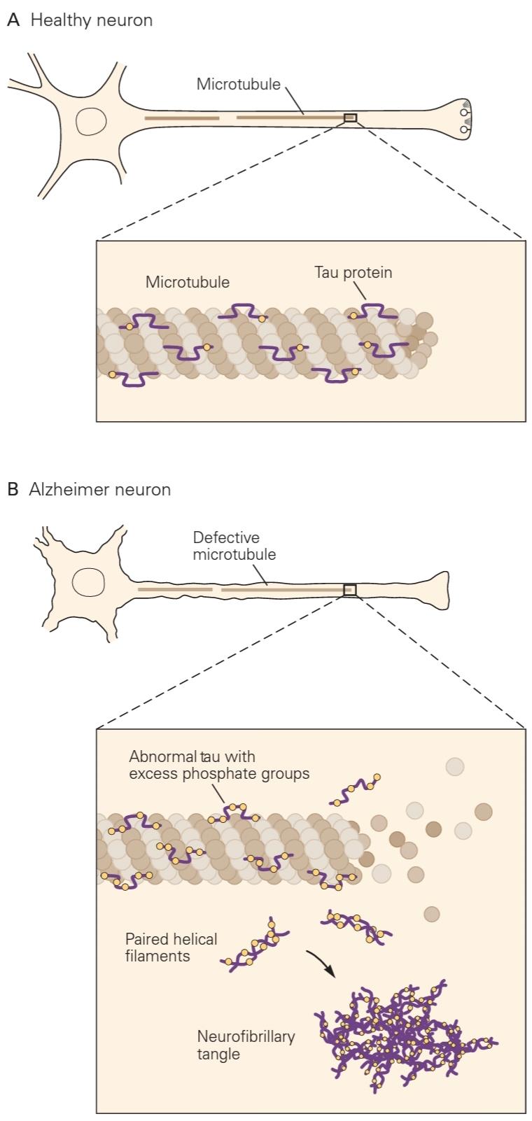
Figure 64-13 Formulation of neurofibrillary tangles.
شکل 64-13 فرمولاسیون درهم تنیدههای نوروفیبریلاری.
A. In healthy neurons, tau protein associates with normal microtubules but not as paired helical filaments, and contributes to the structural integrity of the neuron.
الف. در نورونهای سالم، پروتئین تاو با میکروتوبولهای طبیعی مرتبط است، اما نه بهصورت رشتههای مارپیچی جفت، و به یکپارچگی ساختاری نورون کمک میکند.
B. In a diseased neuron, the tau protein becomes hyperphosphorylated and loses its association with normal microtubules, which begin to disassemble. It then forms paired helical filaments, which become sequestered in neurofibrillary tangles.
ب. در یک نورون بیمار، پروتئین تاو هیپرفسفریله میشود و ارتباط خود را با میکروتوبولهای معمولی از دست میدهد که شروع به از هم پاشیدن میکنند. سپس رشتههای مارپیچی جفتی را تشکیل میدهد که در پیچ و تابهای نوروفیبریلاری جدا میشوند.
For many years, controversy raged between those who believe that Aβ is the main causal agent of AD and those who believe that tau-rich tangles play a major role. These partisans have been called “Baptists” and “Tauists,” respectively. Baptists pointed to the fact that during the development of AD pathology, which begins about 15 years before symptom onset, accumulation of neocortical Aβ precedes the development of neocortical tau pathology. More recent evidence suggests, however, that Aβ accumulation appears to some- how drive tau aggregation and spreading in the brain. Thus, Aβ aggregation probably instigates the disease and tau aggregation and spreading likely contribute in a major way to neurodegeneration. For example, transgenic mice that express both mutant APP and mutant tau develop much worse tau pathology.
برای سالهای متمادی، بین کسانی که معتقد بودند Aβ عامل اصلی بیماری پس از میلاد است و کسانی که معتقد بودند گرههای غنی از تاو نقش اصلی را ایفا میکنند، بحث و جدل وجود داشت. این پارتیزانها به ترتیب «باپتیست» و «تاوئیست» نامیده شده اند. باپتیستها به این واقعیت اشاره کردند که در طول توسعه آسیب شناسی AD، که حدود 15 سال قبل از شروع علائم شروع میشود، تجمع Aβ نئوکورتیکال قبل از ایجاد آسیب شناسی تاو نئوکورتتیکال است. با این حال، شواهد جدیدتر نشان میدهد که تجمع Aβ به نظر میرسد تا حدودی باعث تجمع و انتشار تاو در مغز میشود. بنابراین، تجمع Aβ احتمالاً بیماری را تحریک میکند و تجمع tau و گسترش احتمالاً به روش عمده ای به تخریب عصبی کمک میکند. به عنوان مثال، موشهای تراریخته که هم APP جهش یافته و هم تاو جهش یافته را بیان میکنند، آسیب شناسی تاو بسیار بدتری دارند.
There appears to be interplay between plaques and tangles. Injection of Aβ42 into specific brain regions of transgenic mice that express a mutant tau protein increases the number of tangles in nearby neurons. Further, a manipulation that reduces the number and size of plaques leads to a decrease in levels of hyperphosphorylated tau. Importantly, recent experiments suggest that Aβ deposition in some way promotes spreading of tau aggregates from one brain region to another, possibly transsynaptically in a prion-like fashion. The details of this process remain to be worked out and are likely to be extremely important.
به نظر میرسد بین پلاکها و گرهها تداخلی وجود دارد. تزریق Aβ42 به مناطق خاصی از مغز موشهای تراریخته که پروتئین تاو جهش یافته را بیان میکنند، تعداد گرهها را در نورونهای مجاور افزایش میدهد. علاوه بر این، دستکاری که تعداد و اندازه پلاکها را کاهش میدهد منجر به کاهش سطح تائو هیپرفسفریله میشود. نکته مهم این است که آزمایشهای اخیر نشان میدهد که رسوب Aβ به نحوی باعث گسترش تودههای تاو از یک ناحیه مغز به منطقه دیگر میشود، احتمالاً به صورت فراسیناپسی به شکل پریون مانند. جزییات این فرآیند باید بررسی شود و احتمالاً بسیار مهم خواهد بود.
There is now abundant evidence from cell culture and studies in animal models that several proteins that aggregate in neurodegenerative diseases, including tau and synuclein, can spread from cell to cell in a prion-like manner. This is particularly important as a potential disease mechanism. For example, if the cell-to-cell spreading of misfolded proteins occurs in the extracellular space, this process might be interrupted with antibodies directed against the appropriate disease-associated protein. In fact, this now serves as the basis of several clinical trials in humans targeting tau and synuclein.
در حال حاضر شواهد فراوانی از کشت سلولی و مطالعات در مدلهای حیوانی وجود دارد که نشان میدهد چندین پروتئین که در بیماریهای تخریبکننده عصبی تجمع مییابند، از جمله تاو و سینوکلئین، میتوانند از سلولی به سلول دیگر به شیوهای پریون مانند پخش شوند. این به ویژه به عنوان یک مکانیسم بالقوه بیماری مهم است. به عنوان مثال، اگر گسترش سلول به سلول پروتئینهای نادرست تا شده در فضای خارج سلولی اتفاق بیفتد، ممکن است این فرآیند با آنتی بادیهایی که علیه پروتئین مرتبط با بیماری مناسب هدایت میشوند، قطع شود. در واقع، این اکنون به عنوان پایه چندین آزمایش بالینی در انسان با هدف قرار دادن تاو و سینوکلئین عمل میکند.
»» فصل قبل: فصل شصت و سه: مکانیسمهای ژنتیکی در بیماریهای نورودژنراتیو سیستم عصبی
»» فصل اول: فصل مغز و رفتار
»» تمامی کتاب