
دیدکلی
پروتئینها اعمال مهم و متنوعی در ساختارهای زنده انجام میدهند. مثلا آنزیمها تعداد زیادی از تبدیلهای شیمیایی را کاتالیز میکنند. این تبدیلها میتواند شامل واکنشهای سادهای از قبیل آبدار شدن دیاکسید کربن تا تبدیلهای پیچیدهای از قبیل همانند سازی کروموزومها، رشتههای بلند و حلقهای DNA، یعنی مواد ژنتیکی موجود در سلولهای زنده باشد.
آنزیمها میتوانند واکنشهای خاصی را میلیونها بار تسریع کنند. پروتئینهای دیگری به عنوان شبکه حمل و نقل و ذخیره ساز عمل میکنند. هموگلوبین انتقالگر اکسیژن و هزاران پروتئین دیگر که نقش حیاتی و اساسی در عملکرد بخشهای مختلف بدن دارند، میباشد. در اینجا با عملکرد برخی از این پروتئینها از دیدگاه شیمی آشنا میشویم.
میوگلوبین
میوگلوبین، در ماهیچه فعال است، جایی که اکسیژن را ذخیره میکند و هنگامی که مورد لزوم باشد، اکسیژن را رها مینماید.
هموگلوبین
هموگلوبین، در گلبولهای قرمز وجود دارد و انتقال اکسیژن را تسهیل میکند. بدون هموگلوبین، خون میتواند فقط جزئی (تقریبا ۲۱ درصد) از اکسیژن مورد لزوم بدن را جذب نماید.
چگونگی اتصال اکسیژن به این پروتئینها
راز توانایی حمل اکسیژن بوسیله میوگلوبین و هموگلوبین، در وجود واحد ویژه غیر پلی پپتیدی موسوم به گروه هِم، متصل به پروتئین نهفته است. هِم ، یک لیگاند حلقوی آلی (موسوم به پورفیرین) است که در آن، واحدهای استخلاف شده پیرول با چهار اتصال، اتم آهن را احاطه میکنند. این کمپلکس، قرمز است و به خون ، رنگ قرمز خاص خود را میبخشد. اتم آهن در هِم، به چهار نیتروژن متصل است. لیکن میتواند دو گروه اضافی دیگر را در بالا و پایین سطح حلقه پورفیرین اسکان دهد.
در میوگلوبین، یکی از این گروهها، حلقه ایمیدازول یک واحد هیستیدین است که به یکی از بخشهای مارپیچ پروتئین متصل میباشد. عامل مهم دیگر برای عمل پروتئین، اکسیژن است. در نزدیکی موضع اتصال اکسیژن، دومین ایمیدازول یک واحد هیستیدین قرار دارد و به نظر میرسد که این واحد، این موضع از هم را بوسیله ممانعت فضایی محافظت میکند.
مسمومیت با مونواکسیدکربن
مونواکسیدکربن که آن نیز درگروه هِم به آهن متصل میگردد. بدین ترتیب از انتقال اکسیژن جلوگیری مینماید. به علت حضور دومین گروه ایمیدازول پیوندی نه چندان قوی ایجاد خواهد کرد. در نتیجه مسمومیت بوسیله منوکسید کربن در بیماری که در معرض گاز قرار میگیرد را میتوان با وارد کردن اکسیژن به بیمار از بین برد.
ساختار میوگلوبین
ساختمان اولیه میوگلوبین، شامل ۱۵۳ باقیمانده آمینو اسیدی با ترتیبی شناخته شده است. میوگلوبین، دارای هشت بخش مارپیچی α است که ساختمان ثانوی آن را شامل میشود. طولانیترین این بخشها، شامل ۲۳ باقیمانده است. ساختمان سوم دارای خمشی است که به میوگلوبین شکل سه بعدی میدهد.
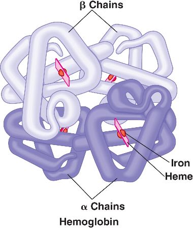
ساختار هموگلوبین
هموگلوبین دارای چهار زنجیره پروتئینی است. دو زنجیره α هر یک با ۱۴۱ باقیمانده و دو زنجیر β هر یک با ۱۴۷ باقیمانده. هر زنجیر دارای گروه هِم خاص خود میباشد و ساختمان سوم مشابهی با ساختمان سوم میوگلوبین دارد. گرچه برهمکنش ضعیفی بین دو زنجیر α و دو زنجیر β وجود دارد، اما تماسهای زیادی بین آمینها ایجاد میگردد.
تشابه ساختار هموگلوبین و میوگلوبین
چین خوردن هموگلوبین و میوگلوبین در چندین نمونه زنده، علیرغم ترتیبهای مختلف آمینو اسید، بسیار مشابه است. این وضعیت نشان میدهد که این ساختمان سوم ویژه، یک کنفیگوراسیون بهینه در اطراف گروه هِم است.
ساختار ویژه هِم و عملکرد در ششها
ساختار ویژه به هِم اجازه میدهد تا از طریق ششها، اکسیژن جذب کند و برای یک انتقال امن، تا جایی که لازم باشد، اکسیژن را نگه دارد و هنگامی که ضروری است، اکسیژن را آزاد کند.
تفاوتهای هموگلوبین و میوگلوبین
۳. میوگلوبین تنها یک گروه آهن دار هم دارد ولی هموگلوبین دارای چهار گروه آهن دار هم میباشد.
۴. میوگلوبین تنها با یک اکسیژن پیوند هیدروژنی برقرار میکند در حالی که هموگلوبین با چهار مولکول اکسیژن پیوند هیدروژنی برقرار میکند.
۵. میوگلوبین به دستور یک ژن ساخته میشود ولی هموگلوبین توسط دو ژن ساخته میشود.
یادآوری۱: هر دو پروتئین انتقال دهنده هستند.
یادآوری۲: میزان بالای میوگلوبین در ماهیچه ها یک سازگاری مهم برای بالابردن کارآیی تنفس در پرندگان بلندپرواز نظیر غازهای وحشی (نه تمامی پرندگان) است.
دیگر مهره داران نظیر پستانداران (از جمله آدمی) در ماهیچه های خود دارای میوگلوبین هستند.
یادآوری۳: مولکول هِم مسئول رنگ قرمز هموگلوبین و میوگلوبین است. یک اتم آهن دو ظرفیتی در مرکز آن قرار گرفته است. اگر اتم آهن دو ظرفیتی(+Fe²) به آهن سه ظرفیتی(+Fe³) اکسید شود، عملکرد مولکول از بین خواهد رفت.
هموگلوبین به علت داشتن آهن که در حالت احیا شده میباشد، میتواند با اکسیژن و دیاکسید کربن ترکیب شده و به ترتیب اکسی هموگلوبین (oxyhemoglobin) و کربو آمینو هموگلوبین (carbaminohemoglobin) تشکیل دهد.
با توجه به بالابودن فشار اکسیژن در ششها، اکسی هموگلوبین در ریهها تشکیل شده و پس از رسیدن به بافتها، اکسیژن جدا شده و دیاکسید کربن به آن متصل میگردد. به این ترتیب امکان حمل اکسیژن از ریهها به بافتها و دیاکسیدکربن از بافتها به ریهها امکانپذیر میگردد. از طرف دیگر سطح بسیار زیاد گویچههای قرمز نسبت به حجم آنها (به علت داشتن شکل مقعرالطرفین) سبب تسریع و تسهیل اشباع هموگلوبین با اکسیژن در شش ها میشود.
علاوه بر انتقال اکسیژن توسط هموگلوبین، این مولکول عمل دیگری نیز انجام میدهد و آن عبارت از تثبیت فشار اکسیژن در بافت ها است.
۱۵ Difference Between Hemoglobin And Myoglobin (With Pictures &Similarities)
Haemoglobin
Hemoglobin is the protein molecule in red blood cells that carries oxygen from the lungs to the body’s tissues and returns carbon dioxide from the tissues back to the lungs.
Hemoglobin is made up of four protein molecules (globulin chains) that are connected together. The normal adult hemoglobin (abbreviated Hgb or Hb) molecule contains two alpha-globulin chains and two beta-globulin chains. In fetuses and infants, beta chains are not common and the hemoglobin molecule is made up of two alpha chains and two gamma chains. As the infant grows, the gamma chains are gradually replaced by beta chains, forming the adult hemoglobin structure.
Each globulin chain contains an important iron-containing porphyrin compound termed heme. Embedded within the heme compound is an iron atom that is vital in transporting oxygen and carbon dioxide in our blood. The iron contained in hemoglobin is also responsible for the red color of blood.

Hemoglobin also plays an important role in maintaining the shape of the red blood cells. In their natural shape, red blood cells are round with narrow centers resembling a donut without a hole in the middle. Abnormal hemoglobin structure can, therefore, disrupt the shape of red blood cells and impede their function and flow through blood vessels.
Haemoglobin levels vary from person to person. Men usually have higher levels than women. A haemoglobin “cut-off” level is set for blood donation to ensure that your haemoglobin will not drop below normal after you have donated blood. Normal ranges for haemoglobin differ between ethnic populations, and males and females, and are also affected by age, especially in women. Individuals with haemoglobin levels below the normal range are, by definition, anaemic. There are many causes of anaemia and anaemia due to iron deficiency is common.
Facts About Hemoglobin
- Hemoglobin is an iron and oxygen-binding protein in blood, specifically red blood cell.
- Hemoglobin is abbreviated as Hb.
- Hemoglobin has a molecular weight of 64 kDa.
- Hemoglobin has 4 polypeptide chains: alpha, Beta, Delta and Gamma or epsilon (Depending on the type of hemoglobin).
- Systematically present all over the body.
- Hemoglobin occurs as a tetrameric protein and binds molecular oxygen on Red blood cells thereby ensuring that they are distributed throughout the body.
- Types of hemoglobin include Hemoglobin A, Hemoglobin A2 and Haemoglobin F.
- Takes oxygen from the lungs and transport to the rest of the body.
- Oxygen binding affected by PH and CO2.
- Haemoglobin exhibits a sigmoid curve in terms of oxygen dissociation (oxygen binding to hemoglobin at neutral PH).
- Exhibits quaternary structure which is a characteristic of many multi-subunit globular proteins.
- Haemoglobin has a low affinity to bind with oxygen.
- In human, hemoglobin is found throughout the bloodstream.
Myoglobin
Myoglobin is a small, monomeric protein which serves as an intracellular oxygen storage site. It is found in abundance in the skeletal muscle of vertebrates, and is responsible for the characteristic red color of muscle tissue. It traps oxygen within muscle cells, allowing the cells to produce the energy required for muscles to contract.
High concentrations of myoglobin in muscle cells allow organisms to hold their breath for a longer period of time. Diving mammals such as whales and seals have muscles with particularly high abundance of myoglobin.
When heart or skeletal muscle is injured, myoglobin is released into the blood. Elevated levels can be measured within a few hours following an injury.
Myoglobin is filtered from the blood by the kidneys and is released into the urine. Large quantities of myoglobin are toxic to the kidneys. If significant amounts of myoglobin are released into the bloodstream, which can happen after severe trauma or muscle injuries, the excess myoglobin may cause damage to the kidneys and eventually result in kidney failure. Measurement of myoglobin in urine helps to detect this condition.
There is a close chemical similarity between myoglobin and hemoglobin, the oxygen-binding protein of red blood cells. Both proteins contain a molecular constituent called heme, which enables them to combine reversibly with oxygen. The heme group, which contains iron, imparts a red-brown colour to the proteins. The bond between oxygen and hemoglobin is more complex than that between oxygen and myoglobin and accounts for the dual ability hemoglobin has to transport oxygen as well as to store it.
In contact with venous blood, oxygen combines more readily with myoglobin than it does with hemoglobin, favouring the transfer of oxygen from blood to muscle cells. Thus, the oxygen that the working muscle requires for the energy-producing biochemical reactions is provided.

Facts About Myoglobin
- Myoglobin is an iron and oxygen-binding protein found in the muscle tissue of vertebrates in general and in almost all mammals.
- Myoglobin is abbreviated as mb
- Myoglobin has a molecular weight of 16.7 kDa
- Myoglobin has a single polypeptide chains.
- Only present in heart and muscles.
- Myoglobin occurs as a monomeric protein and binds molecular oxygen and distributes it to the muscles.
- A single type of myoglobin is found in all cells.
- Stores oxygen in the muscle cells and releases it when it is needed.
- Oxygen binding in myoglobin is not affected by CO2 and PH.
- Myoglobin exhibits a hyperbolic curve in terms of oxygen dissociation.
- Exhibits tertiary structure.
- Has a high affinity to bind with oxygen, which does not depend on oxygen concentration.
- In humans, myoglobin is found in the bloodstream after muscle injury.

Differences Between Hemoglobin And Myoglobin In Tabular Form
| BASIS OF COMPARISON | HEMOGLOBIN | MYOGLOBIN |
| Definition | Hemoglobin is a red protein which is responsible for transporting oxygen in the blood of vertebrates. | Myoglobin is a red protein with haem which carries and stores oxygen in muscle cells. |
| Molecular weight | Molecular weight is 64 kDa | Molecular weight is 16.7 kDa |
| Number of Chains | Haemoglobin has 4 polypeptide chains: alpha, Beta, Delta and Gamma or epsilon (Depending on the type of haemoglobin). | Myoglobin has a single polypeptide chains. |
| Location | Systematically present all over the body. | Only present in heart and muscles. |
| Also known as | Hb | Mb |
| Occurrence | Haemoglobin occurs as a tetrameric protein. | Myoglobin occurs as a monomeric protein. |
| Types | Types of haemoglobin include Haemoglobin A, Hemoglobin A2 and Haemoglobin F. | A single type of myoglobin is found in all cells. |
| Roles | Takes oxygen from the lungs and transport to the rest of the body. | Stores oxygen in the muscle cells and releases it when it is needed. |
| Oxygen Binding | Oxygen binding affected by PH and CO2. | Oxygen binding not affected by CO2 and PH. |
| Curves | Haemoglobin exhibits a sigmoid curve in terms of oxygen dissociation. | Myoglobin exhibits a hyperbolic curve in terms of oxygen dissociation. |
| Level of structure | Exhibits quaternary structure. | Exhibits tertiary structure. |
| Binding Affinity | Haemoglobin has a low affinity to bind with oxygen. | Has a high affinity to bind with oxygen, which does not depend on oxygen concentration. |
Similarities Between Hemoglobin And Myoglobin
- Both myoglobin and hemoglobin give red color to the blood and muscles.
- Both contain oxygen-binding haem
- Both are globular proteins.

جالب بود درود بر شما