نوروآناتومی بالینی؛ هستههای قاعدهای: گانگلیونهای قاعدهای مغز


ترجمه و بازنویسی علمی فصول کتاب «نوروآناتومی بالینی» | کتاب درسی جامع ساختارهای عصبی (ویرایش دوم)
کتاب درسی نوروآناتومی بالینی، ویرایش دوم. Textbook of Clinical Neuroanatomy (2nd ed.) یکی از منابع معتبر در حوزه نوروآناتومی بالینی (Clinical Neuroanatomy) است که با رویکردی کاربردی، ساختارها و مسیرهای سیستم عصبی (Nervous System) را بهصورت نظاممند و بالینی شرح میدهد.
این ترجمه آموزشی، حاصل تلاش تیم علمی آیندهنگاران مغز به سرپرستی داریوش طاهری است و با هدف ارتقای آموزش علوم اعصاب (Neuroscience) در زبان فارسی، با حفظ دقت علمی، به زبانی روان تدوین شده است.
در ۲۰ فصل این مجموعه، ساختارهایی چون مغز (Brain)، نخاع (Spinal Cord)، اعصاب مغزی (Cranial Nerves)، سیستم لیمبیک (Limbic System)، مسیرهای حسی و حرکتی (Sensory and Motor Pathways)، مخچه (Cerebellum)، دیانسفالون (Diencephalon) و سیستم عصبی خودمختار (Autonomic Nervous System) بررسی میشوند.
این مجموعه برای دانشجویان پزشکی، رزیدنتها و متخصصان علاقهمند به نوروساینس بالینی طراحی شده و تلفیقی از مفاهیم بنیادی، نکات بالینی و نمودارهای عملکردی را ارائه میدهد.
با سپاس از همراهی شما
تیم آیندهنگاران مغز | سرپرستی: داریوش طاهری
13. Basal Nuclei (Basal Ganglia)
۱۳. هستههای قاعدهای (عقدههای قاعدهای)
The basal ganglia are the large masses of grey matter situated within the white core of each cerebral hemisphere and form essential constituents of the extrapyramidal system. The basal ganglia are now recognized as basal nuclei, but the former term is still commonly used.
عقدههای پایه تودههای بزرگی از ماده خاکستری هستند که در هسته سفید هر نیمکره مغزی قرار دارند و اجزای اساسی سیستم خارج هرمی را تشکیل میدهند. عقدههای پایه اکنون به عنوان هستههای پایه شناخته میشوند، اما اصطلاح قبلی هنوز هم معمولاً استفاده میشود.
Anatomically, the term basal ganglia include:
• Corpus striatum,
• Claustrum, and
• Amygdaloid body.
از نظر آناتومیکی، اصطلاح عقدههای قاعدهای شامل موارد زیر است:
• جسم مخطط،
• جسم کلاستروم، و
• جسم آمیگدال.
Functionally, the term basal ganglia also include substantia nigra and subthalamus. Some workers also include red nucleus.
از نظر عملکردی، اصطلاح عقدههای قاعدهای شامل جسم سیاه و ساب تالاموس نیز میشود. برخی از محققان هسته قرمز را نیز در این تعریف قرار میدهند.
The basal nuclei are important in organizing and coordinating somatic motor activities especially willed movements. They are also involved in automatic stereotyped postural and reflex motor activities such as normal individuals swing their arms when they walk.
هستههای قاعدهای در سازماندهی و هماهنگی فعالیتهای حرکتی سوماتیک، به ویژه حرکات ارادی، اهمیت دارند. آنها همچنین در فعالیتهای حرکتی کلیشهای و رفلکسی خودکار مانند چرخاندن بازوها توسط افراد عادی هنگام راه رفتن، نقش دارند.
Corpus Striatum
The corpus striatum is situated lateral to the thalamus. Topographically it is almost completely divided into the caudate nucleus and the lentiform nucleus by a band of nerve fibres, the internal capsule. However, anteroinferior ends of these nuclei remain connected by a few bands of grey matter across the anterior limb of internal capsule. These bands give it a striated appearance, hence the name corpus striatum (Figs 13.1–13.3).
جسم مخطط
جسم مخطط در کنار تالاموس قرار دارد. از نظر توپوگرافی، تقریباً به طور کامل توسط نواری از الیاف عصبی، کپسول داخلی، به هسته دمدار و هسته عدسی شکل تقسیم میشود. با این حال، انتهای قدامی-تحتانی این هستهها توسط چند نوار ماده خاکستری در امتداد اندام قدامی کپسول داخلی به هم متصل میمانند. این نوارها ظاهری مخطط به آن میدهند، از این رو نام جسم مخطط را به خود گرفته است (شکلهای ۱۳.۱ تا ۱۳.۳).

FIG. 13.1 Lateral aspect of the left corpus striatum.
شکل ۱۳.۱ نمای جانبی جسم مخطط چپ.

FIG. 13.2 Relationship of the corpus striatum to the internal capsule.
شکل 13.2 رابطه جسم مخطط با کپسول داخلی.

FIG. 13.3 Corpus striatum, thalamus, claustrum and internal capsule as seen in horizontal section of the cerebral hemisphere.
شکل ۱۳.۳ جسم مخطط، تالاموس، کلاستروم و کپسول داخلی همانطور که در برش افقی نیمکره مغزی دیده میشود.
The lentiform nucleus consists of two parts: a darker lateral part putamen and a medial paler part globus pallidus.
هسته عدسی شکل از دو بخش تشکیل شده است: یک بخش جانبی تیرهتر به نام پوتامن و یک بخش میانی کمرنگتر به نام گلوبوس پالیدوس.
Phylogenetically corpus striatum forms two distinct functional units, the paleostriatum and the neostriatum.
از نظر فیلوژنتیکی، جسم مخطط دو واحد عملکردی مجزا، پالئوستریاتوم و نئواستریاتوم، را تشکیل میدهد.
The globus pallidus is relatively ancient and termed paleostriatum/pallidum. The caudate nucleus and putamen being recent in development, together form the neostriatum/ striatum. The striatum is largely afferent whereas pallidum is largely efferent structure.
گلوبوس پالیدوس نسبتاً قدیمی است و پالئوستریاتوم/پالیدوم نامیده میشود. هسته دمدار و پوتامن که اخیراً در حال توسعه هستند، با هم نئواستریاتوم/استریاتوم را تشکیل میدهند. جسم مخطط عمدتاً آوران است در حالی که پالیدوم عمدتاً وابران است.
These features of corpus striatum may be summarized as follows:
این ویژگیهای جسم مخطط را میتوان به شرح زیر خلاصه کرد:

Caudate Nucleus
Caudate nucleus is a large comma-shaped mass of grey matter, which surrounds the thalamus and is itself surrounded by the lateral ventricle (Fig. 13.4). Its whole length of convexity projects into the cavity of lateral ventricle.
هسته دمی
هسته دمی یک توده بزرگ ویرگول شکل از ماده خاکستری است که تالاموس را احاطه کرده و خود توسط بطن جانبی احاطه شده است (شکل ۱۳.۴). تمام طول تحدب آن به داخل حفره بطن جانبی امتداد دارد.

FIG. 13.4 Relationship of caudate nucleus with the cavity of the fourth ventricle and thalamus. Note that the stria terminalis, the main efferent tract of amygdaloid body projects to the septal area, anterior perforated substance, and anterior hypothalamus.
شکل ۱۳.۴ ارتباط هسته دمدار با حفره بطن چهارم و تالاموس. توجه داشته باشید که استریا ترمینالیس، مسیر اصلی وابران جسم آمیگدالوئید، به ناحیه سپتوم، ماده سوراخدار قدامی و هیپوتالاموس قدامی امتداد دارد.
Its rounded anterior part in front of interventricular foramen is called its head. The head gradually and imperceptibly tapers caudally into the body and then into a tail which merges at its anterior extremity with an almond-shaped mass of grey matter called amygdaloid body (Fig. 13.2).
قسمت قدامی گرد آن در جلوی سوراخ بین بطنی، سر نامیده میشود. سر به تدریج و به طور نامحسوس به سمت پایین به سمت جسم و سپس به دمی باریک میشود که در انتهای قدامی خود با تودهای بادامی شکل از ماده خاکستری به نام جسم آمیگدالوئید ادغام میشود (شکل ۱۳.۲).
The head is large and rounded, and forms the floor and lateral wall of the anterior horn of lateral ventricle. The bands of grey matter connect it to the putamen across the anterior limb of internal capsule (Fig. 13.3).
سر بزرگ و گرد است و کف و دیواره جانبی شاخ قدامی بطن جانبی را تشکیل میدهد. نوارهای ماده خاکستری آن را از طریق بازوی قدامی کپسول داخلی به پوتامن متصل میکنند (شکل ۱۳.۳).
The body is long and narrow, and forms the floor of the central part of lateral ventricle. It is separated from thalamus by stria terminalis and thalamostriate vein.
بدن دراز و باریک است و کف قسمت مرکزی بطن جانبی را تشکیل میدهد. توسط استریا ترمینالیس و ورید تالاموستریا از تالاموس جدا میشود.
The tail is long and slender, and forms the roof of inferior horn of lateral ventricle. It terminates anteriorly (topographically) in the amygdaloid body.
دم بلند و باریک است و سقف شاخ تحتانی بطن جانبی را تشکیل میدهد. از جلو (از نظر توپوگرافی) در جسم آمیگدال خاتمه مییابد.
Lentiform Nucleus
Lentiform nucleus is a large lens-shaped (biconvex) mass of grey matter beneath the insula forming the lateral boundary of internal capsule. In horizontal section of cerebrum, it appears wedge-shaped with broad convex base directed laterally.
هسته عدسی شکل
هسته عدسی شکل، تودهای بزرگ (دو محدب) از ماده خاکستری است که در زیر اینسولا قرار دارد و مرز جانبی کپسول داخلی را تشکیل میدهد. در برش افقی مغز، به شکل گوه با پایه محدب پهن به سمت طرفین دیده میشود.
It has three surfaces and divides into two parts:
سه سطح دارد و به دو بخش تقسیم میشود:
Surfaces
سطوح
• The lateral surface is convex and related to thin sheet of white matter, the external capsule. It is grooved by lateral striate arteries (the central branches of middle cerebral artery).
• سطح جانبی محدب است و به صفحه نازکی از ماده سفید، کپسول خارجی، مرتبط است. این صفحه توسط شریانهای مخطط جانبی (شاخههای مرکزی شریان مغزی میانی) شیاردار شده است.
• Medial surface is more convex and related to internal capsule (limbs and genu). In transverse sections, the medial surface is angulated at the genu.
• سطح داخلی محدبتر است و به کپسول داخلی (اندامها و زانو) مرتبط است. در برشهای عرضی، سطح داخلی در زانو زاویهدار است.
• Inferior surface is related to sublentiform part of internal capsule and lies close to the anterior perforated substance.
• سطح تحتانی به قسمت زیر عدسی شکل کپسول داخلی مرتبط است و نزدیک به ماده سوراخدار قدامی قرار دارد.
Parts
A vertical plate of white matter (external medullary lamina) divides the lentiform nucleus into two parts, the putamen and the globus pallidus. These are clearly seen on a cut section.
اجزا
یک صفحه عمودی از ماده سفید (لایه خارجی مغز) هسته عدسی شکل را به دو قسمت پوتامن و گلوبوس پالیدوس تقسیم میکند. این دو قسمت در یک برش به وضوح دیده میشوند.
The putamen is larger lateral part and consists of densely packed small cells. It is darker in colour, and structurally similar to the caudate nucleus.
پوتامن قسمت جانبی بزرگتر است و از سلولهای کوچک و متراکم تشکیل شده است. رنگ آن تیرهتر است و از نظر ساختاری شبیه به هسته دمدار است.
The globus pallidus is smaller medial part. It is lighter in colour and consists of large (motor) cells. It is also known as pallidum as it appears pale in section (pallid = pale). The globus pallidus is further subdivided by an internal medullary lamina of white matter into outer and inner segments (Fig. 13.3).
گلوبوس پالیدوس قسمت میانی کوچکتر است. رنگ آن روشنتر است و از سلولهای بزرگ (حرکتی) تشکیل شده است. همچنین به عنوان پالیدوم شناخته میشود زیرا در برش رنگ پریده به نظر میرسد (پالید = رنگ پریده). گلوبوس پالیدوس توسط یک لایه داخلی مغز از ماده سفید به بخشهای بیرونی و داخلی تقسیم میشود (شکل 13.3).
Connections of corpus striatum (Fig. 13.5)
The striatum (caudate nucleus and putamen) is the receptive part while globus pallidus is the efferent part (outflow centre) of the corpus striatum.
اتصالات جسم مخطط (شکل ۱۳.۵)
جسم مخطط (هسته دمدار و پوتامن) بخش گیرنده است در حالی که گلوبوس پالیدوس بخش وابران (مرکز خروجی) جسم مخطط است.
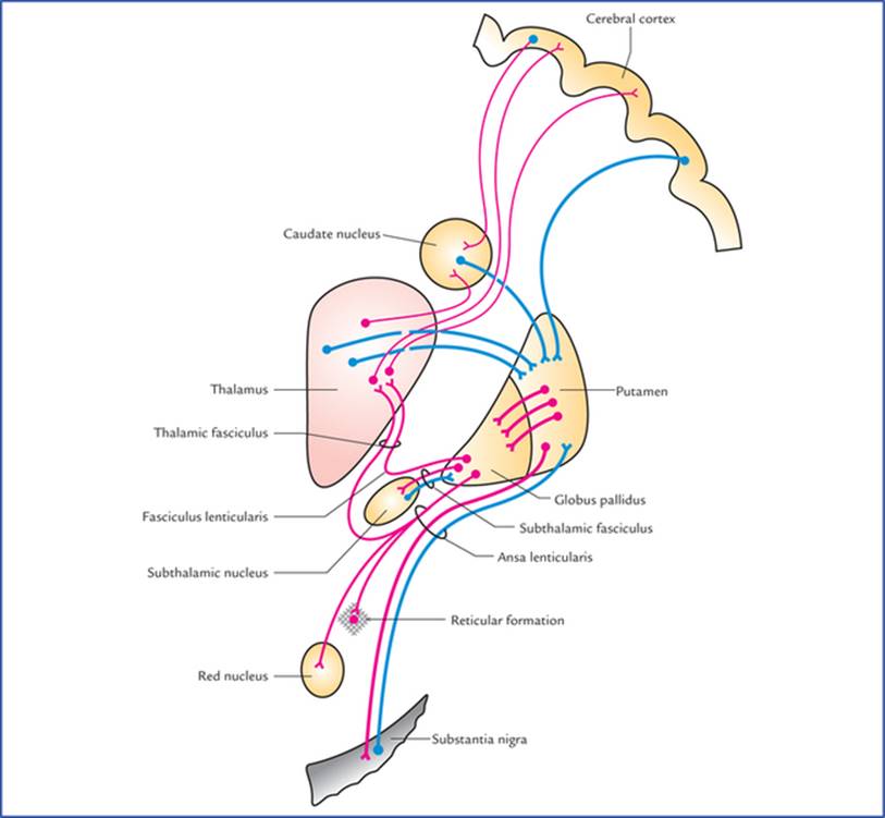
FIG. 13.5 Chief afferent and efferent connections of corpus striatum.
شکل ۱۳.۵ اتصالات آوران و وابران اصلی جسم مخطط.
Afferent connections
The striatum receives afferents chiefly from cerebral cortex, thalamus and substantia nigra.
اتصالات آوران
جسم مخطط عمدتاً آورانها را از قشر مغز، تالاموس و جسم سیاه دریافت میکند.
• Corticostriate fibres arise from a wide area of the ipsilateral cerebral cortex and reach the striatum through both internal and external capsules.
• فیبرهای کورتیکواستریات از ناحیه وسیعی از قشر مغز همان طرف منشأ میگیرند و از طریق کپسولهای داخلی و خارجی به جسم مخطط میرسند.
• Thalamostriate fibres arise from mediodorsal, intralaminar and midline nuclei of the thalamus. The majority of these fibres end in the caudate nucleus, the remaining pass through the internal capsule to reach the putamen.
• فیبرهای تالاموستریا از هستههای مدیودورسال، اینترالامینار و خط میانی تالاموس منشأ میگیرند. اکثر این فیبرها به هسته دمدار ختم میشوند و بقیه از کپسول داخلی عبور میکنند تا به پوتامن برسند.
• Nigrostriate fibres arise from substantia nigra and ascend up to terminate in the corpus striatum, mainly in putamen and caudate nucleus.
• فیبرهای نیگرواستریات از جسم سیاه منشأ میگیرند و به سمت بالا صعود میکنند تا در جسم مخطط، عمدتاً در پوتامن و هسته دمدار، خاتمه یابند.
The nigrostriate fibres carry dopamine synthesized by nerve cells of substantia nigra to striatum (melanin normally present in the substantia nigra is a byproduct of dopamine metabolism). It is believed that these fibres have an inhibitory effect on corpus striatum.
فیبرهای نیگرواستریات، دوپامین سنتز شده توسط سلولهای عصبی جسم سیاه را به جسم مخطط منتقل میکنند (ملانین که معمولاً در جسم سیاه وجود دارد، محصول جانبی متابولیسم دوپامین است). اعتقاد بر این است که این فیبرها اثر مهاری بر جسم مخطط دارند.
Efferent connections
Most of the output of striatum goes to the globus pallidus, however, some efferents also go to substantia nigra.
اتصالات وابران
بیشتر خروجی جسم مخطط به گلوبوس پالیدوس میرود، با این حال، برخی از وابران نیز به جسم سیاه میروند.
The globus pallidus is the main efferent component of corpus striatum.
گلوبوس پالیدوس جزء اصلی وابران جسم مخطط است.
The outflow from globus pallidus goes mainly to the thalamus (ventrolateral and ventroanterior nuclei) (Figs 13.5, 13.6). This occurs by way of two fasciculi: (a) ansa lenticularis looping round the posterior limb of internal capsule, and (b) fasciculus lenticularis traversing the internal capsule.
خروجی گلوبوس پالیدوس عمدتاً به تالاموس (هستههای شکمی-جانبی و شکمی-قدامی) میرود (شکلهای ۱۳.۵، ۱۳.۶). این امر از طریق دو فاسیکول رخ میدهد: (الف) آنسا لنتیکولاریس که دور اندام خلفی کپسول داخلی حلقه زده است، و (ب) فاسیکولوس لنتیکولاریس که از کپسول داخلی عبور میکند.

FIG. 13.6 Outflow tracts from globus pallidus (paleostriatum). Also, note the interconnection between subthalamic nucleus and globus pallidus.
شکل ۱۳.۶ مسیرهای خروجی از گلوبوس پالیدوس (پالئوستریاتوم). همچنین، به ارتباط بین هسته سابتالاموس و گلوبوس پالیدوس توجه کنید.
These fasciculi enter the region between the red nucleus and thalamus, where they unite with the dentatorubro-thalamic tract to form the thalamic fasciculus, which ends in the ventrolateral and ventroanterior nuclei of thalamus which in turn project to the motor and premotor areas of cerebral cortex.
این فاسیکولها وارد ناحیه بین هسته قرمز و تالاموس میشوند، جایی که با راه دنتاتوربرو-تالاموس متحد میشوند تا فاسیکول تالاموس را تشکیل دهند، که در هستههای شکمی-جانبی و شکمی-قدامی تالاموس ختم میشود که به نوبه خود به نواحی حرکتی و پیش حرکتی قشر مغز منتهی میشوند.
The subthalamic fasciculus passes through the internal capsule and forms direct reciprocal connections between subthalamic nucleus and globus pallidus.
فاسیکول ساب تالاموس از کپسول داخلی عبور میکند و ارتباطات متقابل مستقیمی بین هسته ساب تالاموس و گلوبوس پالیدوس تشکیل میدهد.
Some fibres from globus pallidus also pass to the substantia nigra (pallidonigral fibres).
برخی از فیبرهای گلوبوس پالیدوس نیز به جسم سیاه (فیبرهای پالیدونیگرال) منتقل میشوند.
Functions of corpus striatum
In submammalian vertebrates, the corpus striatum is the sensory and motor integrating and control centre. In later evolution, the activity of corpus striatum has become subordinate to those of cerebral cortex but remain essential for muscle tone, quality of movement, posture and locomotion.
عملکردهای جسم مخطط
در مهرهداران زیر پستاندار، جسم مخطط مرکز کنترل و یکپارچهسازی حسی و حرکتی است. در تکامل بعدی، فعالیت جسم مخطط تابع قشر مغز شده است، اما برای تون عضلانی، کیفیت حرکت، وضعیت بدن و حرکت ضروری است.
• Corpus striatum controls the automatic associated movements like, swinging of arms during walking.
• It helps in smoothening the voluntary motor activity of the body.
• جسم مخطط حرکات خودکار مرتبط مانند چرخاندن بازوها هنگام راه رفتن را کنترل میکند.
• به روان شدن فعالیت حرکتی ارادی بدن کمک میکند.
Claustrum
Claustrum is a thin saucer-shaped mass of grey matter situated between the putamen and insula. It is considered as a detached part of the insula (Fig. 13.6). Its connections and functions are not known.
کلاستروم
کلاستروم یک توده نازک نعلبکی شکل از ماده خاکستری است که بین پوتامن و اینسولا قرار دارد. این بخش به عنوان بخشی جدا از اینسولا در نظر گرفته میشود (شکل ۱۳.۶). اتصالات و عملکردهای آن مشخص نیست.
Amygdaloid Body
Amygdaloid body is an almond-shaped mass of grey matter in the temporal lobe, lying anterosuperior to the tip of inferior horn of lateral ventricle. It is situated deep to uncus which serves as a surface landmark for its location.
جسم آمیگدالوئید
جسم آمیگدالوئید تودهای بادامی شکل از ماده خاکستری در لوب گیجگاهی است که در قسمت قدامی-فوقانی نوک شاخ تحتانی بطن جانبی قرار دارد. این جسم در عمق آنکوس قرار دارد که به عنوان یک نقطه عطف سطحی برای موقعیت آن عمل میکند.
Developmentally it is related to basal nuclei but functionally it is included in the limbic system and therefore shares its functions. For details see limbic system (page 229).
از نظر تکاملی به هستههای قاعدهای مرتبط است اما از نظر عملکردی در سیستم لیمبیک قرار دارد و بنابراین عملکردهای آن را به اشتراک میگذارد. برای جزئیات بیشتر به سیستم لیمبیک (صفحه 229) مراجعه کنید.
Substantia Nigra
Substantia nigra is described in detail in Chapter 8.
جسم سیاه
جسم سیاه به تفصیل در فصل 8 توضیح داده شده است.
Subthalamus (Subthalamic Nucleus)
This small nucleus in the ventral part of the diencephalon looks like a biconvex lens in coronal section. It is located caudal to the lateral half of the thalamus and inferomedial to the globus pallidus. It is separated from thalamus by a smaller nucleus, called zona inserta. The subthalamic nucleus and globus pallidus are interconnected by the subthalamic fasciculus which traverses the internal capsule (Fig. 13.6).
ساب تالاموس (هسته ساب تالاموس)
این هسته کوچک در قسمت شکمی دیانسفالون، در برش کرونال مانند یک عدسی دو محدب به نظر میرسد. این هسته در قسمت خلفی نیمه جانبی تالاموس و در قسمت تحتانی داخلی گلوبوس پالیدوس قرار دارد. این هسته توسط یک هسته کوچکتر به نام زونا اینسرتا از تالاموس جدا میشود. هسته ساب تالاموس و گلوبوس پالیدوس توسط دسته ساب تالاموس که از کپسول داخلی عبور میکند، به هم متصل هستند (شکل 13.6).
Functions of Basal Nuclei
وظایف هستههای قاعدهای
• Concerned with planning and programming of voluntary movements.
• مربوط به برنامهریزی و برنامهریزی حرکات ارادی.
• Determine how rapidly a movement is to be performed and how large the movement must be.
• مشخص کنید که یک حرکت با چه سرعتی قرار است انجام شود و اندازه آن حرکت چقدر باید باشد.
• Decrease muscle tone and inhibit unwanted muscular activity.
• کاهش تون عضلانی و مهار فعالیت عضلانی ناخواسته.
• Regulate the muscle tone and thus helps in smoothening the voluntary motor activities of the body.
• تنظیم تون عضلانی و در نتیجه کمک به روانسازی فعالیتهای حرکتی ارادی بدن.
• Control automatic associated movements, like swinging of arms during walking.
• کنترل حرکات خودکار مرتبط، مانند تاب دادن بازوها هنگام راه رفتن.
• Control group of movements for emotional expression.
• کنترل گروهی از حرکات برای بیان احساسات.
• Control reflex muscular activity.
• کنترل فعالیت رفلکسی عضلات.
Neural Circuitry Involved in Functioning of Basal Nuclei (Flowchart 13.1)
The striatum receives information from three major sources: (a) the cerebral cortex, (b) the thalamus, and (c) the substantia nigra. The striatum projects to the globus pallidus and substantia nigra. The globus pallidus (the outflow nucleus of striatum) projects to the thalamus and subthalamic nucleus. The substantia nigra also projects to the thalamus. The thalamus projects to the cerebral cortex. This information is integrated within the corpus striatum and outflow passes back to the above areas. This circular pathway functions as follows:
مدارهای عصبی دخیل در عملکرد هستههای قاعدهای (فلوچارت ۱۳.۱)
جسم مخطط اطلاعات را از سه منبع اصلی دریافت میکند: (الف) قشر مغز، (ب) تالاموس، و (ج) جسم سیاه. جسم مخطط به گلوبوس پالیدوس و جسم سیاه امتداد مییابد. گلوبوس پالیدوس (هسته خروجی جسم مخطط) به تالاموس و هسته ساب تالاموس امتداد مییابد. جسم سیاه نیز به تالاموس امتداد مییابد. تالاموس به قشر مغز امتداد مییابد. این اطلاعات در جسم مخطط ادغام شده و جریان خروجی به نواحی فوق بازمیگردد. این مسیر دایرهای به شرح زیر عمل میکند:

FLOWCHART 13.1 Neural circuitry of basal nuclei.
The activity of the basal ganglia is initiated by information received from the cerebral cortex, the thalamus, and the brainstem (substantia nigra and red nucleus). The outflow from the basal ganglia is channelled through the globus pallidus, which then influences the activities of the motor areas of the cerebral cortex. The motor cortex then controls the motor activities through corticospinal and corticonuclear fibres. Thus, basal ganglia can control muscular movements by influencing the cerebral cortex rather than through direct descending pathways to the brainstem and spinal cord.
نمودار جریان ۱۳.۱ مدار عصبی هستههای قاعدهای.
فعالیت عقدههای قاعدهای توسط اطلاعات دریافتی از قشر مغز، تالاموس و ساقه مغز (جسم سیاه و هسته قرمز) آغاز میشود. جریان خروجی عقدههای قاعدهای از طریق گلوبوس پالیدوس هدایت میشود که سپس بر فعالیتهای نواحی حرکتی قشر مغز تأثیر میگذارد. سپس قشر حرکتی فعالیتهای حرکتی را از طریق فیبرهای قشری-نخاعی و قشری-هستهای کنترل میکند. بنابراین، عقدههای قاعدهای میتوانند حرکات عضلانی را با تأثیر بر قشر مغز کنترل کنند، نه از طریق مسیرهای نزولی مستقیم به ساقه مغز و نخاع.
Clinical Correlation
Disorders of the Basal Ganglia
The lesions of basal ganglia result in various forms of unwanted involuntary movements and disturbance in muscle tone. These disorders include parkinsonism, chorea, athetosis and ballismus.
همبستگی بالینی
اختلالات عقدههای قاعدهای
ضایعات عقدههای قاعدهای منجر به اشکال مختلف حرکات ناخواسته غیرارادی و اختلال در تون عضلانی میشوند. این اختلالات شامل پارکینسونیسم، کره، آتتوز و بالیسموس است.
• Parkinsonism (also called Parkinson’s disease/paralysis agitans)
This disease usually occurs after 50 years of age due to deficiency of the neurotransmitter dopamine in the corpus striatum following a lesion in substantia nigra and/or its projections (i.e. nigrostriate fibres). Dopamine is synthesized in the melanin containing pigmented cells of substantia nigra and transported to the corpus striatum through nigrostriate fibres. Dopamine causes inhibition of cells within the corpus striatum. The neurological changes in the Parkinsonism appear to be a release phenomenon due to lack of inhibitory influences following dopamine deficiency.
• پارکینسونیسم (همچنین به عنوان بیماری پارکینسون/فلج آژیتانس شناخته میشود)
این بیماری معمولاً پس از 50 سالگی به دلیل کمبود انتقالدهنده عصبی دوپامین در جسم مخطط پس از ضایعه در جسم سیاه و/یا برآمدگیهای آن (یعنی فیبرهای نیگرواستریات) رخ میدهد. دوپامین در سلولهای رنگدانهای حاوی ملانین جسم سیاه سنتز میشود و از طریق فیبرهای نیگرواستریات به جسم مخطط منتقل میشود. دوپامین باعث مهار سلولهای درون جسم مخطط میشود. به نظر میرسد تغییرات عصبی در پارکینسونیسم یک پدیده آزادسازی به دلیل عدم وجود تأثیرات مهاری پس از کمبود دوپامین باشد.
Characteristic features of Parkinsonism (Fig. 13.7)
ویژگیهای بارز پارکینسونیسم (شکل 13.7)

FIG. 13.7 Presenting features of patient suffering from Parkinsonism.
شکل ۱۳.۷. ارائه ویژگیهای بیمار مبتلا به پارکینسون.
– Resting tremors, i.e. a slight shaking of hands when person is not performing a task. The tremors are diminished with movement and exaggerated by emotional excitation.
– لرزش در حالت استراحت، یعنی لرزش خفیف دستها زمانی که فرد در حال انجام کاری نیست. این لرزشها با حرکت کاهش یافته و با تحریک عاطفی تشدید میشوند.
– Lead-pipe or cogwheel type of muscular rigidity due to increased muscle tone (in contrast to clasp-knife rigidity of upper motor neuron lesions).
– سفتی عضلانی از نوع لوله سربی یا چرخدنده به دلیل افزایش تون عضلانی (در مقایسه با سفتی چاقویی شکل ضایعات نورون حرکتی فوقانی).
– Pill-rolling movements of hands, consisting of circular movements of the opposed thumb and index fingertips is a kind of resting tremor).
– حرکات غلتاندن قرص دستها، شامل حرکات دایرهای انگشت شست و اشاره در جهت مخالف، نوعی لرزش در حالت استراحت است.
– Mask-like face or loss of facial expression, due to lack of control of group of muscles of movements for emotional expression.
– صورت ماسک مانند یا از دست دادن حالت چهره، به دلیل عدم کنترل گروهی از عضلات حرکتی برای ابراز احساسات.
– Stiff, shuffling gait due to rigidity of joints.
– راه رفتن سفت و کشیده به دلیل سفتی مفاصل.
– Stooped-posture. The stance of the patient is affected by rigidity. The back is flexed, the arms adducted and flexed, and knees bent.
– حالت خمیده. وضعیت ایستادن بیمار تحت تأثیر سفتی قرار میگیرد. کمر خم شده، بازوها به سمت داخل خم شده و جمع میشوند و زانوها خم میشوند.
– General slowing-down of movements and absence of associated movements, such as arm-swinging during walking.
– کند شدن عمومی حرکات و عدم وجود حرکات مرتبط، مانند تاب دادن بازوها هنگام راه رفتن.
To summarize the Parkinson’s disease is characterized by a triad of symptoms, viz. tremor, rigidity, and akinesia.
به طور خلاصه، بیماری پارکینسون با سه علامت مشخص میشود: لرزش، سفتی و آکینزی.
Treatment
درمان
– The administration of L-dopa, a precursor of dopamine, or
– تجویز L-dopa، پیشساز دوپامین، یا
– By placing small lesions (stereotactic surgery) in the globus pallidus and thalamus to diminish the cortical effects of abnormal discharges from corpus striatum, or
– با قرار دادن ضایعات کوچک (جراحی استریوتاکتیک) در گلوبوس پالیدوس و تالاموس برای کاهش اثرات قشری ترشحات غیرطبیعی از جسم مخطط، یا
– By striatal implants of dopamine containing neurons of fetal origin.
– با کاشت نورونهای حاوی دوپامین با منشأ جنینی در جسم مخطط.
• Chorea
This is characterized by quick, jerky, irregular purposeless involuntary movements, which involve primarily, the tongue, face and limbs. Swift grimaces and sudden movements of the head or limbs are good examples. The chorea is of two types: (a) Sydenham’s chorea, and (b) Huntington’s chorea (Table 13.1).
• کره
این بیماری با حرکات سریع، تند، نامنظم و بیهدف مشخص میشود که عمدتاً زبان، صورت و اندامها را درگیر میکند. شکلکهای سریع و حرکات ناگهانی سر یا اندامها نمونههای خوبی از آن هستند. کره دو نوع است: (الف) کره سیدنهام و (ب) کره هانتینگتون (جدول ۱۳.۱).
Table 13.1
Differences between Sydenham’s chorea and Huntington’s chorea
جدول ۱۳.۱
تفاوتهای بین کره سیدنهام و کره هانتینگتون
Sydenham’s chorea (St. Vitus’ dance) | Huntington’s chorea | |
Age | Occurs in children (between the ages of 5 and 15 years) primarily affecting girls and is associated with rheumatic fever | Occurs in adults (between the ages of 30 and 45 years) and is an autosomal dominant inherited disease |
Pathology | Antibodies produced by the antigens of streptococcal bacteria combine with the proteins of the membranes of neurons of striatum (caudate nucleus and putamen) producing inflammatory changes in the striatum | Degeneration of GABA secreting neurons of striatum (caudate nucleus and putamen). Thus, striatonigral pathway is not able to inhibit the dopamine/secreting neurons of the substantia nigra. As a result, there occurs an over activity of nigrostriate fibres |
Involuntary movements | Relatively less rapid and jerky | Relatively more rapid and jerky |
CT scan of brain | Shows no significant finding | Shows enlarged lateral ventricle due to degeneration of caudate nucleus |
Recovery | Full recovery (as a rule) | No recovery |
• Athetosis
This consists of slow, sinuous, writhing movements (called athetoid movements) that commonly involve distal segments of the limbs, i.e. movements occur more in the muscles of the fingers and toes than in the proximal muscles of the limbs. The athetosis results due to lesions in neo-striatum and globus pallidus, breaking neuronal circuitry involving basal nuclei and cerebral cortex.
• آتتوز
این شامل حرکات آهسته، سینوسی و پیچشی (به نام حرکات آتتوئید) است که معمولاً بخشهای انتهایی اندامها را درگیر میکند، یعنی حرکات بیشتر در عضلات انگشتان دست و پا نسبت به عضلات پروگزیمال اندامها رخ میدهد. آتتوز به دلیل ضایعات در نئو-استریاتوم و گلوبوس پالیدوس ایجاد میشود که مدارهای عصبی شامل هستههای پایه و قشر مغز را مختل میکند.
• Ballismus
It usually results due to vascular lesion of the subthalamic nucleus where normal smooth movements of different parts of the body are integrated.
• بالیسموس
این بیماری معمولاً به دلیل ضایعه عروقی هسته ساب تالاموس ایجاد میشود که در آن حرکات نرم و طبیعی قسمتهای مختلف بدن یکپارچه میشوند.
The ballismus is characterized by violent burst of irregular movements in the trunk, girdles and proximal extremities. It usually involves the proximal musculature of opposite extremity, and limb suddenly flies about in all directions out of control, i.e. contralateral flinging (ballistic) movements of one or both extremities.
بالیسموس با حرکات نامنظم و شدید ناگهانی در تنه، کمربند و اندامهای پروگزیمال مشخص میشود. این بیماری معمولاً عضلات پروگزیمال اندام مقابل را درگیر میکند و اندام ناگهان در تمام جهات خارج از کنترل پرواز میکند، یعنی حرکات پرتابی (بالستیک) یک یا هر دو اندام.
The disease may be restricted to one limb (monoballismus), but usually involves both upper and lower limbs on the contralateral side of the lesion (hemiballismus). This is because the subthalamic nucleus of one side projects mainly to the ipsilateral cortex through globus pallidus and thalamus.
این بیماری ممکن است به یک اندام محدود شود (مونوبالیسموس)، اما معمولاً هر دو اندام فوقانی و تحتانی را در سمت مقابل ضایعه درگیر میکند (همی بالیسموس). دلیل این امر این است که هسته ساب تالاموس یک طرف عمدتاً از طریق گلوبوس پالیدوس و تالاموس به قشر همان طرف امتداد مییابد.
Clinical Problems
مشکلات بالینی
1. A patient aged 65, presents with unintentional or resting tremors of hands (i.e. tremors occur at rest and disappear during voluntary movement), and slow voluntary movements. On examination, the following signs are elicited:
1. بیماری 65 ساله با لرزش غیرعمدی یا در حال استراحت دستها (یعنی لرزش در حالت استراحت رخ میدهد و در حین حرکت ارادی از بین میرود) و حرکات ارادی کند مراجعه میکند. در معاینه، علائم زیر مشاهده میشود:
(a) Pill-rolling movements of hands, (b) face is expressionless (mask-like), (c) speech is slurred and monotonous, (d) cannot stand upright (a stooped posture), (e) short, shuffling gait, (f) increased muscle tone, and (g) cogwheel rigidity of joints. Make the clinical diagnosis.
(الف) حرکات غلتاندن قرص دستها، (ب) چهره بیحالت (ماسک مانند)، (ج) گفتار نامفهوم و یکنواخت، (د) عدم توانایی در ایستادن (قامت خمیده)، (ه) راه رفتن کوتاه و کشیده، (و) افزایش تون عضلانی، و (ز) سفتی مفاصل چرخدندهای. تشخیص بالینی را انجام دهید.
2. What do you understand by the term ‘chorea’ and tell the differences between the Sydenham’s chorea and Huntington’s chorea?
2. منظور از اصطلاح «کره» چیست و تفاوتهای بین «کره سیدنهام» و «کره هانتینگتون» را بیان کنید؟
3. A patient aged 55 has suddenly developed involuntary movements in his trunk and the left upper limb. His limb abruptly, vigorously and aimlessly would be thrown about in all directions, knocking over anything that comes in its path. Make the clinical diagnosis.
3. بیماری 55 ساله به طور ناگهانی دچار حرکات غیرارادی در تنه و اندام فوقانی چپ خود شده است. اندام او به طور ناگهانی، با شدت و بیهدف به هر سو پرتاب میشد و هر چیزی را که سر راهش قرار میگرفت، واژگون میکرد. تشخیص بالینی را مطرح کنید.
Clinical Problem Solving
حل مسئله بالینی
1. This is a typical case of Parkinsonism (see page 159).
1. این یک مورد معمول پارکینسونیسم است (به صفحه 159 مراجعه کنید).
2. The term chorea stands for a syndrome in which the patient exhibits involuntary, quick, jerky, irregular movements, which are non-repetitive, viz. swift grimaces (grimace = distortion of the face). The differences between Sydenham’s chorea and Huntington’s chorea are presented in Table 13.1.
2. اصطلاح کره به سندرمی اشاره دارد که در آن بیمار حرکات غیرارادی، سریع، تند و نامنظمی را نشان میدهد که غیرتکراری هستند، یعنی شکلکهای سریع (شکلک = اعوجاج صورت). تفاوتهای بین کره سیدنهام و کره هانتینگتون در جدول 13.1 ارائه شده است.
3. It is a typical case of left sided hemiballismus due to damage of right subthalamic nucleus (also see page 160).
3. این یک مورد معمول از همیبالیسموس سمت چپ به دلیل آسیب هسته سابتالاموس راست است (همچنین به صفحه 160 مراجعه کنید).
Textbook of Clinical Neuroanatomy, 2 ed
Chapter 1. Development of the Nervous System
Chapter 2. Organization and Functions of the Nervous System
Chapter 3. Peripheral Nerves and Ganglia
Chapter 4. Receptors and Effectors
Chapter 5. Dermatomes and Muscular Activity
Chapter 6. Central Nervous System: an Overview
Chapter 7. Spinal Cord
Chapter 8. Brainstem
Chapter 9. Nuclei, Functional Components and Distribution of Cranial Nerves
Chapter 10. Cerebellum and Fourth Ventricle
Chapter 11. Diencephalon and Third Ventricle
Chapter 12. Cerebrum
Chapter 13. Basal Nuclei (Basal Ganglia)
Chapter 14. White Matter of the Cerebrum and Lateral Ventricles
Chapter 15. Blood Supply of the Brain
Chapter 16. Meninges And Cerebrospinal Fluid
Chapter 17. Somatic Motor and Sensory Pathways
Chapter 18. Special Senses and their Neural Pathways
Chapter 19. Reticular Formation and Limbic System
Chapter 20. Autonomic Nervous System