
علوم اعصاب پروس؛ فراتر از قشر حسی پیکری اولیه: مسیرهای قشری-قشری و نزولی


دعای مطالعه [ نمایش ]
بِسْمِ الله الرَّحْمنِ الرَّحیمِ
اَللّهُمَّ اَخْرِجْنى مِنْ ظُلُماتِ الْوَهْمِ
خدايا مرا بيرون آور از تاريكىهاى وهم،
وَ اَكْرِمْنى بِنُورِ الْفَهْمِ
و به نور فهم گرامى ام بدار،
اَللّهُمَّ افْتَحْ عَلَيْنا اَبْوابَ رَحْمَتِكَ
خدايا درهاى رحمتت را به روى ما بگشا،
وَانْشُرْ عَلَيْنا خَزائِنَ عُلُومِكَ بِرَحْمَتِكَ يا اَرْحَمَ الرّاحِمينَ
و خزانههاى علومت را بر ما باز كن به امید رحمتت اى مهربانترين مهربانان.
آیندهنگاران مغز: «ما مغز را میشناسیم، تا آینده را بسازیم.»
کتاب «علوم اعصاب» اثر پروس و همکاران بهعنوان یکی از جامعترین و معتبرترین منابع در حوزه علوم اعصاب، همچنان مرجع کلیدی برای درک پیچیدگیهای مغز و سیستم عصبی است. این اثر با بهرهگیری از تازهترین پژوهشها و توضیحات دقیق درباره سازوکارهای عصبی، پلی میان دانش پایه علوم اعصاب و کاربردهای بالینی ایجاد میکند و نقشی بیبدیل در آموزش، پژوهش و ارتقای دانش مغز و اعصاب ایفا مینماید.
ترجمه دقیق و علمی این شاهکار توسط برند علمی «آیندهنگاران مغز» به مدیریت داریوش طاهری، دسترسی فارسیزبانان به مرزهای نوین دانش علوم اعصاب را ممکن ساخته و رسالتی علمی برای ارتقای آموزش، فهم عمیقتر عملکرد مغز و سیستم عصبی و توسعه روشهای نوین در حوزه سلامت عصبی فراهم آورده است.
» کتاب علوم اعصاب پروس
» » فصل ۹: سیستم حسی-پیکری: حس لامسه و حس عمقی؛ قسمت هفتم
» Neuroscience; Dale Purves, et al.
»» Chapter 9: The Somatosensory System: Touch and Proprioception
در حال ویرایش
Beyond Sl: Corticocortical and Descending Pathways
Somatosensory information is distributed from the primary somatosensory cortex to “higher order” cortical fields. One of these higher-order cortical centers, the secondary somatosensory cortex, lies in the upper bank of the lateral sulcus (see Figures 9.10 and 9.11). SII receives convergent projections from all subdivisions of SI, and these inputs are necessary for the function of SII; lesions of SI eliminate the somatosensory responses of SII neurons. SII sends projections in turn to limbic structures such as the amygdala and hippocampus (see Chapters 30 and 31). This latter pathway is believed to play an important role in tactile learning and memory.
فراتر از Sl: مسیرهای قشری-قشری و نزولی
اطلاعات حسی-پیکری از قشر حسی-پیکری اولیه به میدانهای قشری “مرتبه بالاتر” توزیع میشود. یکی از این مراکز قشری مرتبه بالاتر، قشر حسی-پیکری ثانویه، در قسمت بالایی شیار جانبی قرار دارد (به شکلهای ۹.۱۰ و ۹.۱۱ مراجعه کنید). SII تصاویر همگرا را از تمام زیربخشهای SI دریافت میکند و این ورودیها برای عملکرد SII ضروری هستند. ضایعات SI پاسخهای حسی-پیکری نورونهای SII را از بین میبرد. SII به نوبه خود تصاویر را به ساختارهای لیمبیک مانند آمیگدال و هیپوکامپ ارسال میکند (به فصلهای ۳۰ و ۳۱ مراجعه کنید). اعتقاد بر این است که این مسیر اخیر نقش مهمی در یادگیری و حافظه لمسی دارد.
Neurons in SI also project to parietal areas posterior to area 2, especially areas 5a and 7b. These areas receive direct projections from area 2 and, in turn, supply inputs to neurons in motor and premotor areas of the frontal lobe. This is a major route by which information derived from proprioceptive afferents signaling the current state of muscle contraction gains access to circuits that initiate voluntary movements. More generally, the projections from parietal cortex to motor cortex are critical for the integration of sensory and motor information (see Chapters 17, 27, and 29 for discussion of sensorimotor integration in the parietal and frontal lobes).
نورونهای ناحیه SI همچنین به نواحی آهیانهای خلفی ناحیه ۲، به ویژه نواحی ۵a و ۷b، پرتو میافکنند. این نواحی، پرتوها را مستقیماً از ناحیه ۲ دریافت میکنند و به نوبه خود، ورودیهایی را به نورونهای نواحی حرکتی و پیش حرکتی لوب پیشانی تأمین میکنند. این یک مسیر اصلی است که از طریق آن، اطلاعات مشتق شده از آورانهای حس عمقی که وضعیت فعلی انقباض عضله را نشان میدهند، به مدارهایی که حرکات ارادی را آغاز میکنند، دسترسی پیدا میکنند. به طور کلیتر، پرتوها از قشر آهیانهای به قشر حرکتی برای ادغام اطلاعات حسی و حرکتی حیاتی هستند (برای بحث در مورد ادغام حسی-حرکتی در لوبهای آهیانه و پیشانی به فصلهای ۱۷، ۲۷ و ۲۹ مراجعه کنید).
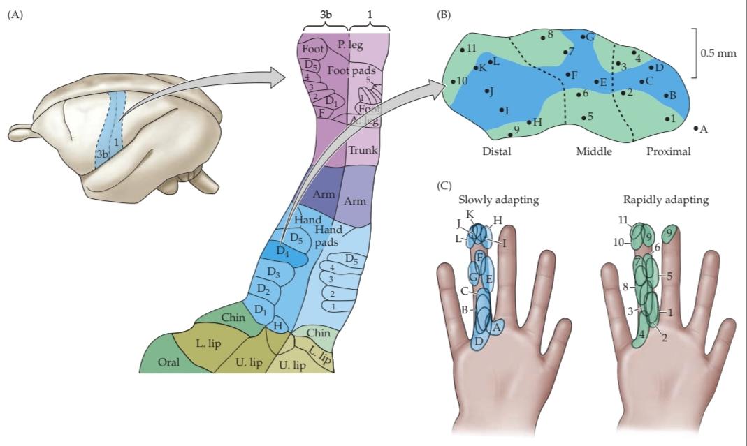
FIGURE 9.13 Neurons in the primary somatosensory cortex form functionally distinct columns. (A) Primary somatosensory map in the owl monkey based, as for the human in Figure 9.11, on the electrical responsiveness of the cortex to peripheral stimulation. The enlargement on the right shows Brodmann’s areas 3b and 1, which process most cutaneous mechanosensory information. The arrangement is generally similar to that determined in humans. Note the presence of regions that are devoted to the representation of individual digits. (B) Modular organization of responses within the representation of a single digit, showing the location of electrode penetrations that encountered rapidly adapting (green) and slowly adapting (blue) responses within the representation of digit 4. (C) Distribution of slowly adapting and rapidly adapting receptive fields used to derive the plot in (B). Although the receptive fields of these different classes of afferents overlap on the skin surface, they are partially segregated within the cortical representation (A after Kaas, 1993; C after Sur et al., 1980.)
شکل ۹.۱۳ نورونها در قشر حسی-پیکری اولیه، ستونهای عملکردی متمایزی را تشکیل میدهند. (الف) نقشه حسی-پیکری اولیه در میمون جغدی، همانند انسان در شکل ۹.۱۱، بر اساس پاسخدهی الکتریکی قشر به تحریک محیطی است. بزرگنمایی در سمت راست، نواحی ۳b و ۱ برودمن را نشان میدهد که بیشتر اطلاعات حسگرهای مکانیکی پوستی را پردازش میکنند. این ترتیب عموماً مشابه ترتیب تعیینشده در انسان است. به وجود مناطقی که به نمایش ارقام منفرد اختصاص داده شدهاند، توجه کنید. (ب) سازماندهی مدولار پاسخها در نمایش یک رقم واحد، محل نفوذ الکترودهایی را که با پاسخهای سریع تطبیقی (سبز) و آهسته تطبیقی (آبی) در نمایش رقم ۴ مواجه شدهاند، نشان میدهد. (ج) توزیع میدانهای پذیرنده آهسته تطبیقی و سریع تطبیقی که برای استخراج نمودار در (ب) استفاده شده است. اگرچه میدانهای پذیرندهی این طبقات مختلف آورانها روی سطح پوست همپوشانی دارند، اما تا حدی در نمایش قشری از هم جدا هستند (A پس از Kaas، ۱۹۹۳؛ C پس از Sur و همکاران، ۱۹۸۰).
Finally, a fundamental but often neglected feature of the somatosensory system is the presence of massive descending projections. These pathways originate in sensory cortical fields and run to the thalamus, brainstem, and spinal cord. Indeed, descending projections from the somatosensory cortex outnumber ascending somatosensory pathways. Although their physiological role is not well understood, it is generally thought that descending projections modulate the ascending flow of sensory information at the level of the thalamus and brainstem.
در نهایت، یک ویژگی اساسی اما اغلب نادیده گرفته شده از سیستم حسی-پیکری، وجود مسیرهای نزولی عظیم است. این مسیرها از میدانهای قشر حسی سرچشمه میگیرند و به تالاموس، ساقه مغز و نخاع میروند. در واقع، مسیرهای نزولی از قشر حسی-پیکری از مسیرهای حسی-پیکری صعودی بیشتر هستند. اگرچه نقش فیزیولوژیکی آنها به خوبی درک نشده است، اما عموماً تصور میشود که مسیرهای نزولی جریان صعودی اطلاعات حسی را در سطح تالاموس و ساقه مغز تنظیم میکنند.
Plasticity in the Adult Cerebral Cortex
The analysis of maps of the body surface in primary somatosensory cortex and the responses to altered patterns of activity in peripheral afferents has been instrumental in understanding the potential for the reorganization of cortical circuits in adults. Jon Kaas and Michael Merzenich were the first to explore this issue, by examining the impact of peripheral lesions (e.g., cutting a nerve that innervates the hand, or amputation of a digit) on the topographic maps in somatosensory cortex. Immediately after the lesion, the corresponding region of the cortex was found to be unresponsive. After a few weeks, however, the unresponsive area became responsive to stimulation of neighboring regions of the skin (Figure 9.14). For example, if digit 3 was amputated, cortical neurons that formerly responded to stimulation of digit 3 now responded to stimulation of digits 2 or 4. Thus, the central representation of the remaining digits had expanded to take over the cortical territory that had lost its main input. Such “functional remapping” also occurs in the somatosensory nuclei in the thalamus and brainstem; indeed, some of the reorganization of cortical circuits may depend on this concurrent subcortical plasticity. This sort of adjustment in the somatosensory system may contribute to the altered sensation of phantom limbs after amputation (see Chapter 10, Clinical Applications). Similar plastic changes have been demonstrated in the visual, auditory, and motor cortices, suggesting that some ability to reorganize after peripheral deprivation or injury is a general property of the mature neocortex.
انعطافپذیری در قشر مغز بزرگسالان
تحلیل نقشههای سطح بدن در قشر حسی-پیکری اولیه و پاسخها به الگوهای تغییر یافته فعالیت در آورانهای محیطی، در درک پتانسیل سازماندهی مجدد مدارهای قشری در بزرگسالان نقش مهمی داشته است. جان کاس و مایکل مرزنیچ اولین کسانی بودند که با بررسی تأثیر ضایعات محیطی (مثلاً قطع عصبی که دست را عصبدهی میکند یا قطع انگشت) بر روی نقشههای توپوگرافی در قشر حسی-پیکری، این موضوع را بررسی کردند. بلافاصله پس از ضایعه، ناحیه مربوطه از قشر مغز بیپاسخ ماند. با این حال، پس از چند هفته، ناحیه بیپاسخ به تحریک مناطق مجاور پوست پاسخ داد (شکل 9.14). به عنوان مثال، اگر انگشت 3 قطع شود، نورونهای قشر مغز که قبلاً به تحریک انگشت 3 پاسخ میدادند، اکنون به تحریک انگشت 2 یا 4 پاسخ میدهند. بنابراین، نمایش مرکزی انگشتهای باقی مانده گسترش یافته و قلمرو قشر مغز را که ورودی اصلی خود را از دست داده بود، به دست گرفته است. چنین «بازنگاشت عملکردی» همچنین در هستههای حسی-پیکری در تالاموس و ساقه مغز رخ میدهد؛ در واقع، برخی از سازماندهی مجدد مدارهای قشری ممکن است به این انعطافپذیری همزمان زیرقشری بستگی داشته باشد. این نوع تنظیم در سیستم حسی-پیکری ممکن است در تغییر حس اندامهای خیالی پس از قطع عضو نقش داشته باشد (به فصل 10، کاربردهای بالینی مراجعه کنید). تغییرات انعطافپذیری مشابهی در قشرهای بینایی، شنوایی و حرکتی نشان داده شده است، که نشان میدهد نوعی توانایی سازماندهی مجدد پس از محرومیت یا آسیب محیطی، یک ویژگی کلی نئوکورتکس بالغ است.

FIGURE 9.14 Functional changes in the somatosensory cortex following amputation of a digit. (A) Diagram of the somatosensory cortex in the owl monkey, showing the approximate location of the hand representation. (B) The hand representation in the animal before amputation; the numbers correspond to different digits. (C) The cortical map determined in the same animal 2 months after amputation of digit 3. The map has changed substantially; neurons in the area formerly responding to stimulation of digit 3 now respond to stimulation of digits 2 and 4. (After Merzenich et al., 1984.)
شکل ۹.۱۴ تغییرات عملکردی در قشر حسی-پیکری پس از قطع انگشت. (الف) نمودار قشر حسی-پیکری در میمون جغدی، که محل تقریبی نمایش دست را نشان میدهد. (ب) نمایش دست در حیوان قبل از قطع عضو؛ اعداد مربوط به ارقام مختلف هستند. (ج) نقشه قشری که در همان حیوان ۲ ماه پس از قطع انگشت ۳ تعیین شده است. نقشه به طور قابل توجهی تغییر کرده است؛ نورونهای ناحیهای که قبلاً به تحریک انگشت ۳ پاسخ میدادند، اکنون به تحریک ارقام ۲ و ۴ پاسخ میدهند. (به نقل از مرزنیچ و همکاران، ۱۹۸۴.)
Appreciable changes in cortical representation also can occur in response to physiological changes in sensory or motor experience. For instance, if a monkey is trained to use a specific digit for a particular task that is repeated many times, the functional representation of that digit, determined by electrophysiological mapping, can expand at the expense of the other digits (Figure 9.15). In fact, significant changes in receptive fields of somatosensory neurons can be detected. when a peripheral nerve is blocked temporarily by a local anesthetic. The transient loss of sensory input from a small area of skin induces a reversible reorganization of the receptive fields of both cortical and subcortical neurons. During this period, the neurons assume new receptive fields that respond to tactile stimulation of the skin surrounding the anesthetized region. Once the effects of the local anesthetic subside, the receptive fields of cortical and subcortical neurons return to their usual size. The common experience of an anesthetized area of skin feeling disproportionately large as experienced, for example, following dental anesthesia may be a consequence of this temporary change.
تغییرات قابل توجهی در بازنمایی قشری همچنین میتواند در پاسخ به تغییرات فیزیولوژیکی در تجربه حسی یا حرکتی رخ دهد. به عنوان مثال، اگر یک میمون آموزش ببیند که از یک انگشت خاص برای یک کار خاص که بارها تکرار میشود استفاده کند، بازنمایی عملکردی آن انگشت، که توسط نقشهبرداری الکتروفیزیولوژیکی تعیین میشود، میتواند به قیمت از دست رفتن سایر انگشتان گسترش یابد (شکل 9.15). در واقع، هنگامی که یک عصب محیطی به طور موقت توسط یک بیحسکننده موضعی مسدود میشود، میتوان تغییرات قابل توجهی در میدانهای پذیرنده نورونهای حسی-پیکری تشخیص داد. از دست دادن گذرای ورودی حسی از یک ناحیه کوچک از پوست، باعث سازماندهی مجدد برگشتپذیر میدانهای پذیرنده نورونهای قشری و زیرقشری میشود. در طول این دوره، نورونها میدانهای پذیرنده جدیدی را به خود میگیرند که به تحریک لمسی پوست اطراف ناحیه بیحس شده پاسخ میدهند. به محض اینکه اثرات بیحسکننده موضعی فروکش کند، میدانهای پذیرنده نورونهای قشری و زیرقشری به اندازه معمول خود باز میگردند. تجربه رایج احساس نامتناسب بزرگ شدن ناحیه بیحسشده پوست نسبت به آنچه مثلاً پس از بیحسی دندانپزشکی تجربه میشود، ممکن است نتیجه این تغییر موقت باشد.
Despite these intriguing observations, the mechanism, purpose, and significance of the reorganization of sensory and motor maps that occurs in adult cortex are not known. Clearly, changes in cortical circuitry occur in the adult brain. Centuries of clinical observations, however, indicate that these changes may be of limited value for recovery of function following brain injury, and they may well lead to symptoms that detract from rather than enhance the quality of life following neural damage. Given their rapid and reversible character, most of these changes in cortical function probably reflect alterations in the strength of synapses already present. Indeed, finding ways to prevent or redirect the synaptic events that underlie injury induced plasticity could reduce the long-term impact of acute brain damage.
علیرغم این مشاهدات جذاب، مکانیسم، هدف و اهمیت سازماندهی مجدد نقشههای حسی و حرکتی که در قشر مغز بزرگسالان رخ میدهد، مشخص نیست. واضح است که تغییرات در مدارهای قشر مغز در مغز بزرگسالان رخ میدهد. با این حال، قرنها مشاهدات بالینی نشان میدهد که این تغییرات ممکن است برای بازیابی عملکرد پس از آسیب مغزی ارزش محدودی داشته باشند و ممکن است منجر به علائمی شوند که به جای افزایش کیفیت زندگی پس از آسیب عصبی، از آن بکاهند. با توجه به ویژگی سریع و برگشتپذیر آنها، بیشتر این تغییرات در عملکرد قشر مغز احتمالاً منعکس کننده تغییرات در قدرت سیناپسهای موجود هستند. در واقع، یافتن راههایی برای جلوگیری یا تغییر مسیر رویدادهای سیناپسی که زمینهساز انعطافپذیری ناشی از آسیب هستند، میتواند تأثیر طولانی مدت آسیب حاد مغزی را کاهش دهد.

FIGURE 9.15 Functional expansion of a cortical representation by a repet- itive behavioral task. (A) An owl monkey was trained in a task that required heavy usage of digits 2, 3, and occasionally 4. (B) The map of the digits in the primary somatosensory cortex prior to training. (C) After several months of “practice,” a larger region of the cortex contained neurons activated by the digits used in the task. Note that the specific arrangements of the digit representations are somewhat different from those for the monkey shown in Figure 9.14, indicating the variability of the cortical representation in individual animals. (After Jenkins et al., 1990.)
شکل ۹.۱۵ گسترش عملکردی یک بازنمایی قشری توسط یک تکلیف رفتاری تکراری. (الف) یک میمون جغدی در تکلیفی آموزش دید که نیاز به استفاده زیاد از ارقام ۲، ۳ و گاهی ۴ داشت. (ب) نقشه ارقام در قشر حسی-پیکری اولیه قبل از آموزش. (ج) پس از چند ماه «تمرین»، ناحیه بزرگتری از قشر مغز حاوی نورونهایی بود که توسط ارقام استفاده شده در تکلیف فعال میشدند. توجه داشته باشید که ترتیب خاص بازنمایی ارقام تا حدودی با ترتیب میمون نشان داده شده در شکل ۹.۱۴ متفاوت است، که نشان دهنده تنوع بازنمایی قشری در حیوانات مختلف است. (بعد از جنکینز و همکاران، ۱۹۹۰.)
Summary
The components of the somatosensory system process information conveyed by mechanical stimuli that either impinge on the body surface (cutaneous mechanoreception) or are generated within the body itself (proprioception). Somatosensory processing is performed by neurons distributed across several brain structures that are connected by both ascending and descending pathways. Transmission of afferent mechanosensory information from the periphery to the brain begins with a variety of receptor types that initiate action potentials. This activity is then conveyed centrally via a chain of nerve cells organized into distinct gray matter structures and white matter tracts. First order neurons in this chain are the primary sensory neurons located in the dorsal root and cranial nerve ganglia. The next set of neurons conveying ascending mechanosensory signals is located in brainstem nuclei (although there are also projection neurons located in the spinal cord that project to the brainstem). The final link in the pathway from periphery to cerebral cortex consists of neurons found in the thalamus, which in turn project to the postcentral gyrus. These pathways are topographically arranged throughout the system, with the amount of cortical and subcortical space allocated to various body parts being proportional to the density of peripheral receptors. Studies of nonhuman primates show that specific cortical regions correspond to each functional submodality; area 3b, for example, processes information from low threshold cutaneous receptors, while area 3a processes inputs from proprioceptors. Thus, at least two broad criteria operate in the organization of the somatosensory system: modality and somatotopy. The end result of this complex interaction is the unified perceptual representation of the body and its ongoing interaction with the environment.
خلاصه
اجزای سیستم حسی-پیکری، اطلاعات منتقل شده توسط محرکهای مکانیکی را پردازش میکنند که یا به سطح بدن برخورد میکنند (حس مکانیکی پوستی) یا در داخل خود بدن تولید میشوند (حس عمقی). پردازش حس پیکری توسط نورونهایی انجام میشود که در چندین ساختار مغز توزیع شدهاند و توسط مسیرهای صعودی و نزولی به هم متصل هستند. انتقال اطلاعات حسی-پیکری آوران از محیط به مغز با انواع مختلفی از گیرندهها آغاز میشود که پتانسیلهای عمل را آغاز میکنند. سپس این فعالیت به صورت مرکزی از طریق زنجیرهای از سلولهای عصبی که در ساختارهای متمایز ماده خاکستری و مسیرهای ماده سفید سازماندهی شدهاند، منتقل میشود. نورونهای مرتبه اول در این زنجیره، نورونهای حسی اولیه هستند که در ریشه پشتی و گانگلیونهای عصبی جمجمهای قرار دارند. مجموعه بعدی نورونهایی که سیگنالهای حسی-پیکری صعودی را منتقل میکنند، در هستههای ساقه مغز قرار دارند (اگرچه نورونهای پروجکشن نیز در نخاع وجود دارند که به ساقه مغز امتداد مییابند). حلقه نهایی در مسیر از محیط به قشر مغز شامل نورونهایی است که در تالاموس یافت میشوند و به نوبه خود به شکنج پس مرکزی امتداد مییابند. این مسیرها از نظر توپوگرافی در سراسر سیستم مرتب شدهاند و میزان فضای قشری و زیرقشری اختصاص داده شده به قسمتهای مختلف بدن متناسب با تراکم گیرندههای محیطی است. مطالعات روی نخستیسانان غیرانسانی نشان میدهد که نواحی قشری خاصی با هر زیر مدالیته عملکردی مطابقت دارند؛ به عنوان مثال، ناحیه 3b اطلاعات دریافتی از گیرندههای پوستی با آستانه پایین را پردازش میکند، در حالی که ناحیه 3a ورودیهای دریافتی از گیرندههای عمقی را پردازش میکند. بنابراین، حداقل دو معیار کلی در سازماندهی سیستم حسی-پیکری عمل میکنند: مدالیته و سوماتوتوپی. نتیجه نهایی این تعامل پیچیده، نمایش ادراکی یکپارچه از بدن و تعامل مداوم آن با محیط است.
ADDITIONAL READING
کلیک کنید «Reviews»
Abraira, V. E. and D. D. Ginty (2013) The sensory neurons of touch. Neuron 79: 618-639.
Barnes, S. J. and G. T. Finnerty (2010) Sensory experience and cortical rewiring. Neuroscientist 16: 186-198.
Chapin, J. K. (1987) Modulation of cutaneous sensory transmission during movement: Possible mechanisms and biological significance. In Higher Brain Function: Recent Explorations of the Brain’s Emergent Properties, S. P. Wise (ed.). New York: John Wiley and Sons, pp. 181-209.
Darian-Smith, I. (1982) Touch in primates. Annu. Rev. Psychol. 33: 155-194.
Johansson, R. S. and J. R. Flanagan (2009) Coding and use of tactile signals from the fingertips in object manipulation tasks. Nat. Rev. Neurosci. 10: 345-359.
Johansson, R. S. and A. B. Vallbo (1983) Tactile sensory coding in the glabrous skin of the human. Trends Neurosci. 6: 27-32.
Johnson, K. O. (2002) Neural basis of haptic perception. In Seven’s Handbook of Experimental Psychology, 3rd Edition, H. Pash- ler and S. Yantis (eds.). Vol 1: Sensation and Perception. New York: Wiley, pp. 537-583.
Kaas, J. H. (1990) Somatosensory system. In The Human Nervous System, G Paxinos (ed.). San Diego: Academic Press, pp. 813-844.
Kaas, J. H. (1993) The functional organization of somatosensory cortex in primates. Ann. Anat. 175: 509-518.
Kaas, J. H. and C. E. Collins (2003) The organization of somatosensory cortex in anthropoid primates. Adv. Neurol. 93: 57-67.
Mountcastle, V. B. (1975) The view from within: Pathways to the study of perception. Johns Hopkins Med. J. 136: 109-131.
Nicolelis, M. A. and E. E. Fanselow (2002) Thalamocortical optimization of tactile processing according to behavioral state. Nature Neurosci. 5 (6): 517-523.
Petersen, R. S., S. Panzeri and M. E. Diamond (2002) Population coding in somatosensory cortex. Curr. Opin. Neurobiol. 12: 441-447.
Ranade, S. S., R. Syeda and A. Patapoutian (2015) Mechanically activated ion channels. Neuron. 87: 1162-1179.
Saal, H. P. and S. J. Bensmaia (2014) Touch is a team effort: interplay of submodalities in cutaneous sensibility. Trends Neurosci. 37: 689-697.
Woolsey, C. (1958) Organization of somatosensory and motor areas of the cerebral cortex. In Biological and Biochemical Bases of Behavior, H. F. Harlow and C. N. Woolsey (eds.). Madison: University of Wisconsin Press, pp. 63-82.
کلیک کنید «Important original papers»
Adrian, E. D. and Y. Zotterman (1926) The impulses produced by sensory nerve endings. II. The response of a single end or- gan. J. Physiol. 61: 151-171.
Friedman, R. M., L. M. Chen and A. W. Roe (2004) Modality maps within primate somatosensory cortex. Proc. Natl. Acad. Sci. USA 101: 12724-12729.
Johansson, R. S. (1978) Tactile sensibility of the human hand: Receptive field characteristics of mechanoreceptive units in the glabrous skin. J. Physiol. (Lond.) 281: 101-123.
Johnson, K. O. and G. D. Lamb (1981) Neural mechanisms of spatial tactile discrimination: Neural patterns evoked by Braille-like dot patterns in the monkey. J. Physiol. (Lond.) 310: 117-144.
Jones, E. G. and D. P. Friedman (1982) Projection pattern of functional components of thalamic ventrobasal complex on monkey somatosensory cortex. J. Neurophysiol. 48: 521-544.
Jones, E. G. and T. P. S. Powell (1969) Connexions of the somatosensory cortex of the rhesus monkey. I. Ipsilateral connex- ions. Brain 92: 477-502.
Lamotte, R. H. and M. A. Srinivasan (1987) Tactile discrimination of shape: Responses of rapidly adapting mechanoreceptive afferents to a step stroked across the monkey fingerpad. J. Neurosci. 7: 1672-1681.
Laubach, M., J. Wessber and M. A. L. Nicolelis (2000) Cortical ensemble activity increasingly predicts behavior outcomes during learning of a motor task. Nature 405: 567-571.
Moore, C. I. and S. B. Nelson (1998) Spatiotemporal subthreshold receptive fields in the vibrissa representation of rat primary somatosensory cortex. J. Neurophysiol. 80: 2882-2892.
Moore, C. I., S. B. Nelson and M. Sur (1999) Dynamics of neuronal processing in rat somatosensory cortex. Trends Neurosci. 22: 513-520.
Nicolelis, M. A. L., L. A. Baccala, R. C. S. Lin and J. K. Chapin (1995) Sensorimotor encoding by synchronous neural ensem- ble activity at multiple levels of the somatosensory system. Science 268: 1353-1359.
Ranade, S. S. and 16 others (2014) Piezo2 is the major transducer of mechanical forces for touch sensation in mice. Nature 516: 121-125.
Sur, M. (1980) Receptive fields of neurons in areas 3b and 1 of somatosensory cortex in monkeys. Brain Res. 198: 465-471.
Wall, P. D. and W. Noordenhos (1977) Sensory functions which remain in man after complete transection of dorsal columns. Brain 100: 641-653.
Weber, A. I. and 6 others (2013) Spatial and temporal codes mediate the tactile perception of natural textures. Proc. Natl. Acad. Sci. USA 110: 17107-17112.
Woo, S. H. and 11 others (2014) Piezo2 is required for Merkelcell mechanotransduction. Nature 509: 622-626.
Zhu, J. J. and B. Connors (1999) Intrinsic firing patterns and whisker evoked synaptic responses of neurons in the rat barrel cortex. J. Neurophysiol. 81: 1171-1183.
کلیک کنید «Books»
Hertenstein, M. J. and S. J. Weiss (eds.) (2011) The Handbook of Touch: Neuroscience, Behavioral, and Health Perspectives. New York: Springer.
Linden, D. J. (2015) Touch: The Science of Hand, Heart, and Mind. New York: Viking Penguin.
Mountcastle, V. B. (1998) Perceptual Neuroscience: The Cerebral Cortex. Cambridge, MA: Harvard University Press.
Go to the NEUROSCIENCE 6e Companion Website at oup-arc.com/access/purves-6e for Web Topics, Animations, Flashcards, and more. Go to DASHBOARD for additional resources and assessments.
«فصل نهم کتاب علوم اعصاب پروس به زبان انگلیسی»
🚀 با ما همراه شوید!
تازهترین مطالب و آموزشهای مغز و اعصاب را از دست ندهید. با فالو کردن کانال تلگرام آیندهنگاران مغز، از ما حمایت کنید!



