
علوم اعصاب پروس؛ اجزای حسی-پیکری تالاموس و قشر حسی-پیکری اولیه


دعای مطالعه [ نمایش ]
بِسْمِ الله الرَّحْمنِ الرَّحیمِ
اَللّهُمَّ اَخْرِجْنى مِنْ ظُلُماتِ الْوَهْمِ
خدايا مرا بيرون آور از تاريكىهاى وهم،
وَ اَكْرِمْنى بِنُورِ الْفَهْمِ
و به نور فهم گرامى ام بدار،
اَللّهُمَّ افْتَحْ عَلَيْنا اَبْوابَ رَحْمَتِكَ
خدايا درهاى رحمتت را به روى ما بگشا،
وَانْشُرْ عَلَيْنا خَزائِنَ عُلُومِكَ بِرَحْمَتِكَ يا اَرْحَمَ الرّاحِمينَ
و خزانههاى علومت را بر ما باز كن به امید رحمتت اى مهربانترين مهربانان.
آیندهنگاران مغز: «ما مغز را میشناسیم، تا آینده را بسازیم.»
کتاب «علوم اعصاب» اثر پروس و همکاران بهعنوان یکی از جامعترین و معتبرترین منابع در حوزه علوم اعصاب، همچنان مرجع کلیدی برای درک پیچیدگیهای مغز و سیستم عصبی است. این اثر با بهرهگیری از تازهترین پژوهشها و توضیحات دقیق درباره سازوکارهای عصبی، پلی میان دانش پایه علوم اعصاب و کاربردهای بالینی ایجاد میکند و نقشی بیبدیل در آموزش، پژوهش و ارتقای دانش مغز و اعصاب ایفا مینماید.
ترجمه دقیق و علمی این شاهکار توسط برند علمی «آیندهنگاران مغز» به مدیریت داریوش طاهری، دسترسی فارسیزبانان به مرزهای نوین دانش علوم اعصاب را ممکن ساخته و رسالتی علمی برای ارتقای آموزش، فهم عمیقتر عملکرد مغز و سیستم عصبی و توسعه روشهای نوین در حوزه سلامت عصبی فراهم آورده است.
» کتاب علوم اعصاب پروس
» » فصل ۹: سیستم حسی-پیکری: حس لامسه و حس عمقی؛ قسمت ششم
» Neuroscience; Dale Purves, et al.
»» Chapter 9: The Somatosensory System: Touch and Proprioception
در حال ویرایش
Somatosensory Components of the Thalamus
Each of the several ascending somatosensory pathways originating in the spinal cord and brainstem converges on the ventral posterior complex of the thalamus and terminates in an organized fashion (Figure 9.10). One of the organizational features of this complex instantiated by the pattern of afferent terminations is a complete and orderly somatotopic representation of the body and head. As already mentioned, the more laterally located ventral posterior lateral nucleus (VPL) receives projections from the medial lemniscus carrying somatosensory information from the body and posterior head, whereas the more medially located ventral posterior medial nucleus (VPM) receives axons from the trigeminal lemniscus conveying somatosensory information from the face. In addition, inputs carrying different types of somatosensory information for example, those that respond to different types of mechanoreceptors, to muscle spindle afferents, or to Golgi tendon organs terminate on separate populations of relay cells within the ventral posterior complex. Thus, the information supplied by different somatosensory receptors remains segregated in its passage to cortical circuits.
اجزای حسی-پیکری تالاموس
هر یک از چندین مسیر حسی-پیکری صعودی که از نخاع و ساقه مغز سرچشمه میگیرند، در کمپلکس خلفی-شکمی تالاموس همگرا میشوند و به شکلی سازمانیافته خاتمه مییابند (شکل 9.10). یکی از ویژگیهای سازمانی این کمپلکس که با الگوی پایانههای آوران مشخص میشود، نمایش کامل و منظم سوماتوتوپیک بدن و سر است. همانطور که قبلاً ذکر شد، هسته جانبی-خلفی-شکمی (VPL) که در سمت چپتر قرار دارد، تصاویر را از لمنیسکوس میانی که اطلاعات حسی-پیکری را از بدن و سر خلفی حمل میکند، دریافت میکند، در حالی که هسته داخلی-خلفی-شکمی (VPM) که در سمت چپتر قرار دارد، آکسونها را از لمنیسکوس سه قلو که اطلاعات حسی-پیکری را از صورت منتقل میکنند، دریافت میکند. علاوه بر این، ورودیهایی که انواع مختلفی از اطلاعات حسی-پیکری را حمل میکنند، به عنوان مثال، آنهایی که به انواع مختلف گیرندههای مکانیکی، آورانهای دوک عضلانی یا اندامهای تاندونی گلژی پاسخ میدهند، در جمعیتهای جداگانهای از سلولهای رله در کمپلکس خلفی شکمی خاتمه مییابند. بنابراین، اطلاعات ارائه شده توسط گیرندههای حسی-پیکری مختلف در عبور خود به مدارهای قشری به صورت جداگانه باقی میمانند.
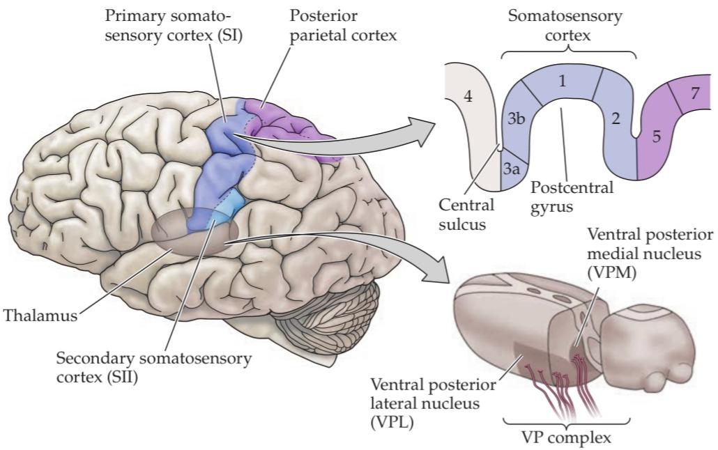
FIGURE 9.10 Somatosensory portions of the thalamus and their cortical targets in the postcentral gyrus. The ventral posterior nuclear complex comprises the VPM, which relays somatosensory information carried by the trigeminal system from the face, and the VPL, which relays somatosensory information from the rest of the body. The diagram at the upper right shows the organization of the primary somatosensory cortex in the postcentral gyrus, shown here in a section cutting across the gyrus from anterior to posterior. (After Brodal, 1992 and Jones et al., 1982.)
شکل ۹.۱۰ بخشهای حسی-پیکری تالاموس و اهداف قشری آنها در شکنج پسمرکزی. کمپلکس هستهای خلفی-شکمی شامل VPM است که اطلاعات حسی-پیکری حملشده توسط سیستم سهقلو را از صورت رله میکند و VPL که اطلاعات حسی-پیکری را از بقیه بدن رله میکند. نمودار بالا سمت راست، سازماندهی قشر حسی-پیکری اولیه در شکنج پسمرکزی را نشان میدهد که در اینجا در بخشی که از جلو به عقب شکنج را قطع میکند، نشان داده شده است. (After Brodal, 1992 and Jones et al., 1982.)
Primary Somatosensory Cortex
The majority of the axons arising from neurons in the ventral posterior complex of the thalamus project to cortical neurons located in layer 4 of the primary somatosensory cortex (see Box 27A for a description of cortical lamination). The primary somatosensory cortex in humans is located in the postcentral gyrus of the parietal lobe and comprises four distinct regions, or fields, known as Brodmann’s areas 3a, 3b, 1, and 2 (Figure 9.11A). Mapping studies in humans and other primates show further that each of these four cortical areas contains a separate and complete representation of the body. In these somatotopic maps, the foot, leg, trunk, forelimbs, and face are represented in a medial to lateral arrangement, as shown in Figure 9.11B.
قشر حسی-پیکری اولیه
اکثر آکسونهای ناشی از نورونهای کمپلکس شکمی-خلفی تالاموس به نورونهای قشری واقع در لایه ۴ قشر حسی-پیکری اولیه متصل میشوند (برای توصیف لایهبندی قشری به کادر ۲۷A مراجعه کنید). قشر حسی-پیکری اولیه در انسان در شکنج پس مرکزی لوب آهیانه قرار دارد و شامل چهار ناحیه یا میدان مجزا است که به عنوان نواحی برودمن ۳a، ۳b، ۱ و ۲ شناخته میشوند (شکل ۹.۱۱A). مطالعات نقشهبرداری در انسان و سایر نخستیها همچنین نشان میدهد که هر یک از این چهار ناحیه قشری شامل یک نمایش جداگانه و کامل از بدن است. در این نقشههای سوماتوتوپیک، پا، ساق پا، تنه، اندامهای جلویی و صورت به صورت میانی-جانبی نمایش داده میشوند، همانطور که در شکل ۹.۱۱B نشان داده شده است.
A salient feature of somatotopic maps, recognized soon after their discovery, is their failure to represent the human body in its actual proportions. When neurosurgeons determined the representation of the human body in the primary sensory (and motor) cortex, the homunculus (“little man”) defined by such mapping procedures had a grossly enlarged face and hands compared with the torso and proximal limbs (Figure 9.11C). These anomalies arise because manipulation, facial expression, and speech are extraordinarily important for humans and require a great deal of circuitry, both central and peripheral, to govern them. Thus, in humans the cervical spinal cord is enlarged to accommodate the extra circuitry related to the hand and upper limb, and as stated earlier, the density of receptors is greater in regions such as the hands and lips.
یکی از ویژگیهای برجسته نقشههای سوماتوتوپیک، که اندکی پس از کشف آنها مشخص شد، عدم توانایی آنها در نمایش بدن انسان در نسبتهای واقعی آن است. هنگامی که جراحان مغز و اعصاب، نمایش بدن انسان را در قشر حسی (و حرکتی) اولیه تعیین کردند، هومونکولوس (“مرد کوچک”) که توسط چنین رویههای نقشهبرداری تعریف میشد، در مقایسه با تنه و اندامهای پروگزیمال، صورت و دستهای بسیار بزرگتری داشت (شکل 9.11C). این ناهنجاریها به این دلیل ایجاد میشوند که دستکاری، بیان صورت و گفتار برای انسان فوقالعاده مهم هستند و برای کنترل آنها به مدار زیادی، چه مرکزی و چه محیطی، نیاز است. بنابراین، در انسان، نخاع گردنی برای تطبیق با مدارهای اضافی مربوط به دست و اندام فوقانی بزرگ میشود و همانطور که قبلاً گفته شد، تراکم گیرندهها در مناطقی مانند دستها و لبها بیشتر است.
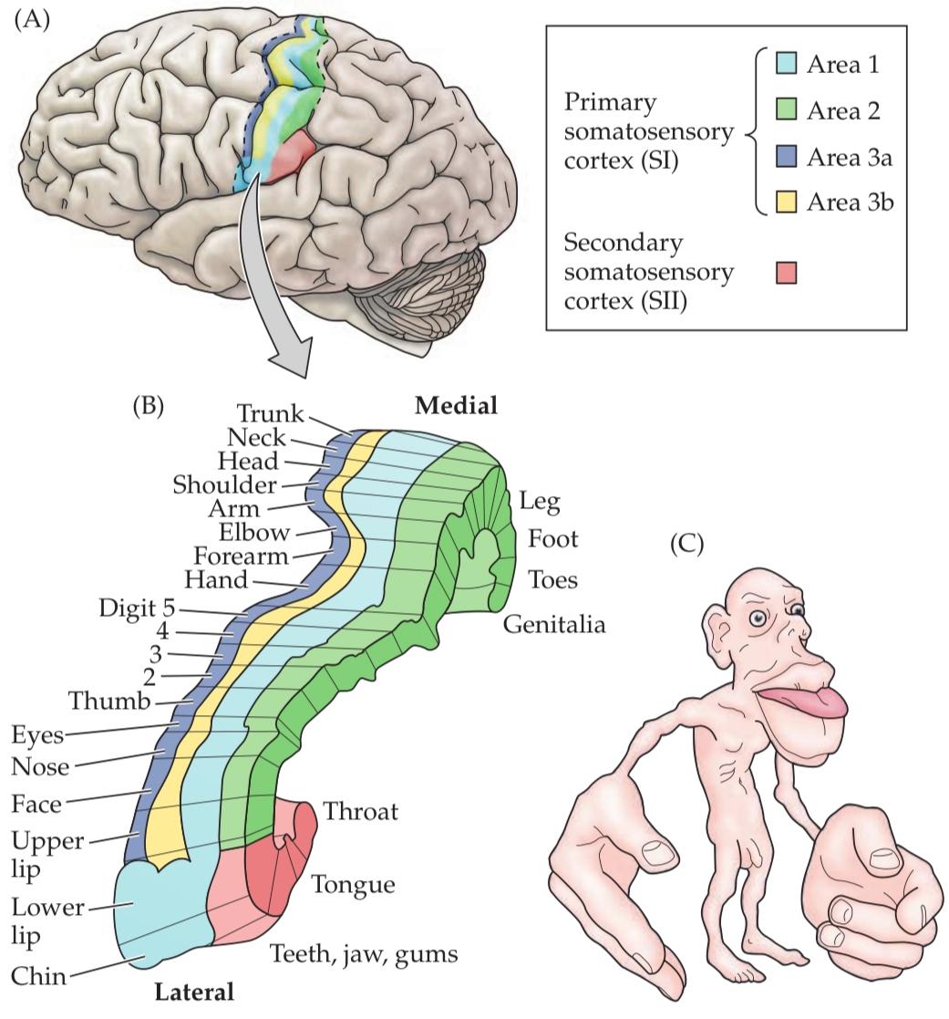
FIGURE 9.11 Somatotopic order in the human primary somatosensory cortex. (A) Diagram showing the region of the human cortex from which electrical activity is recorded following mechanosensory stimulation of different parts of the body. (The patients in the study were undergoing neurosurgical procedures for which such mapping was required.) Although modern imaging methods are now refining these classical data, the human somatotopic map defined in the 1930s has remained generally valid. (B) Diagram showing the somatotopic representation of body parts from medial to lateral. (C) Cartoon of the homunculus constructed on the basis of such mapping. Note that the amount of somatosensory cortex devoted to the hands and face is much larger than the relative amount of body surface in these regions. A similar disproportion is apparent in the primary motor cortex, for much the same reasons (see Chapter 17). (After Penfield and Rasmussen, 1950, and Corsi, 1991.)
شکل ۹.۱۱ ترتیب سوماتوتوپیک در قشر حسی-پیکری اولیه انسان. (الف) نموداری که ناحیهای از قشر مغز انسان را نشان میدهد که فعالیت الکتریکی از آن پس از تحریک حسگرهای مکانیکی قسمتهای مختلف بدن ثبت میشود. (بیماران مورد مطالعه تحت عمل جراحی مغز و اعصاب بودند که برای آنها چنین نقشهبرداری لازم بود.) اگرچه روشهای تصویربرداری مدرن اکنون در حال اصلاح این دادههای کلاسیک هستند، نقشه سوماتوتوپیک انسان که در دهه ۱۹۳۰ تعریف شده بود، به طور کلی معتبر باقی مانده است. (ب) نموداری که نمایش سوماتوتوپیک قسمتهای بدن را از میانی تا جانبی نشان میدهد. (ج) کارتون هومونکولوس که بر اساس چنین نقشهبرداری ساخته شده است. توجه داشته باشید که مقدار قشر حسی-پیکری اختصاص داده شده به دستها و صورت بسیار بزرگتر از مقدار نسبی سطح بدن در این مناطق است. عدم تناسب مشابهی در قشر حرکتی اولیه، به دلایل تقریباً مشابه، آشکار است (به فصل ۱۷ مراجعه کنید). (بعد از پنفیلد و راسموسن، ۱۹۵۰، و کورسی، ۱۹۹۱.)
BOX 9A Patterns of Organization within the Sensory Cortices: Brain Modules
Observations over the last 45 years have made it clear that there is an iterated substructure within the somatosensory (and many other) cortical maps. This substructure takes the form of units called modules, each involving hundreds or thousands of nerve cells in repeating patterns. The advantages of these iterated patterns for brain function remain largely mysterious; for the neurobiologist, however, such iterated arrangements have provided important clues about cortical connectivity and the mechanisms by which neural activity influences brain development (see Chapter 25).
کادر ۹ الف الگوهای سازماندهی در قشرهای حسی: ماژولهای مغز
مشاهدات طی ۴۵ سال گذشته روشن کرده است که یک زیرساختار تکرارشونده در نقشههای قشری حسی-پیکری (و بسیاری دیگر) وجود دارد. این زیرساختار به شکل واحدهایی به نام ماژول است که هر کدام شامل صدها یا هزاران سلول عصبی در الگوهای تکرارشونده هستند. مزایای این الگوهای تکرارشونده برای عملکرد مغز تا حد زیادی مرموز باقی مانده است. با این حال، برای متخصص نوروبیولوژی، چنین ترتیبات تکرارشوندهای سرنخهای مهمی در مورد اتصال قشری و مکانیسمهایی که فعالیت عصبی از طریق آنها بر رشد مغز تأثیر میگذارد، ارائه داده است (به فصل ۲۵ مراجعه کنید).
The observation that the somatosensory cortex comprises elementary units of vertically linked cells was first noted in the 1920s by the Spanish neuroanatomist Rafael Lorente de Nó, based on his studies in the rat. The potential importance of cortical modularity remained largely unexplored until the 1950s, however, when electrophysiological experiments indicated an arrangement of repeating units in the brains of cats and, later, monkeys. Vernon Mountcastle, a neurophysiologist at Johns Hopkins University School of Medicine, found that vertical microelectrode penetrations in the primary somatosensory cortex of these animals encountered cells that responded to the same sort of mechanical stimulus presented at the same location on the body surface. Soon after Mountcastle’s pioneering work, David Hubel and Torsten Wiesel discovered a similar arrangement in the cat primary visual cortex. These and other observations led Mountcastle to the general view that “the elementary pattern of organization of the cerebral cortex is a vertically oriented column or cylinder of cells capable of input output functions of considerable complexity.” Since these discoveries in the late 1950s and early 1960s, the view that modular circuits represent a fundamental feature of the mammalian cerebral cortex has gained wide acceptance, and many such entities have now been described in various cortical regions (see figure).
این مشاهده که قشر حسی-پیکری از واحدهای ابتدایی سلولهای متصل به صورت عمودی تشکیل شده است، اولین بار در دهه ۱۹۲۰ توسط رافائل لورنته د نو، متخصص نوروآناتومی اسپانیایی، بر اساس مطالعاتش روی موش صحرایی، مورد توجه قرار گرفت. با این حال، اهمیت بالقوه مدولاریتی قشر مغز تا دهه ۱۹۵۰ تا حد زیادی ناشناخته باقی ماند، تا اینکه آزمایشهای الکتروفیزیولوژیکی، چیدمانی از واحدهای تکرارشونده را در مغز گربهها و بعدها میمونها نشان دادند. ورنون مونتکسل، متخصص نوروفیزیولوژی در دانشکده پزشکی دانشگاه جانز هاپکینز، دریافت که نفوذهای میکروالکترود عمودی در قشر حسی-پیکری اولیه این حیوانات با سلولهایی مواجه میشود که به همان نوع محرک مکانیکی ارائه شده در همان محل روی سطح بدن پاسخ میدهند. کمی پس از کار پیشگامانه مونتکسل، دیوید هوبل و تورستن ویزل چیدمان مشابهی را در قشر بینایی اولیه گربه کشف کردند. این مشاهدات و سایر مشاهدات، مونتکسل را به این دیدگاه کلی رساند که “الگوی ابتدایی سازماندهی قشر مغز، یک ستون یا استوانه عمودی از سلولهایی است که قادر به توابع ورودی-خروجی با پیچیدگی قابل توجه هستند.” از زمان این اکتشافات در اواخر دهه ۱۹۵۰ و اوایل دهه ۱۹۶۰، این دیدگاه که مدارهای مدولار نشاندهنده یک ویژگی اساسی قشر مغز پستانداران هستند، پذیرش گستردهای یافته است و اکنون بسیاری از این موجودیتها در مناطق مختلف قشر مغز توصیف شدهاند (شکل را ببینید).
This wealth of evidence for such patterned circuits has led many neuroscientists to conclude, like Mountcastle, that modules are a fundamental feature of the cerebral cortex, essential for perception, cognition, and perhaps even consciousness. Despite the prevalence of iterated modules, there are some problems with the view that modular units are universally important in cortical function. First, although modular circuits of a given class are readily seen in the brains of some species, they have not been found in the same brain regions of other, sometimes closely related, animals. Second, not all regions of the mammalian cortex are organized in a modular fashion. And third, no clear function of such modules has been discerned, much effort and speculation notwithstanding. This salient feature of the organization of the somatosensory cortex and other cortical (and some subcortical) regions therefore remains a tantalizing puzzle.
این شواهد فراوان برای چنین مدارهای الگودار، بسیاری از دانشمندان علوم اعصاب را مانند مونتکسل به این نتیجه رسانده است که ماژولها یک ویژگی اساسی قشر مغز هستند که برای ادراک، شناخت و شاید حتی آگاهی ضروری هستند. علیرغم شیوع ماژولهای تکرارشونده، مشکلاتی در این دیدگاه وجود دارد که واحدهای مدولار به طور جهانی در عملکرد قشر مغز مهم هستند. اول، اگرچه مدارهای مدولار از یک دسته خاص به راحتی در مغز برخی از گونهها دیده میشوند، اما در همان مناطق مغزی سایر حیوانات، که گاهی اوقات ارتباط نزدیکی با هم دارند، یافت نشدهاند. دوم، همه مناطق قشر پستانداران به صورت مدولار سازماندهی نشدهاند. و سوم، با وجود تلاش و گمانهزنیهای زیاد، هیچ عملکرد واضحی از چنین ماژولهایی تشخیص داده نشده است. بنابراین، این ویژگی برجسته سازماندهی قشر حسی-پیکری و سایر مناطق قشری (و برخی از مناطق زیرقشری) همچنان یک معمای وسوسهانگیز است.
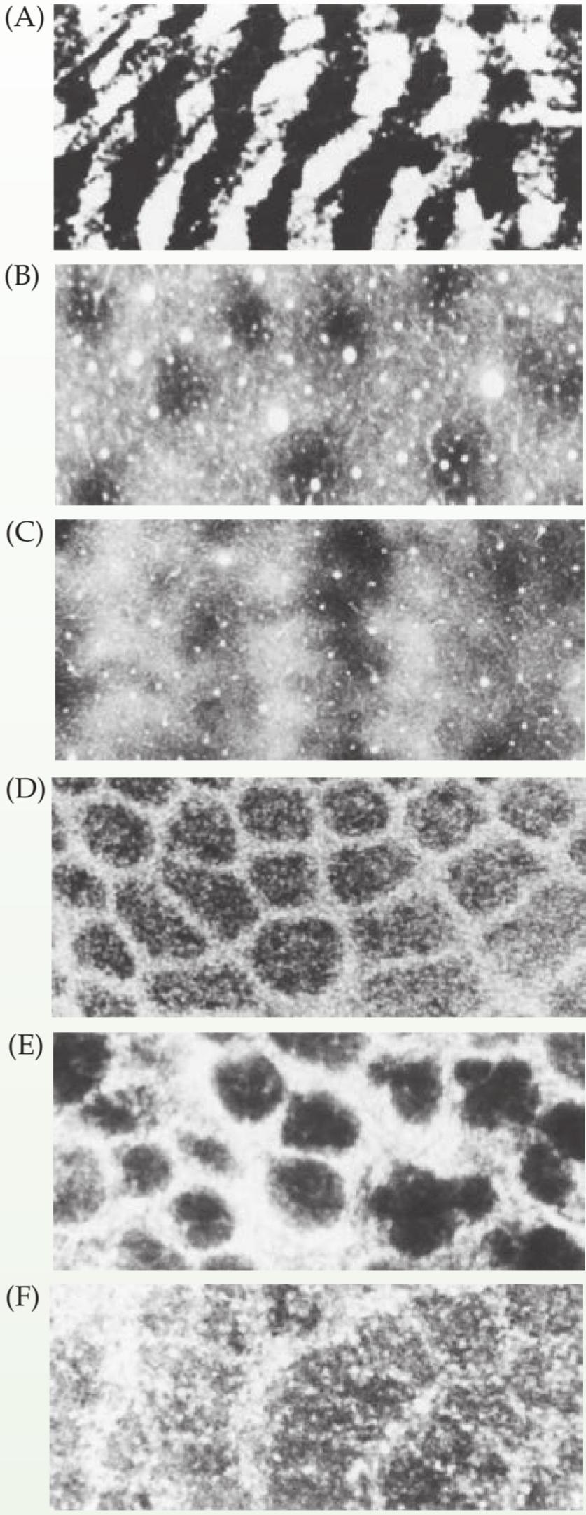
Examples of iterated modular substructures in the mammalian brain. Ocular dominance columns in layer IV in the primary visual cortex (V1) of a rhesus monkey. (B) Repeating units called blobs in layers II and III in V1 of a squirrel monkey. (C) Stripes in layers II and III in V2 of a squirrel monkey. (D) Barrels in layer IV in primary somatosensory cortex of a rat. (E) Glomeruli in the olfactory bulb of a mouse. (F) Iterated units called barreloids in the thalamus of a rat. These and other examples indicate that modular organization is commonplace in the brain. These units are on the order of 100 to several hundred microns across. (From Purves et al., 1992.)
نمونههایی از زیرساختارهای مدولار تکرارشونده در مغز پستانداران. ستونهای تسلط چشمی در لایه IV در قشر بینایی اولیه (V1) یک میمون رزوس. (ب) واحدهای تکرارشونده به نام حبابها در لایههای II و III در V1 یک میمون سنجابی. (ج) نوارها در لایههای II و III در V2 یک میمون سنجابی. (د) بشکهها در لایه IV در قشر حسی-پیکری اولیه یک موش صحرایی. (ه) گلومرولها در پیاز بویایی یک موش صحرایی. (و) واحدهای تکرارشونده به نام بشکههای استوانهای در تالاموس یک موش صحرایی. اینها و نمونههای دیگر نشان میدهند که سازماندهی مدولار در مغز امری عادی است. این واحدها در حدود ۱۰۰ تا چند صد میکرون عرض دارند. (از Purves و همکاران، ۱۹۹۲.)
Such distortions are also apparent when topographical maps are compared across species. In the rat brain, for example, an inordinate amount of the somatosensory cortex is devoted to representing the large facial whiskers that are key components of the somatosensory input for rats and mice. (Box 9A), while raccoons overrepresent their paws and the platypus its bill. In short, the sensory input (or motor out- put) that is particularly significant to a given species gets relatively more cortical representation.
چنین تحریفهایی همچنین هنگام مقایسه نقشههای توپوگرافی بین گونهها آشکار میشوند. به عنوان مثال، در مغز موش صحرایی، مقدار بیش از حدی از قشر حسی-پیکری به نمایش سبیلهای بزرگ صورت اختصاص داده شده است که اجزای کلیدی ورودی حسی-پیکری برای موشهای صحرایی و موشهای خانگی هستند. (کادر 9A)، در حالی که راکونها پنجههای خود و پلاتیپوس منقار خود را بیش از حد نمایش میدهند. به طور خلاصه، ورودی حسی (یا خروجی حرکتی) که به طور خاص برای یک گونه خاص مهم است، نمایش قشری نسبتاً بیشتری پیدا میکند.
Although the topographic organization of the several somatosensory areas is similar, the functional properties. of the neurons in each region are distinct. Experiments carried out in nonhuman primates indicate that neurons in areas 3b and 1 respond primarily to cutaneous stimuli, whereas neurons in 3a respond mainly to stimulation of proprioceptors; area 2 neurons process both tactile and proprioceptive stimuli. These differences in response properties reflect, at least in part, parallel sets of inputs from functionally distinct classes of neurons in the ventral posterior complex. In addition, a rich pattern of corticocortical connections between SI areas contributes significantly to the elaboration of SI response properties. Area 3b receives the bulk of the input from the ventral posterior complex and provides a particularly dense projection to areas 1 and 2. This arrangement of connections establishes a functional hierarchy in which area 3b serves as an obligatory first step in cortical processing of somatosensory information (Figure 9.12). Consistent with this view, lesions of area 3b in non-human primates result in profound deficits in all forms of tactile sensations mediated by cutaneous mecha- noreceptors, while lesions limited to areas 1 or 2 result in partial deficits and an inability to use tactile information to discriminate either the texture of objects (area 1 deficit) or the size and shape of objects (area 2 deficit).
اگرچه سازماندهی توپوگرافی چندین ناحیه حسی-پیکری مشابه است، اما خواص عملکردی نورونها در هر ناحیه متمایز است. آزمایشهای انجام شده در نخستیسانان غیرانسانی نشان میدهد که نورونهای نواحی 3b و 1 در درجه اول به محرکهای پوستی پاسخ میدهند، در حالی که نورونهای ناحیه 3a عمدتاً به تحریک گیرندههای عمقی پاسخ میدهند؛ نورونهای ناحیه 2 هم محرکهای لمسی و هم محرکهای عمقی را پردازش میکنند. این تفاوتها در خواص پاسخ، حداقل تا حدی، مجموعههای موازی ورودیها را از طبقات عملکردی متمایز نورونها در کمپلکس شکمی-خلفی منعکس میکنند. علاوه بر این، الگوی غنی از اتصالات قشری-قشری بین نواحی SI به طور قابل توجهی در توسعه خواص پاسخ SI نقش دارد. ناحیه 3b بخش عمدهای از ورودیها را از کمپلکس شکمی-خلفی دریافت میکند و یک تصویر متراکم ویژه به نواحی 1 و 2 ارائه میدهد. این ترتیب اتصالات یک سلسله مراتب عملکردی ایجاد میکند که در آن ناحیه 3b به عنوان اولین گام اجباری در پردازش قشری اطلاعات حسی-پیکری عمل میکند (شکل 9.12). مطابق با این دیدگاه، ضایعات ناحیه 3b در نخستیسانان غیرانسانی منجر به نقصهای عمیق در تمام اشکال حس لامسه میشود که توسط گیرندههای مکانیکی پوستی ایجاد میشوند، در حالی که ضایعات محدود به نواحی 1 یا 2 منجر به نقصهای جزئی و ناتوانی در استفاده از اطلاعات لمسی برای تمایز بافت اشیاء (نقص ناحیه 1) یا اندازه و شکل اشیاء (نقص ناحیه 2) میشوند.
Even finer parcellations of functionally distinct neuronal populations exist within single cortical areas. Based on his analysis of electrode penetrations in primary somatosensory cortex, Vernon Mountcastle was the first to suggest that neurons with similar response properties might be clustered together into functionally distinct “columns” that traverse the depth of the cortex. Subsequent studies of finely spaced electrode penetrations in area 3b provided strong evidence in support of this idea, demonstrating that neurons with rapidly and slowly adapting properties were clustered into separate zones within the representation of a single digit (Figure 9.13). In the past, it was assumed that the rapidly and slowly adapting cortical neurons receive segregated inputs from rapidly and slowly adapting mechanoreceptors, respectively. However, the cortical slowly adapting neurons all show a large touch OFF response in addition to the sustained firing during contact. Such OFF responses are signaled only by rapidly adapting afferents in fingers. Furthermore, the rapidly adapting cortical neurons sometimes show sustained firing in response to stimuli of preferred directions. Thus, the cortical rapidly and slowly adapting columns reflect differential processing of convergent inputs from different peripheral receptors, rather than the strict segregation of afferent inputs that convey distinct physiological signals. This columnar organization of cortical areas, a fundamental feature of cortical organization throughout the neocortex (see Box 27A), is especially pronounced in visual cortical areas in primates (see Chapter 12). Slowly and rapidly adapting columns in somatosensory cortex are therefore more analogous to orientation columns in the visual cortex (reflecting cortical computations derived from converging input) than ocular dominance columns. (which reflect strictly segregated thalamocortical inputs).
حتی دستههای ظریفتری از جمعیتهای نورونی با عملکرد متمایز در نواحی قشری منفرد وجود دارند. ورنون مونتکسل، بر اساس تجزیه و تحلیل خود از نفوذ الکترودها در قشر حسی-پیکری اولیه، اولین کسی بود که پیشنهاد کرد نورونهایی با ویژگیهای پاسخ مشابه ممکن است در “ستونهای” عملکردی متمایز که در عمق قشر مغز امتداد دارند، با هم خوشهبندی شوند. مطالعات بعدی در مورد نفوذ الکترودهای با فاصله دقیق در ناحیه 3b شواهد محکمی در حمایت از این ایده ارائه داد و نشان داد که نورونهایی با ویژگیهای تطبیق سریع و آهسته در مناطق جداگانهای در نمایش یک رقم واحد خوشهبندی شدهاند (شکل 9.13). در گذشته، فرض بر این بود که نورونهای قشری با تطبیق سریع و آهسته، به ترتیب ورودیهای جداگانهای را از گیرندههای مکانیکی با تطبیق سریع و آهسته دریافت میکنند. با این حال، نورونهای قشری با تطبیق آهسته، علاوه بر شلیک مداوم در طول تماس، همگی یک پاسخ خاموش شدن لمسی بزرگ نشان میدهند. چنین پاسخهای خاموش شدن فقط توسط آورانهای تطبیق سریع در انگشتان سیگنال میشوند. علاوه بر این، نورونهای قشری با تطبیق سریع گاهی اوقات در پاسخ به محرکهای جهتهای ترجیحی، شلیک مداوم نشان میدهند. بنابراین، ستونهای قشری که به سرعت و به آرامی تطبیق مییابند، پردازش افتراقی ورودیهای همگرا از گیرندههای محیطی مختلف را منعکس میکنند، نه تفکیک دقیق ورودیهای آوران که سیگنالهای فیزیولوژیکی متمایزی را منتقل میکنند. این سازماندهی ستونی نواحی قشری، که یک ویژگی اساسی سازماندهی قشری در سراسر نئوکورتکس است (به کادر 27A مراجعه کنید)، به ویژه در نواحی قشر بینایی در نخستیسانان برجسته است (به فصل 12 مراجعه کنید). بنابراین، ستونهای تطبیق آهسته و سریع در قشر حسی-پیکری بیشتر شبیه ستونهای جهتیابی در قشر بینایی هستند (که محاسبات قشری مشتق شده از ورودی همگرا را منعکس میکنند) تا ستونهای تسلط چشمی (که ورودیهای تالاموکورتیکال کاملاً تفکیک شده را منعکس میکنند).
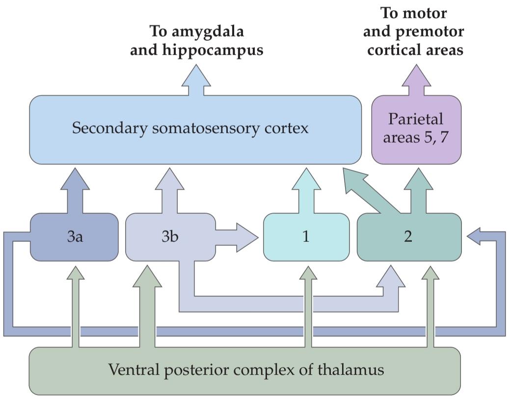
FIGURE 9.12 Connections within the somatosensory cortex establish functional hierarchies. Inputs from the ventral posterior complex of the thalamus terminate in Brod- mann’s areas 3a, 3b, 1, and 2, with the greatest density of projections in area 3b. Area 3b in turn projects heavily to ar- eas 1 and 2, and the functions of these areas are dependent on the activity of area 3b. All subdivisions of primary somatosensory cortex project to secondary somatosensory cortex; the functions of SII are dependent on the activity of SI.
شکل ۹.۱۲ اتصالات درون قشر حسی-پیکری، سلسله مراتب عملکردی را ایجاد میکنند. ورودیهای کمپلکس شکمی-خلفی تالاموس در نواحی ۳a، ۳b، ۱ و ۲ برودمن خاتمه مییابند که بیشترین تراکم پروجکشنها در ناحیه ۳b است. ناحیه ۳b به نوبه خود به شدت به نواحی ۱ و ۲ تصویر میدهد و عملکردهای این نواحی به فعالیت ناحیه ۳b وابسته است. تمام زیربخشهای قشر حسی-پیکری اولیه به قشر حسی-پیکری ثانویه تصویر میدهند. عملکردهای SII به فعالیت SI وابسته است.
Although these cortical patterns reflect specificity in the underlying patterns of thalamocortical and corticocortical connections, the functional significance of columns remains unclear (see Box 9A).
اگرچه این الگوهای قشری، نشاندهندهی اختصاصی بودن الگوهای زیربنایی ارتباطات تالاموکورتیکال و کورتیکوکورتیکال هستند، اهمیت عملکردی ستونها هنوز مشخص نیست (به کادر 9A مراجعه کنید).
Beyond Sl: Corticocortical and Descending Pathways
Somatosensory information is distributed from the primary somatosensory cortex to “higher order” cortical fields. One of these higher-order cortical centers, the secondary somatosensory cortex, lies in the upper bank of the lateral sulcus (see Figures 9.10 and 9.11).
«فصل نهم کتاب علوم اعصاب پروس به زبان انگلیسی»
🚀 با ما همراه شوید!
تازهترین مطالب و آموزشهای مغز و اعصاب را از دست ندهید. با فالو کردن کانال تلگرام آیندهنگاران مغز، از ما حمایت کنید!



