ترجمه و بازنویسی علمی فصول کتاب «نوروآناتومی بالینی» | کتاب درسی جامع ساختارهای عصبی (ویرایش دوم)
کتاب درسی نوروآناتومی بالینی، ویرایش دوم. Textbook of Clinical Neuroanatomy (2nd ed.) یکی از منابع معتبر در حوزه نوروآناتومی بالینی (Clinical Neuroanatomy) است که با رویکردی کاربردی، ساختارها و مسیرهای سیستم عصبی (Nervous System) را بهصورت نظاممند و بالینی شرح میدهد.
این ترجمه آموزشی، حاصل تلاش تیم علمی آیندهنگاران مغز به سرپرستی داریوش طاهری است و با هدف ارتقای آموزش علوم اعصاب (Neuroscience) در زبان فارسی، با حفظ دقت علمی، به زبانی روان تدوین شده است.
در ۲۰ فصل این مجموعه، ساختارهایی چون مغز (Brain)، نخاع (Spinal Cord)، اعصاب مغزی (Cranial Nerves)، سیستم لیمبیک (Limbic System)، مسیرهای حسی و حرکتی (Sensory and Motor Pathways)، مخچه (Cerebellum)، دیانسفالون (Diencephalon) و سیستم عصبی خودمختار (Autonomic Nervous System) بررسی میشوند.
این مجموعه برای دانشجویان پزشکی، رزیدنتها و متخصصان علاقهمند به نوروساینس بالینی طراحی شده و تلفیقی از مفاهیم بنیادی، نکات بالینی و نمودارهای عملکردی را ارائه میدهد.
با سپاس از همراهی شما
تیم آیندهنگاران مغز | سرپرستی: داریوش طاهری
19. Reticular Formation and Limbic System
۱۹. تشکیلات مشبک و سیستم لیمبیک
Reticular Formation
The reticular formation is defined as diffuse ill-defined mass of intermingled neurons and nerve fibres occupying the entire core of brainstem (Fig. 19.1). The reticular formation has derived its name from its light microscopic appearance of a vague network of nerve cells and nerve fibres. It has been defined to include all areas within the brainstem (except the named nuclei and tracts) which when stimulated will produce arousal.
تشکیلات مشبک
تشکیلات مشبک به عنوان تودهای پراکنده و نامشخص از نورونها و فیبرهای عصبی در هم آمیخته تعریف میشود که کل هسته ساقه مغز را اشغال میکند (شکل ۱۹.۱). تشکیلات مشبک نام خود را از ظاهر میکروسکوپی نوری خود که شبکهای مبهم از سلولهای عصبی و فیبرهای عصبی است، گرفته است. تعریف شده است که شامل تمام نواحی درون ساقه مغز (به جز هستهها و مسیرهای نامگذاری شده) میشود که هنگام تحریک، باعث برانگیختگی میشوند.

FIG. 19.1 Location of reticular formation in the brainstem.
شکل ۱۹.۱ محل تشکیلات مشبک در ساقه مغز.
Phylogenetically it represents the old reticular core of brain and contains within it the vital cardiac and respiratory centres which control respiration, heart rate and blood pressure. In primitive vertebrates, the diffuse arrangement of neurons was named ‘reticular’.
از نظر فیلوژنتیکی، این بخش نمایانگر هسته شبکهای قدیمی مغز است و در درون خود مراکز حیاتی قلب و تنفس را که تنفس، ضربان قلب و فشار خون را کنترل میکنند، جای داده است. در مهرهداران اولیه، آرایش پراکنده نورونها «مشبک» نامیده میشد.
The reticular formation receives data from most of the sensory systems and has efferent (direct or indirect) connections with all the levels of neuraxis.
تشکیلات مشبک، دادهها را از اکثر سیستمهای حسی دریافت میکند و با تمام سطوح نوراکسیس (محور عصبی) ارتباط وابران (مستقیم یا غیرمستقیم) دارد.
The knowledge of reticular system is important, because:
(a) it regulates levels of consciousness, and alertness,
(b) i t regulates respiration, blood pressure, heart rate and other vegetative functions,
(c) it regulates tone of skeletal muscles, and
(d) it modulates the impulses in the pain pathways.
شناخت سیستم مشبک مهم است، زیرا:
(الف) سطح هوشیاری و بیداری را تنظیم میکند،
(ب) تنفس، فشار خون، ضربان قلب و سایر عملکردهای نباتی را تنظیم میکند،
(ج) تون عضلات اسکلتی را تنظیم میکند، و
(د) تکانههای موجود در مسیرهای درد را تعدیل میکند.
Clinical Correlation
Damage of the reticular activating system in the core of the brainstem leads to progressive loss of consciousness, followed by stupor, coma and death.
همبستگی بالینی
آسیب سیستم فعالکننده مشبک در هسته ساقه مغز منجر به از دست دادن تدریجی هوشیاری و به دنبال آن بیحسی، کما و مرگ میشود.
Anatomical Extension
The reticular formation extends cranially to the dienceph-alon (subthalamus, hypothalamus and thalamus) and caudally to the spinal cord in the cervical region. These extensions are either actual or projectional. According to some authorities some centres of cerebrum and cerebellum are also closely related functionally to the reticular formation of brainstem.
گسترش آناتومیک
تشکیلات مشبک از سمت جمجمه تا دیانسفالون (ساب تالاموس، هیپوتالاموس و تالاموس) و از سمت خلف تا نخاع در ناحیه گردنی امتداد دارد. این امتدادها یا واقعی هستند یا فرافکنی. به گفته برخی از متخصصان، برخی از مراکز مخ و مخچه نیز از نظر عملکردی ارتباط نزدیکی با تشکیلات مشبک ساقه مغز دارند.
Although reticular formation is described to be consisting of network of nerve fibres and scattered neurons, among them a number of regions with fairly localized cell groups called reticular nuclei, have been recognised.
اگرچه تشکیلات مشبک به عنوان شبکهای از الیاف عصبی و نورونهای پراکنده توصیف میشود، در میان آنها تعدادی ناحیه با گروههای سلولی نسبتاً موضعی به نام هستههای مشبک شناسایی شدهاند.
Reticular nuclei in the brainstem
The reticular nuclei in brainstem are arranged into three longitudinal columns (Fig. 19.2).
هستههای مشبک در ساقه مغز
هستههای مشبک در ساقه مغز به صورت سه ستون طولی چیده شدهاند (شکل ۱۹.۲).
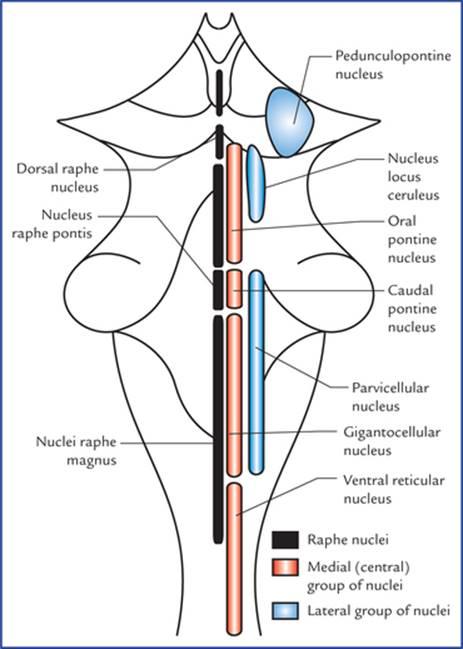
FIG. 19.2 Schematic diagram to show the reticular nuclei in the brainstem.
شکل ۱۹.۲ نمودار شماتیک برای نشان دادن هستههای مشبک در ساقه مغز.
• Median column lies in the midline and consists of intermediate size neurons. The nuclei of this column are termed raphe nuclei.
• ستون میانی در خط وسط قرار دارد و از نورونهای با اندازه متوسط تشکیل شده است. هستههای این ستون، هستههای رافه نامیده میشوند.
• Medial column consists of nuclei which are made up large-size neurons, hence this column is also termed magnocellular column.
• ستون میانی از هستههایی تشکیل شده است که نورونهای با اندازه بزرگ را تشکیل میدهند، از این رو این ستون ستون مگنوسلولار نیز نامیده میشود.
• Lateral column consists of nuclei which are made up of small neurons, hence this column is also termed parvo-cellular column (parvus = little, small).
• ستون جانبی از هستههایی تشکیل شده است که از نورونهای کوچک تشکیل شدهاند، از این رو این ستون ستون پارو-سلولی (parvus = کوچک، ریز) نیز نامیده میشود.
The nuclei belonging to these columns are shown in Figure 19.2. Since it is not advisable for the student to burden his memory with the names of all the nuclei, only those which have a functional or descriptive value are labelled.
هستههای متعلق به این ستونها در شکل ۱۹.۲ نشان داده شدهاند. از آنجایی که توصیه نمیشود دانشآموز حافظه خود را با نام همه هستهها پر کند، فقط آنهایی که ارزش عملکردی یا توصیفی دارند، برچسبگذاری شدهاند.
The reticular nuclei as seen in transverse sections of the midbrain, pons and medulla are shown in Figure 19.3.
هستههای مشبک همانطور که در مقاطع عرضی مغز میانی، پل مغزی و بصل النخاع دیده میشوند، در شکل ۱۹.۳ نشان داده شدهاند.
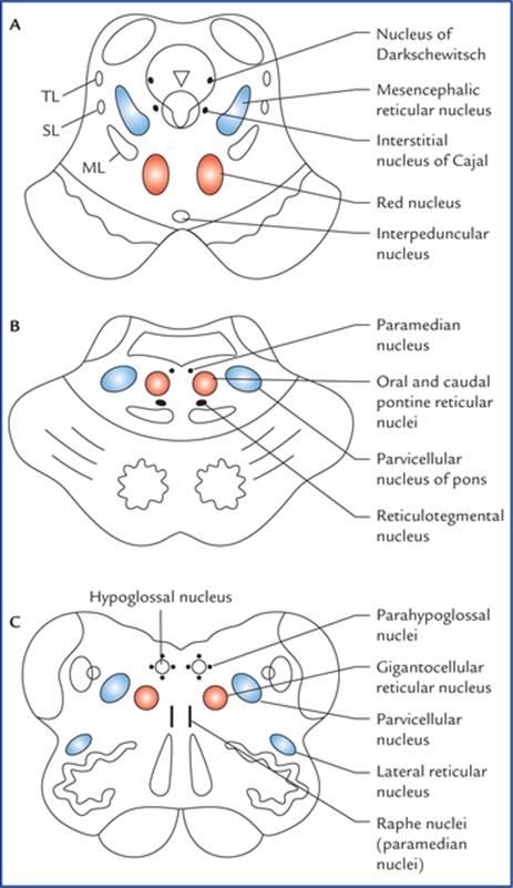
FIG. 19.3 The transverse sections of: midbrain (A), pons (B), and medulla (C) showing the location of reticular nuclei. (ML = medial lemniscus, SL = spinal lemniscus, TL = trigeminal lemniscus.)
شکل ۱۹.۳ برشهای عرضی: مغز میانی (A)، پل مغزی (B) و بصل النخاع (C) که محل هستههای مشبک را نشان میدهند. (ML = لمنیسکوس داخلی، SL = لمنیسکوس نخاعی، TL = لمنیسکوس سه قلو.)
Raphe nuclei (median group of nuclei)
The raphe nuclei form a contiguous column in the mid-line. The neurons of raphe nuclei produce serotonin, a substance that they use as a neurotransmitter. The dorsal raphe nucleus located in the midbrain projects to the spinal cord and forms the pain controlling pathway.
هستههای رافه (گروه میانی هستهها)
هستههای رافه یک ستون پیوسته در خط میانی تشکیل میدهند. نورونهای هستههای رافه، سروتونین تولید میکنند، مادهای که به عنوان انتقالدهنده عصبی از آن استفاده میکنند. هسته رافه پشتی که در مغز میانی قرار دارد، به نخاع امتداد یافته و مسیر کنترل درد را تشکیل میدهد.
The nucleus raphe magnus in medulla projects to the caudal part of the spinal nucleus of the trigeminal nerve and influences perception of pain through spinal nucleus.
هسته رافه بزرگ در بصل النخاع به قسمت انتهایی هسته نخاعی عصب سه قلو امتداد یافته و از طریق هسته نخاعی بر درک درد تأثیر میگذارد.
In fact once the raphe nuclei (vide supra) are activated, the serotogenic neurons inhibit the transmission of pain impulses from dorsal grey horns and spinal nucleus of trigeminal nerve respectively.
در واقع، هنگامی که هستههای رافه (وید سوپرا) فعال میشوند، نورونهای سروتوژنیک به ترتیب انتقال تکانههای درد از شاخهای خاکستری پشتی و هسته نخاعی عصب سه قلو را مهار میکنند.
Clinical Correlation
The electrical stimulation of either the dorsal raphe nucleus (the periaqueductal grey matter) or the nucleus raphe magnus results in loss of the ability to experience pain from sites of injury or disease. The former procedure has been used clinically in the management of otherwise intractable pain.
همبستگی بالینی
تحریک الکتریکی هسته رافه پشتی (ماده خاکستری اطراف مجرا) یا هسته رافه ماگنوس منجر به از دست دادن توانایی احساس درد از محل آسیب یا بیماری میشود. روش اول به صورت بالینی در مدیریت دردهای غیرقابل تحمل استفاده شده است.
Medial group of nuclei
The medial group of nuclei includes ventral reticular nucleus (in medulla), gigantocellular nucleus (in medulla and pons) and oral and caudal pontine nuclei (in pons). Nuclei of this group receive afferents from nuclei of lateral group and efferents from these nuclei ascend or descend longitudinally in the brainstem and give collaterals to the other reticular cells, thus forming a polysynaptic pathway—a characteristic of impulse transmission through the reticular formation.
گروه میانی هستهها
گروه میانی هستهها شامل هسته مشبک شکمی (در بصل النخاع)، هسته غولپیکر سلولی (در بصل النخاع و پل مغزی) و هستههای پل مغزی دهانی و دمی (در پل مغزی) است. هستههای این گروه، آورانهایی از هستههای گروه جانبی دریافت میکنند و آورانهای این هستهها به صورت طولی در ساقه مغز بالا یا پایین میروند و به سایر سلولهای مشبک، مسیرهای جانبی میدهند و در نتیجه یک مسیر پلی سیناپسی تشکیل میدهند – مشخصه انتقال تکانه از طریق تشکیلات مشبک.
Lateral group of nuclei
The lateral group of nuclei includes parvicellular nuclei of medulla and pons, nucleus locus ceruleus of pons and pedunculopontine nucleus of the midbrain.
گروه جانبی هستهها
گروه جانبی هستهها شامل هستههای پارویسلولی بصلالنخاع و پل مغزی، هسته لوکوس سرولئوس پل مغزی و هسته پدانکولوپونتین مغز میانی است.
These nuclei receive collaterals from several ascending pathways and project to the medial group of nuclei of the reticular formation. They are regarded as an association region of the reticular formation.
این هستهها از چندین مسیر صعودی، شاخههای جانبی دریافت میکنند و به گروه میانی هستههای تشکیلات مشبک متصل میشوند. آنها به عنوان یک ناحیه ارتباطی تشکیلات مشبک در نظر گرفته میشوند.
Connections of Reticular Formation
The reticular formation receives information from almost all the principal parts of the nervous system and in turn, projects (directly or indirectly) to all these parts (Fig. 19.4).
اتصالات تشکیلات مشبک
تشکیلات مشبک تقریباً از تمام بخشهای اصلی سیستم عصبی اطلاعات دریافت میکند و به نوبه خود (مستقیم یا غیرمستقیم) به تمام این بخشها متصل میشود (شکل ۱۹.۴).
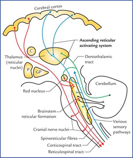
FIG. 19.4 Connections of reticular formation. (P = putamen, and GP = globus pallidus.)
شکل ۱۹.۴ اتصالات تشکیلات مشبک. (P = پوتامن، و GP = گلوبوس پالیدوس.)
The reticular pathways are polysynaptic, both ascending, descending, and crossed and uncrossed. As a result a unilateral stimulation produces bilateral responses.
مسیرهای شبکهای چند سیناپسی هستند، هم صعودی، هم نزولی، و هم متقاطع و غیر متقاطع. در نتیجه، یک تحریک یک طرفه پاسخهای دو طرفه ایجاد میکند.
Afferent connections
The afferents are classified into three types:
اتصالات آوران
آورانها به سه نوع طبقهبندی میشوند:
Afferents from various sensory pathways or systems
– Optic system—through tectoreticular fibres,
– Olfactory and limbic systems—through variety of descending pathways,
– Auditory system—through tectoreticular fibres,
– Gustatory system,
– Spinal pathways—through spinoreticular fibres. A considerable number of fibres of spinothalamic tract terminate in the lateral reticular nucleus of medulla, which in turn project to the cerebellum. Spinoreticulo-cerebellar pathway is an important pathway for carrying exteroceptive sensations to the cerebellum,
– Trigeminal pathways.
آورانها از مسیرها یا سیستمهای حسی مختلف
– سیستم بینایی – از طریق فیبرهای تکتورتیکولار،
– سیستمهای بویایی و لیمبیک – از طریق مسیرهای نزولی متنوع،
– سیستم شنوایی – از طریق فیبرهای تکتورتیکولار،
– سیستم چشایی،
– مسیرهای نخاعی – از طریق فیبرهای نخاعی-شبکهای. تعداد قابل توجهی از فیبرهای راه نخاعی-تالاموسی در هسته مشبک جانبی بصل النخاع ختم میشوند که به نوبه خود به مخچه منتهی میشوند. مسیر نخاعی-مخچهای مسیر مهمی برای انتقال حسهای خارجی به مخچه است،
– مسیرهای سه قلو.
Afferent fibres from other parts of central nervous system
– Cerebellum from both but mainly from contralateral fastigial nucleus.
– Basal ganglia, mainly from corpus striatum.
– Thalamus, hypothalamus and subthalamus.
– Limbic system, mainly from septal areas, amygdaloid nuclei, and hippocampus.
– Cerebral cortex mainly from motor and sensory areas of the cerebral cortex.
– Red nucleus, substantia nigra and habenular nuclei.
الیاف آوران از سایر بخشهای سیستم عصبی مرکزی
– مخچه از هر دو، اما عمدتاً از هسته فاستیژیال طرف مقابل.
– عقدههای قاعدهای، عمدتاً از جسم مخطط.
– تالاموس، هیپوتالاموس و ساب تالاموس.
– سیستم لیمبیک، عمدتاً از نواحی سپتوم، هستههای آمیگدالوئید و هیپوکامپ.
– قشر مغز، عمدتاً از نواحی حرکتی و حسی قشر مغز.
– هسته قرمز، هستههای جسم سیاه و هستههای هابنولار.
Other factors influencing the activity of reticular formation
– Hormones and chemical substances, viz. adrenaline, ace-tylcholine and carbon dioxide.
– Drugs, viz. barbiturates, anaesthetics and tranquillizers.
سایر عوامل مؤثر بر فعالیت تشکیلات مشبک
– هورمونها و مواد شیمیایی، مانند آدرنالین، استیل کولین و دی اکسید کربن.
– داروها، مانند باربیتوراتها، داروهای بیهوشی و آرامبخشها.
Efferent connections
The efferent connections of reticular formation are to all the parts of CNS from which it receives afferents but mainly to:
اتصالات وابران
اتصالات وابران تشکیلات مشبک به تمام قسمتهای سیستم عصبی مرکزی که از آنها آوران دریافت میکند، مربوط میشود، اما عمدتاً به:
• Autonomic and locomotor control centres of brainstem and spinal cord.
• Cranial nerve nuclei, e.g. dorsal nucleus of vagus.
• Cerebral cortex—indirectly through diencephalic nuclei.
• Red nucleus, substantia nigra and tectum of midbrain.
• مراکز کنترل خودکار و حرکتی ساقه مغز و نخاع.
• هستههای اعصاب جمجمهای، مثلاً هسته پشتی واگ.
• قشر مغز – بهطور غیرمستقیم از طریق هستههای دیانسفالیک.
• هسته قرمز، جسم سیاه و تکتوم مغز میانی.
Functional Divisions of Reticular Formation
Functionally the reticular formation is divided into two systems: (a) the ascending reticular activating system (ARAS), and (b) the descending reticular system (DRS). The ascending reticular activating system is commonly termed by the clinicians simply as reticular activating system (RAS).
تقسیمبندیهای عملکردی تشکیلات مشبک
از نظر عملکردی، تشکیلات مشبک به دو سیستم تقسیم میشود: (الف) سیستم فعالکننده مشبک صعودی (ARAS) و (ب) سیستم مشبک نزولی (DRS). سیستم فعالکننده مشبک صعودی معمولاً توسط پزشکان به سادگی سیستم فعالکننده مشبک (RAS) نامیده میشود.
Ascending reticular activating system (Fig. 19.4)
Most of the ascending tracts, viz. spinothalamic tract, trigem-inal lemniscus, lateral lemniscus and central vestibular pathway, while passing through the brainstem give collaterals to the lateral part of the reticular formation which projects to the intralaminar and reticular nuclei of the thalamus. These nuclei in turn project to the widespread areas of the cerebral cortex.
سیستم فعالکننده مشبک صعودی (شکل 19.4)
بیشتر مسیرهای صعودی، یعنی مسیر نخاعی-تالاموسی، لمنیسکوس سهقلو-مینال، لمنیسکوس جانبی و مسیر دهلیزی مرکزی، هنگام عبور از ساقه مغز، به قسمت جانبی تشکیلات مشبک، که به هستههای بین لایهای و مشبک تالاموس منتهی میشود، مسیرهای فرعی میدهند. این هستهها به نوبه خود به نواحی گسترده قشر مغز منتهی میشوند.
When this part of reticular formation is stimulated, the individual becomes alert hence it is termed ascending reticular activating system.
وقتی این بخش از تشکیلات مشبک تحریک میشود، فرد هوشیار میشود، از این رو به آن سیستم فعالکننده شبکهای صعودی میگویند.
The ascending reticular activating system is believed to be responsible for maintaining a state of alertness and consciousness.
اعتقاد بر این است که سیستم فعالکننده شبکهای صعودی مسئول حفظ حالت هوشیاری و آگاهی است.
Clinical Correlation
• The visual and acoustic stimuli can stimulate the reticular activating system to maintain alertness and attention, therefore the stimuli such as sounds of ringing alarm clock or sudden bright light, can arouse consciousness. Conversely, removal of visual and auditory stimuli may lead to drowsiness and sleep.
همبستگی بالینی
• محرکهای بصری و صوتی میتوانند سیستم فعالکنندهی شبکهای را برای حفظ هوشیاری و توجه تحریک کنند، بنابراین محرکهایی مانند صدای زنگ ساعت یا نور ناگهانی شدید میتوانند هوشیاری را بیدار کنند. برعکس، حذف محرکهای بصری و شنیداری ممکن است منجر به خوابآلودگی و خواب شود.
• The functions of reticular activating system (RAS) can be affected by certain drugs. For example, general anaesthetics and tranquillizers cause its suppression. On the other hand, ammonia and other irritants stimulate it.
• عملکرد سیستم فعالکنندهی شبکهای (RAS) میتواند تحت تأثیر داروهای خاصی قرار گیرد. به عنوان مثال، داروهای بیهوشی عمومی و آرامبخشها باعث سرکوب آن میشوند. از سوی دیگر، آمونیاک و سایر محرکها آن را تحریک میکنند.
• A coma is a state of unconsciousness (due to inactivity of RAS). In coma, even the most powerful external stimuli cannot cause arousal.
• کما حالتی از بیهوشی است (به دلیل عدم فعالیت RAS). در کما، حتی قویترین محرکهای خارجی نیز نمیتوانند باعث برانگیختگی شوند.
The sleep is thought to occur because of a decrease in activity within the RAS.
تصور میشود که خواب به دلیل کاهش فعالیت در RAS رخ میدهد.
N.B. The nuclei within the reticular formation, generate a continuous flow of impulses unless they are inhibited by other parts of the brain or drugs.
توجه: هستههای درون تشکیلات مشبک، جریان مداومی از تکانهها را ایجاد میکنند، مگر اینکه توسط سایر قسمتهای مغز یا داروها مهار شوند.
Descending reticular system
Descending reticular system consists of descending pathways from reticular formation to the autonomic centres in the brainstem and, the lateral and anterior horn cells in the spinal cord (see reticulospinal tracts in Chapter 7).
سیستم شبکهای نزولی
سیستم شبکهای نزولی شامل مسیرهای نزولی از تشکیلات مشبک به مراکز خودکار در ساقه مغز و سلولهای شاخ جانبی و قدامی در نخاع است (به مسیرهای شبکهای نخاعی در فصل 7 مراجعه کنید).
Clinical Correlation
The descending fibres from reticular formation constitute one of the most important motor pathways. The fibres from reticular formation to autonomic centres in the brainstem are critical in controlling respiratory and cardiac rhythms and other vital functions.
همبستگی بالینی
الیاف نزولی از تشکیلات مشبک یکی از مهمترین مسیرهای حرکتی را تشکیل میدهند. الیاف از تشکیلات مشبک به مراکز خودکار در ساقه مغز در کنترل ریتمهای تنفسی و قلبی و سایر عملکردهای حیاتی حیاتی هستند.
Functions of Reticular Formation
عملکردهای تشکیلات مشبک
• Maintains the normal state of consciousness or wakefulness through its connections with cerebral cortex by way of ascending reticular activating system.
• از طریق ارتباط با قشر مغز از طریق سیستم فعالکننده شبکهای صعودی، حالت طبیعی هوشیاری یا بیداری را حفظ میکند.
• Regulates respiration, heart rate blood pressure and other vegetative functions through autonomic reflex centres present within it in the brainstem.
• از طریق مراکز رفلکس خودکار موجود در ساقه مغز، تنفس، ضربان قلب، فشار خون و سایر عملکردهای نباتی را تنظیم میکند.
• Controls muscular activity, directly through reticulospi-nal projections to lower motor neurons and indirectly by influencing the activities of cerebellum, red nucleus, substantia nigra, corpus striatum, and cerebral cortex.
• فعالیت عضلانی را مستقیماً از طریق انشعابات شبکهای-نخاعی به نورونهای حرکتی تحتانی و غیرمستقیم با تأثیرگذاری بر فعالیتهای مخچه، هسته قرمز، جسم سیاه، جسم مخطط و قشر مغز کنترل میکند.
• Controls receptivity of sensory end organs.
• پذیرش اندامهای انتهایی حسی را کنترل میکند.
• Controls threshold of central sensory pathways.
• آستانه مسیرهای حسی مرکزی را کنترل میکند.
• Regulates endocrine, visceral and emotional functions, through its connections with hypothalamus and limbic lobe.
• از طریق ارتباط با هیپوتالاموس و لوب لیمبیک، عملکردهای غدد درونریز، احشایی و عاطفی را تنظیم میکند.
Therefore, reticular formation constitutes the one, if not the most important regulatory mechanisms within the CNS.
بنابراین، تشکیلات مشبک یکی از مکانیسمهای تنظیمی، اگر نگوییم مهمترین، در سیستم عصبی مرکزی است.
Limbic System
The word limbus means ring, the term limbic system is applied to the parts of the cortical and subcortical structures that form a ring around the upper end of the brainstem.
سیستم لیمبیک
کلمه لیمبوس به معنای حلقه است، اصطلاح سیستم لیمبیک به بخشهایی از ساختارهای قشری و زیرقشری اطلاق میشود که حلقهای را در اطراف انتهای بالایی ساقه مغز تشکیل میدهند.
The limbic system was formerly called rhinencephalon because of its association to olfaction, but in human beings only a small part of it is actually concerned with smell.
سیستم لیمبیک قبلاً به دلیل ارتباطش با بویایی، رایننسفالون نامیده میشد، اما در انسان تنها بخش کوچکی از آن در واقع با بو مرتبط است.
The limbic cortex is phylogenetically oldest part of the cerebral cortex and made up of primitive type of cortical tissue called allocortex which consists of only three layers and surrounds the hilum of the cerebral hemisphere. There is second ring of transitional cortex called juxta-allocortex between the allocortex and the neocortex. It consists of three to six layers. The cortical tissue of remaining non-limbic portion of the hemisphere is called neocortex which is made up of six layers and most highly developed in man.
قشر لیمبیک از نظر فیلوژنتیکی قدیمیترین بخش قشر مغز است و از نوع اولیهای از بافت قشری به نام آلوکورتکس تشکیل شده است که تنها از سه لایه تشکیل شده و ناف نیمکره مغزی را احاطه کرده است. حلقه دوم قشر انتقالی به نام ژوکستا-آلوکورتکس بین آلوکورتکس و نئوکورتکس وجود دارد. این حلقه از سه تا شش لایه تشکیل شده است. بافت قشری بخش غیر لیمبیک باقی مانده نیمکره، نئوکورتکس نامیده میشود که از شش لایه تشکیل شده و در انسان بسیار تکامل یافته است.
The limbic system plays a vital role in elaboration of emotional behaviour, drive, and memory.
سیستم لیمبیک نقش حیاتی در توسعه رفتارهای عاطفی، انگیزه و حافظه دارد.
Functions of the Limbic System
The limbic system is functionally associated with following neural activities:
عملکردهای سیستم لیمبیک
سیستم لیمبیک از نظر عملکردی با فعالیتهای عصبی زیر مرتبط است:
• Emotional aspects of behaviour together with visceral responses accompanying these emotions, particularly the reactions of fear and anger and emotions associated with sexual behaviour which are necessary for:
– survival of an individual including procuring of food and eating behaviour, and
– survival of the species including the sex behaviour.
• جنبههای عاطفی رفتار همراه با پاسخهای احشایی همراه با این احساسات، به ویژه واکنشهای ترس و خشم و احساسات مرتبط با رفتار جنسی که برای موارد زیر ضروری هستند:
– بقای فرد شامل تهیه غذا و رفتار خوردن، و
– بقای گونه شامل رفتار جنسی.
• Brain mechanisms responsible for recent memory.
• مکانیسمهای مغزی مسئول حافظه اخیر.
• Integration of olfactory, visceral and somatic impulses reaching the brain.
• ادغام تکانههای بویایی، احشایی و جسمی که به مغز میرسند.
N.B. The visceral responses following activities in limbic system are expressed through hypothalamus by way of autonomic nervous system. Because of visceral responses to activities in the limbic system, it is also known as visceral brain.
توجه: پاسخهای احشایی پس از فعالیتهای سیستم لیمبیک از طریق هیپوتالاموس و از طریق سیستم عصبی خودکار بیان میشوند. به دلیل پاسخهای احشایی به فعالیتهای سیستم لیمبیک، به عنوان مغز احشایی نیز شناخته میشود.
Components of the Limbic System
The structures forming the limbic system are interposed between the superolateral surfaces of the diencephalon and the inferomedial surfaces of the two cerebral hemispheres. Many of these structures have highly arched forms.
اجزای سیستم لیمبیک
ساختارهای تشکیل دهنده سیستم لیمبیک بین سطوح فوقانی جانبی دیانسفالون و سطوح تحتانی میانی دو نیمکره مغزی قرار دارند. بسیاری از این ساختارها دارای اشکال قوسی شکل هستند.
A large number of structures of the brain are included in the limbic system. However, a fairly accepted list of these structures is presented here.
تعداد زیادی از ساختارهای مغز در سیستم لیمبیک گنجانده شدهاند. با این حال، فهرستی نسبتاً پذیرفته شده از این ساختارها در اینجا ارائه شده است.
Regions of grey matter in limbic system
Cortical structures
• Limbic lobe, consisting of cingulate gyrus, isthmus, parahippocampal gyrus and uncus (anterior part of the parahippocampal gyrus) (Fig. 19.5).
مناطق ماده خاکستری در سیستم لیمبیک
ساختارهای قشری
• لوب لیمبیک، شامل شکنج سینگولیت، ایسموس، شکنج پاراهیپوکامپ و آنکوس (قسمت قدامی شکنج پاراهیپوکامپ) (شکل 19.5).

FIG. 19.5 Limbic lobe consisting of cingulate gyrus, isthmus, parahippocampal gyrus, and uncus.
شکل ۱۹.۵ لوب لیمبیک شامل شکنج سینگولیت، ایسموس، شکنج پاراهیپوکامپ و آنکوس.
• Hippocampal formation (Fig. 19.6) which includes hippocampus (cornu ammonis), dentate gyrus, gyrus fasciolaris and indusium griseum.
• تشکیلات هیپوکامپ (شکل ۱۹.۶) که شامل هیپوکامپ (cornu ammonis)، شکنج دندانهدار، شکنج فاسیولاریس و ایندوسیوم گریزئوم میشود.

FIG. 19.6 Structures forming hippocampal formation (viz. hippocampus, dentate gyrus, gyrus fasciolaris and indusium griseum) and associated structures. (MB = mammillary body of hypothalamus, AN = anterior nucleus of thalamus.)
شکل ۱۹.۶ ساختارهای تشکیلدهندهی هیپوکامپ (هیپوکامپ، شکنج دندانهای، شکنج فاسیولاریس و ایندوسیوم گریزئوم) و ساختارهای مرتبط. (MB = جسم پستانی هیپوتالاموس، AN = هسته قدامی تالاموس.)
N.B. The cingulate gyrus is a ‘satisfaction centre’ of brain and associated with the feeling of satisfaction after a meal or after sexual intercourse.
توجه: شکنج کمربندی یک «مرکز رضایت» مغز است و با احساس رضایت پس از غذا یا پس از رابطه جنسی مرتبط است.
Subcortical nuclei
• Amygdaloid nuclear complex (also called amygdaloid body).
• Septal region and nuclei.
• Olfactory areas (see Chapter 18).
• Hypothalamus especially the mammillary bodies.
• Anterior nucleus of thalamus.
هستههای زیرقشری
• کمپلکس هستهای آمیگدالوئید (که جسم آمیگدالوئید نیز نامیده میشود).
• ناحیه و هستههای سپتوم.
• نواحی بویایی (به فصل ۱۸ مراجعه کنید).
• هیپوتالاموس، بهویژه اجسام پستانی.
• هسته قدامی تالاموس.
Amygdaloid Nuclear Complex (Also Called Amygdaloid Body or Amygdala)
Amygdaloid nuclear complex is an almond-shaped mass of grey matter underlying the rostral part of the parahippocampal gyrus on the anteriormost part of the roof of the inferior horn of lateral ventricle.
کمپلکس هستهای آمیگدالوئید (که جسم آمیگدالوئید یا آمیگدال نیز نامیده میشود)
کمپلکس هستهای آمیگدالوئید یک توده بادامی شکل از ماده خاکستری است که در زیر قسمت جلویی شکنج پاراهیپوکامپ در قدامیترین قسمت سقف شاخ تحتانی بطن جانبی قرار دارد.
Posteriorly the amygdaloid body becomes continuous with tail of caudate nucleus and stria terminalis (Fig. 13.4).
در قسمت خلفی، جسم آمیگدالوئید با دم هسته دمدار و استریا ترمینالیس ممتد میشود (شکل ۱۳.۴).
Connections (Fig. 19.7)
Afferents: Main afferents to amygdaloid body are from primary olfactory regions.
اتصالات (شکل ۱۹.۷)
آورانها: آورانهای اصلی به جسم آمیگدالوئید از مناطق بویایی اولیه هستند.
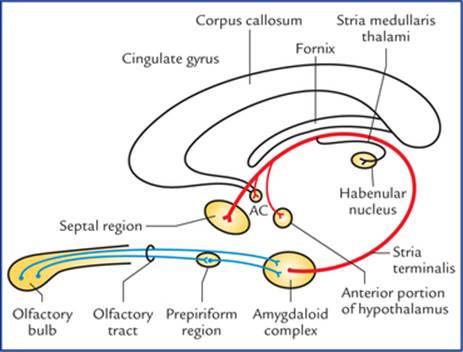
FIG. 19.7 Main afferent and efferent connections of the amygdaloid complex. Note the course of the stria terminalis. (AC = anterior commissure).
شکل ۱۹.۷ اتصالات اصلی آوران و وابران کمپلکس آمیگدالوئید. به مسیر استریا ترمینالیس توجه کنید. (AC = رابط قدامی).
Efferents: Stria terminalis forms the main efferent tract of the amygdaloid body. It takes a circuitous route along with (but not functionally related to) the tail of caudate nucleus in close relation to the lateral ventricle until the level of anterior commissure, where majority of its fibres terminate in the septal area and anterior portion of the hypo-thalamus. The others join the anterior commissure and are distributed to the contralateral amygdaloid body. Some fibres run caudally to reach the habenular nucleus through stria medullaris thalami.
وابران: استریا ترمینالیس مسیر وابران اصلی جسم آمیگدالوئید را تشکیل میدهد. این مسیر به همراه (اما نه از نظر عملکردی مرتبط با) دم هسته دمی در ارتباط نزدیک با بطن جانبی تا سطح رابط قدامی، مسیری دایرهای را طی میکند، جایی که اکثر فیبرهای آن در ناحیه سپتوم و قسمت قدامی هیپوتالاموس خاتمه مییابند. بقیه فیبرها به رابط قدامی میپیوندند و در جسم آمیگدالوئید طرف مقابل توزیع میشوند. برخی از فیبرها به صورت دمی امتداد مییابند تا از طریق استریا مدولا تالامی به هسته هابنولار برسند.
In general the amygdaloid body plays an important role in controlling the somatic responses to internal needs, drives or instincts. Since part of it receives olfactory input, it is believed that the amygdaloid body plays an important role in smell-mediated sexual behaviour.
به طور کلی جسم آمیگدالوئید نقش مهمی در کنترل پاسخهای سوماتیک به نیازهای داخلی، سائقها یا غرایز ایفا میکند. از آنجایی که بخشی از آن ورودی بویایی را دریافت میکند، اعتقاد بر این است که جسم آمیگدال نقش مهمی در رفتار جنسی ناشی از بو ایفا میکند.
Stimulation of amygdaloid body produces excitability, fear and rage. Bilateral damage of amygdaloid body reduces fear and increases sexual activity.
تحریک جسم آمیگدال باعث تحریکپذیری، ترس و خشم میشود. آسیب دو طرفه جسم آمیگدال ترس را کاهش و فعالیت جنسی را افزایش میدهد.
N.B. People in late sixties become pervasive in their sexual behaviour, probably due to atrophy of amygdaloid bodies.
توجه: افراد در اواخر دهه شصت زندگی، احتمالاً به دلیل تحلیل رفتن اجسام آمیگدال، در رفتار جنسی خود بسیار حساس میشوند.
Septal Region
The septal region is on the medial aspect of the frontal lobe beneath the genu and rostrum of corpus callosum and in front of the lamina terminalis. The septal region includes paraterminal and parolfactory gyri. The cerebral cortex in this region is called septal area.
ناحیه سپتال
ناحیه سپتال در قسمت داخلی لوب پیشانی، زیر زانو و روستروم جسم پینهای و در جلوی لایه انتهایی مغز قرار دارد. ناحیه سپتال شامل شکنجهای پاراترمینال و پارولفکتوری است. قشر مغز در این ناحیه، ناحیه سپتال نامیده میشود.
The septal area has been shown to be a pleasure zone of brain in rats.
نشان داده شده است که ناحیه سپتال، ناحیه لذت مغز در موشها است.
Hippocampal Formation
The hippocampal formation consists of: (a) hippocampus, (b) dentate gyrus, (c) subiculum, (d) indusium griseum, and (e) medial and lateral longitudinal striae.
تشکیلات هیپوکامپ
تشکیلات هیپوکامپ شامل موارد زیر است: (الف) هیپوکامپ، (ب) شکنج دندانهای، (ج) سابیکولوم، (د) ایندوسیوم گریزئوم، و (ه) استریای طولی داخلی و جانبی.
Hippocampus (also called ram’s horn or Ammon’s horn)
Hippocampus is an area of cerebral cortex which has rolled into the floor of the inferior horn of the lateral ventricle during fetal life. In adult brain it forms a longitudinal elevation in the floor of inferior horn of the lateral ventricle and is continuous medially with the subiculum and para-hippocampal gyrus.
هیپوکامپ (که شاخ قوچ یا شاخ آمون نیز نامیده میشود)
هیپوکامپ ناحیهای از قشر مغز است که در طول زندگی جنینی به کف شاخ تحتانی بطن جانبی غلتیده است. در مغز بزرگسالان، این ناحیه یک برآمدگی طولی در کف شاخ تحتانی بطن جانبی تشکیل میدهد و از سمت داخل با سابیکولوم و شکنج پاراهیپوکامپ در امتداد است.
The name ‘hippocampus’ meaning ‘sea horse’, is derived from its appearance in coronal section (Fig. 19.8).
نام «هیپوکامپ» به معنی «اسب دریایی» از ظاهر آن در مقطع کرونال گرفته شده است (شکل ۱۹.۸).
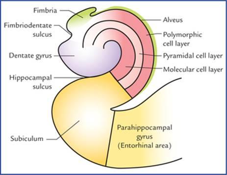
FIG. 19.8 Coronal section of the hippocampus and related structures.
شکل ۱۹.۸ بخش تاجی هیپوکامپ و ساختارهای مرتبط.
In the frontal section the hippocampus is ‘C-shaped’ and its outline bears a resemblance to a ram’s horn, hence the name ram’s horn. It is also called Ammon’s horn after an Egyptian deity with ram’s head. Its anterior extremity is expanded and bears few grooves and intervening ridges. Because of its resemblance to an animal’s paw it is termed pes hippocampi (pes = foot). Traced posteriorly the hippocampus gradually narrows and ultimately ends beneath the splenium of corpus callosum.
در بخش جلویی، هیپوکامپ «C شکل» است و طرح کلی آن شبیه شاخ قوچ است، از این رو نام شاخ قوچ را دارد. همچنین به آن شاخ آمون میگویند که برگرفته از نام یک خدای مصری با سر قوچ است. انتهای قدامی آن گسترش یافته و شیارها و برآمدگیهای کمی دارد. به دلیل شباهت آن به پنجه حیوان، pes hippocampi (pes = پا) نامیده میشود. در امتداد خلف، هیپوکامپ به تدریج باریک میشود و در نهایت در زیر اسپلنیوم جسم پینهای پایان مییابد.
The ventricular surface of hippocampus is covered by a thin layer of white fibres called alveus. The fibres of alveus originate in the hippocampal cortex, course towards the medial border of hippocampus where they converge to form a narrow strip of white matter, the fimbria of hippocampus.
سطح بطنی هیپوکامپ توسط یک لایه نازک از الیاف سفید به نام آلوئوس پوشیده شده است. فیبرهای حفره شکمی از قشر هیپوکامپ سرچشمه میگیرند و به سمت مرز داخلی هیپوکامپ حرکت میکنند، جایی که به هم میرسند و نوار باریکی از ماده سفید، فیمبریا هیپوکامپ، را تشکیل میدهند.
Phylogenetically, hippocampus represents the archicortex and consists of three layers. These are:
• Superficial molecular layer.
• Middle pyramidal cell layer.
• Deep polymorphic cell layer.
از نظر فیلوژنتیکی، هیپوکامپ نمایانگر آرکیکورتکس است و از سه لایه تشکیل شده است. این لایهها عبارتند از:
• لایه مولکولی سطحی.
• لایه سلولی هرمی میانی.
• لایه سلولی چندشکلی عمیق.
N.B. The parahippocampal cortex (neocortex) is made up of six layers. In the region known as subiculum, there is gradual transition from six-layered neocortex to the three-layered archicortex.
توجه: قشر پاراهیپوکامپ (نئوکورتکس) از شش لایه تشکیل شده است. در ناحیهای که به عنوان سوبیکولوم شناخته میشود، گذار تدریجی از نئوکورتکس شش لایه به آرکیکورتکس سه لایه وجود دارد.
Connections (Fig. 19.9)
Afferents: Hippocampus receives fibres mainly from ento-rhinal area (area 28).
اتصالات (شکل 19.9)
آورانها: هیپوکامپ عمدتاً فیبرها را از ناحیه انتورینال (ناحیه 28) دریافت میکند.
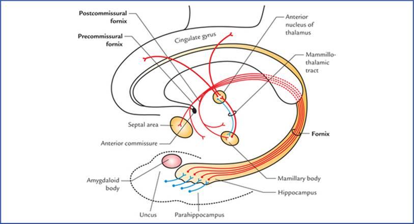
FIG. 19.9 Connections of hippocampus.
شکل ۱۹.۹ اتصالات هیپوکامپ.
Efferents: The fornix is the main efferent tract of the hippocampus.
وابرانها: فورنیکس مسیر اصلی وابران هیپوکامپ است.
The fibres leaving the hippocampus pass:
• To the opposite hippocampus through the commissure of fornix/hippocampal commissure.
• To the septal and anterior hypothalamic regions.
• To the mammillary body which sends impulses to cin-gulate gyrus through anterior nucleus of thalamus.
الیاف خروجی از هیپوکامپ از مسیرهای زیر عبور میکنند:
• از طریق رابط فورنیکس/رابطه هیپوکامپ به هیپوکامپ مقابل.
• به نواحی سپتوم و هیپوتالاموس قدامی.
• به جسم پستانی که از طریق هسته قدامی تالاموس، تکانههایی را به شکنج سینگولیت میفرستد.
Functions of hippocampus
وظایف هیپوکامپ
• Formerly it was regarded as the part of olfactory system but it has no direct connections with the sense of smell in man.
• قبلاً به عنوان بخشی از سیستم بویایی در نظر گرفته میشد، اما هیچ ارتباط مستقیمی با حس بویایی در انسان ندارد.
• In man it is an integrative centre which influences endocrine and visceral functions and emotional states through its connections with hypothalamus, septal nuclei, and the cingulate gyrus. It was once considered as the seat of soul.
• در انسان، این ناحیه یک مرکز یکپارچهسازی است که از طریق ارتباط با هیپوتالاموس، هستههای سپتوم و شکنج کمربندی، بر عملکردهای غدد درونریز و احشایی و حالات عاطفی تأثیر میگذارد. زمانی به عنوان جایگاه روح در نظر گرفته میشد.
• It plays an important role in recent memory.
• نقش مهمی در حافظه اخیر ایفا میکند.
Clinical Correlation
• The hippocampus categorizes the afferent information related to recent memory and forms the new concepts. Then it correlates the new concepts learned for the first time with the pre-existing memory. The conceived facts then become stored in the cerebral cortex as memory. Thus if the hippocampus is damaged or is in a state of shock, the new memory is not formed. As a result the patient cannot tell about, the happenings at the time of accident (loss of recent memory/amnesia) but remembers the past happenings for the old memory is already stored in different parts of the cerebral cortex.
همبستگی بالینی
• هیپوکامپ اطلاعات آوران مربوط به حافظه اخیر را دستهبندی میکند و مفاهیم جدید را شکل میدهد. سپس مفاهیم جدیدی را که برای اولین بار آموخته شده است با حافظه از پیش موجود مرتبط میکند. سپس حقایق درک شده در قشر مغز به عنوان حافظه ذخیره میشوند. بنابراین اگر هیپوکامپ آسیب ببیند یا در حالت شوک باشد، حافظه جدید شکل نمیگیرد. در نتیجه بیمار نمیتواند در مورد اتفاقات زمان حادثه (از دست دادن حافظه اخیر/فراموشی) صحبت کند، اما اتفاقات گذشته را به یاد میآورد زیرا حافظه قدیمی از قبل در قسمتهای مختلف قشر مغز ذخیره شده است.
Emotionally charged memories are affected more than the non-emotional ones.
خاطرات دارای بار احساسی بیشتر از خاطرات غیر احساسی تحت تأثیر قرار میگیرند.
• The hippocampus is the most epileptogenic part of the cerebral hemisphere. Its lesions may cause psychomotor epilepsy.
• هیپوکامپ صرعزاترین قسمت نیمکره مغز است. ضایعات آن ممکن است باعث صرع روانی-حرکتی شود.
Dentate gyrus, indusium griseum, and medial and lateral longitudinal striae
In the fetal brain, the dentate gyrus develops as a further extension of the hippocampus and occupies the interval between the hippocampus and the parahippocampal gyri, lying deep to fimbria. It has a three-layered archicortex. Its surface is toothed hence the name dentate gyrus. When traced anteriorly the dentate gyrus runs medially across the inferior surface of uncus. This part is called tail of dentate gyrus. The posterior end of dentate gyrus is continuous with the splenial gyrus or gyrus fasciolaris, which continues as thin layer of grey matter over the corpus cal-losum called indusium griseum.
شکنج دندانهدار، ایندوسیوم گریزئوم و نوارهای طولی داخلی و خارجی
در مغز جنین، شکنج دندانهدار به عنوان امتداد بیشتر هیپوکامپ توسعه مییابد و فاصله بین هیپوکامپ و شکنجهای پاراهیپوکامپ را اشغال میکند و در عمق فیمبریا قرار دارد. این شکنج دارای یک آرکیکورتکس سه لایه است. سطح آن دندانهدار است، از این رو شکنج دندانهدار نامیده میشود. وقتی از جلو ردیابی شود، شکنج دندانهدار به صورت داخلی در سراسر سطح تحتانی آنکوس امتداد مییابد. این قسمت دم شکنج دندانهدار نامیده میشود. انتهای خلفی شکنج دندانهدار با شکنج طحالی یا شکنج فاسیولاریس ادامه مییابد که به صورت لایه نازکی از ماده خاکستری روی جسم پینهای به نام ایندوسیوم گریزئوم ادامه مییابد.
The indusium griseum is the vestigial grey matter and contains two delicate longitudinal bands of fibres buried in it, the medial and lateral longitudinal striae
ایندوسیوم گریزئوم، ماده خاکستری باقیمانده است و شامل دو نوار طولی ظریف از الیاف است که در آن قرار دارند، به نامهای نوارهای طولی داخلی و خارجی.
Subiculum
It is a transition zone between three-layered archicortex and six-layered neocortex. It receives input from hippocampus and projects through the fornix to the mammillary nuclei and anterior nucleus of the thalamus.
سابیکولوم
این ناحیه، ناحیهای انتقالی بین آرکیکورتکس سه لایه و نئوکورتکس شش لایه است. این ناحیه، اطلاعات را از هیپوکامپ دریافت میکند و از طریق فورنیکس به هستههای پستانی و هسته قدامی تالاموس ارسال میکند.
Fibre bundles of limbic system
• Fornix.
• Mammillothalamic tract.
• Stria medullaris thalami.
• Stria terminalis.
• Medial forebrain bundle.
• Anterior commissure.
• Cingulum.
• Diagonal band (of Broca).
دستههای فیبری سیستم لیمبیک
• فورنیکس.
• مسیر مامیلوتالامیک.
• استریا مدولاریس تالامی.
• استریا ترمینالیس.
• دسته مغز قدامی داخلی.
• رابط قدامی.
• سینگولوم.
• نوار مورب (بروکا).
Several of these components of limbic system are already discussed with olfactory system and diencephalon. The remaining ones are described here.
چندین مورد از این اجزای سیستم لیمبیک قبلاً در مورد سیستم بویایی و مغز میانی مورد بحث قرار گرفتهاند. موارد باقیمانده در اینجا شرح داده شدهاند.
Fornix (Fig. 19.10)
The fornix is a large bundle of mainly projection fibres which connects the hippocampus with the mammillary body. It constitutes the sole efferent system of the hippocampus.
فورنیکس (شکل ۱۹.۱۰)
فورنیکس دسته بزرگی از الیاف عمدتاً برآمده است که هیپوکامپ را به جسم پستانی متصل میکند. این تنها سیستم وابران هیپوکامپ را تشکیل میدهد.
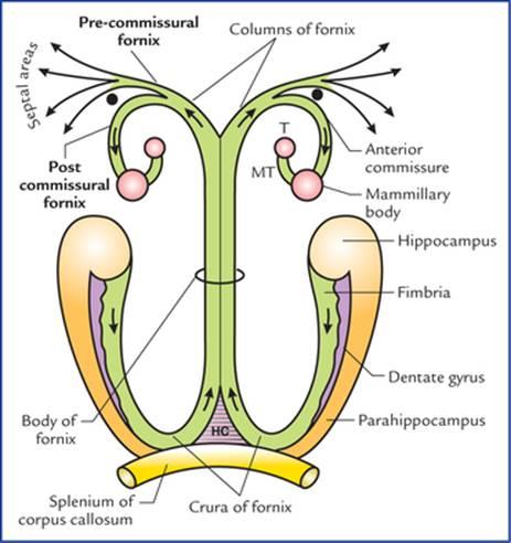
FIG. 19.10 Schematic diagram showing parts of fornix and its connections. (T = anterior nucleus of thalamus, MT = mammillothalamic tract, HC = hippocampal commissure).
شکل ۱۹.۱۰ نمودار شماتیکی که بخشهایی از فورنیکس و اتصالات آن را نشان میدهد. (T = هسته قدامی تالاموس، MT = مسیر مامیلوتالامیک، HC = رابط هیپوکامپ).
On the medial surface of cerebral hemisphere, it is seen as an arched prominent bundle of white fibres below the corpus callosum, along the lower border of septum pellucidum.
در سطح داخلی نیمکره مغزی، به صورت یک دسته برجسته قوسی شکل از الیاف سفید در زیر جسم پینهای، در امتداد مرز پایینی سپتوم پلوسیدوم دیده میشود.
There is one fornix in each cerebral hemisphere but two are so closely related/fused beneath the middle of the body of corpus callosum that they are usually described as a single structure.
در هر نیمکره مغزی یک فورنیکس وجود دارد، اما دو فورنیکس در زیر وسط جسم پینهای آنقدر به هم مرتبط/جوش خورده هستند که معمولاً به عنوان یک ساختار واحد توصیف میشوند.
Origin, course and distribution of its fibres
The fibres of fornix arise mainly from the pyramidal cells of the hippocampus and form a thin layer of white fibres on its ventricular surface called alveus.
منشأ، مسیر و توزیع فیبرهای آن
فیبرهای فورنیکس عمدتاً از سلولهای هرمی هیپوکامپ منشأ میگیرند و یک لایه نازک از فیبرهای سفید به نام آلوئوس را روی سطح بطنی آن تشکیل میدهند.
The fibres of ‘alveus’ collect on the medial margin of hippocampus to form a narrow strip of white matter, the fim-bria, lying flat over the dentate gyrus. The fimbria becomes a rounded band, the crus of fornix as it arches upwards, medially and forwards underneath the splenium of corpus callosum. The two crura, one of each hemisphere, curving over the thalamus, converge and unite in the midline beneath the trunk of corpus callosum to form the body of fornix.
فیبرهای «آلوئوس» در حاشیه داخلی هیپوکامپ جمع میشوند تا یک نوار باریک از ماده سفید، فیمبریا، را تشکیل دهند که به صورت صاف روی شکنج دندانهدار قرار دارد. فیمبریا به یک نوار گرد تبدیل میشود، ساق فورنیکس، زیرا به سمت بالا، داخل و جلو در زیر اسپلنیوم جسم پینهای قوس میگیرد. دو ساق، یکی از هر نیمکره، که روی تالاموس انحنا دارند، در خط میانی زیر تنه جسم پینهای همگرا و متحد میشوند تا جسم فورنیکس را تشکیل دهند.
Anteriorly, the body of fornix divides into two columns, the columns of fornix. Each column of fornix arches downwards towards the anterior commissure, and forms the anterior boundary of interventricular foramen. Then it curves posteriorly through the hypothalamus to end in the mammillary body. These fibres being located posterior to anterior commissure are referred to as postcommissural fornix. For some fibres of column pass in front of anterior commissure to end in the septal area and anterior hypo-thalamic region, etc. to constitute the precommissural fornix.
در جلو، جسم فورنیکس به دو ستون تقسیم میشود، ستونهای فورنیکس. هر ستون از فورنیکس به سمت پایین و به سمت رابط قدامی قوس میزند و مرز قدامی سوراخ بین بطنی را تشکیل میدهد. سپس به سمت عقب از طریق هیپوتالاموس انحنا پیدا میکند تا در جسم پستانی خاتمه یابد. این فیبرها که در خلف رابط قدامی قرار دارند، فورنیکس پسکمیسیور نامیده میشوند. برخی از فیبرهای ستون از جلوی رابط قدامی عبور میکنند تا در ناحیه سپتوم و ناحیه هیپوتالاموس قدامی و غیره خاتمه یابند و فورنیکس پیشکمیسیورال را تشکیل دهند.
N.B.
توجه:
• Hippocampal commissure of fornix. The two crura are interconnected by fibres passing from one to another. These crossing fibres interconnect the two hippocampi and form the commissure of fornix/hippocampa! commissure.
• رابط هیپوکامپی فورنیکس. دو ساقه توسط فیبرهایی که از یکی به دیگری عبور میکنند به هم متصل هستند. این فیبرهای متقاطع، دو هیپوکامپ را به هم متصل کرده و رابط فورنیکس/هیپوکامپ را تشکیل میدهند.
• Some fibres of fornix pass above the splenium of corpus callosum to end in the cingulate gyrus of the same side and constitute the dorsal fornix made up of association fibres.
• برخی از فیبرهای فورنیکس از بالای اسپلنیوم جسم پینهای عبور میکنند تا در شکنج سینگولیت همان طرف خاتمه یابند و فورنیکس پشتی را که از فیبرهای ارتباطی تشکیل شده است، تشکیل میدهند.
• Most of the fibres of fornix is made up of projection fibres connecting hippocampus with the mamillary body
• بیشتر فیبرهای فورنیکس از فیبرهای برآمدگی تشکیل شدهاند که هیپوکامپ را به جسم پستانی متصل میکنند.
Thus, fornix consists of three types of white fibres: (a) projection fibres, (b) commissural fibres, and association fibres.
بنابراین، فورنیکس از سه نوع فیبر سفید تشکیل شده است: (الف) فیبرهای برآمدگی، (ب) فیبرهای ارتباطی و فیبرهای ارتباطی.
Clinical Correlation
Bilateral transection of the fornix may cause a clinical condition called ‘acute amnestic syndrome’ in which an individual is unable to consolidate his short-term memory into long-term memory
همبستگی بالینی
قطع دو طرفه فورنیکس ممکن است باعث ایجاد یک وضعیت بالینی به نام “سندرم فراموشی حاد” شود که در آن فرد قادر به ادغام حافظه کوتاه مدت خود در حافظه بلند مدت نیست.
Mammillothalamic tract (also called bundle of Vicq d’Azyr)
Mammillothalamic tract is a prominent bundle of fibres which carry impulses from mammillary body to the anterior nucleus of thalamus. It also includes some thalamomammary fibres. Mammillothalamic tract is readily demonstrable by gross dissection. The efferents from anterior nucleus of thala-mus are projected mainly to areas 23 and 24 of the cingulate gyrus but some fibres are also shunted to the tegmental nuclei of the midbrain through mammillo-tegmental tract.
راه مامیلوتالامیک (همچنین به عنوان دسته ویک دازیر شناخته میشود)
راه مامیلوتالامیک یک دسته برجسته از فیبرها است که تکانهها را از جسم مامیلاری به هسته قدامی تالاموس منتقل میکند. همچنین شامل برخی از فیبرهای تالاموماماری است. راه مامیلوتالامیک به راحتی با تشریح کلی قابل مشاهده است. اعصاب وابران از هسته قدامی تالاموس عمدتاً به نواحی 23 و 24 شکنج سینگولیت منتقل میشوند، اما برخی از فیبرها نیز از طریق راه مامیلوتالامنتال به هستههای تگمنتوم مغز میانی منتقل میشوند.
Papez Circuit
The Papez circuit includes the following limbic structure (Flowchart 19.1):
مدار پاپز
مدار پاپز شامل ساختار لیمبیک زیر است (فلوچارت 19.1):
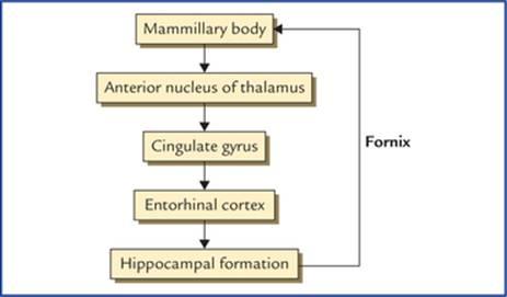
FLOWCHART 19.1 Papez circuit.
1. Hippocampal formation.
2. Mammillary body/nucleus.
3. Anterior nucleus of thalamus.
4. Cingulate gyrus.
5. Entorhinal cortex.
فلوچارت ۱۹.۱ مدار پاپز.
۱. تشکیلات هیپوکامپ.
۲. جسم/هسته پستانی.
۳. هسته قدامی تالاموس.
۴. شکنج سینگولیت.
۵. قشر انتورینال.
The route followed by the circuit of Papez is as under: Hippocampal formation to mammillary nucleus to cingu-lated gyrus to entorhinal cortex to hippocampal formation.
مسیری که مدار پاپز طی میکند به شرح زیر است: تشکیل هیپوکامپ به هسته پستانی، سپس شکنج سینگولیت، سپس قشر انتورینال و در نهایت به تشکیل هیپوکامپ.
The structures included in the Papez circuit and their connections constitute a harmonious mechanism which elaborate central emotion and emotional expressions.
ساختارهای موجود در مدار پاپز و ارتباطات آنها، مکانیسم هماهنگی را تشکیل میدهند که احساسات مرکزی و ابراز احساسات را شکل میدهند.
Clinical Problems
مشکلات بالینی
1. A light slapping or tickling of person’s face or splashing water on it, is a common and effective technique for arousing him from sleep. Similarly cotton-wisp soaked in liquid ammonia is put near the nostrils to arouse an unconscious patient. Why?
۱. سیلی زدن یا قلقلک دادن آرام صورت فرد یا پاشیدن آب روی آن، یک تکنیک رایج و مؤثر برای بیدار کردن او از خواب است. به طور مشابه، یک تکه پنبه آغشته به آمونیاک مایع در نزدیکی سوراخهای بینی قرار داده میشود تا بیمار بیهوش را بیدار کند. چرا؟
2. What is the neuroanatomical basis of the state of consciousness?
۲. اساس نوروآناتومیکی حالت هوشیاری چیست؟
3. A 55-year-old individual met a road-traffic accident and became unconscious thereafter. He was taken to the hospital where he regained consciousness. His CT scan of head did not reveal any brain lesion. When he was enquired—where and how accident took place he could not answer but he could tell the address of his residence and place of work. Provide the anatomical basis.
۳. فردی ۵۵ ساله دچار تصادف رانندگی شد و پس از آن بیهوش شد. او را به بیمارستان منتقل کردند و در آنجا به هوش آمد. سیتیاسکن سر او هیچ ضایعه مغزی را نشان نداد. وقتی از او پرسیدند – کجا و چگونه تصادف رخ داده است – نتوانست پاسخ دهد، اما میتوانست آدرس محل سکونت و محل کار خود را بگوید. اساس آناتومیکی را ارائه دهید.
4. What is the anatomical basis of schizophrenia? Discuss its presenting features.
۴. اساس آناتومیکی اسکیزوفرنی چیست؟ ویژگیهای ظاهری آن را مورد بحث قرار دهید.
5. What is Kiuver-Bucy syndrome?
۵. سندرم کیوور-بوسی چیست؟
Clinical Problem Solving
حل مسئله بالینی
1. This is because these activities stimulate the trigeminal nerve on the face and in the nasal mucosa respectively, which in turn stimulates the reticular activating system (RAS).
1. این به این دلیل است که این فعالیتها به ترتیب عصب سه قلو را در صورت و در مخاط بینی تحریک میکنند که به نوبه خود سیستم فعال کننده مشبک (RAS) را تحریک میکند.
2. The state of consciousness means that the patient should be oriented to time, place, and person. Further, he should be able to appropriately respond to questions and environmental stimuli.
2. وضعیت هوشیاری به این معنی است که بیمار باید نسبت به زمان، مکان و شخص آگاه باشد. علاوه بر این، او باید بتواند به طور مناسب به سوالات و محرکهای محیطی پاسخ دهد.
The consciousness has two facets, namely arousal and awareness, which depend on two brain structures: (a) the brainstem reticular activating system (RAS), and (b) the cerebral cortex.
هوشیاری دو جنبه دارد، یعنی برانگیختگی و آگاهی، که به دو ساختار مغزی بستگی دارند: (الف) سیستم فعال کننده مشبک ساقه مغز (RAS) و (ب) قشر مغز.
The arousal is the phenomenon of being awake, and it is primary function of the RAS, a nonspecific transmission system for sensory inputs which activate the cerebral cortex. The awareness is more sophisticated function requiring intact cortical activity in order to interpret the sensory input and respond accordingly.
برانگیختگی پدیده بیدار بودن است و عملکرد اصلی RAS، یک سیستم انتقال غیر اختصاصی برای ورودیهای حسی است که قشر مغز را فعال میکند. آگاهی عملکرد پیچیدهتری است که به فعالیت قشری سالم نیاز دارد تا ورودی حسی را تفسیر کرده و بر اساس آن پاسخ دهد.
3. The reticular activating system (RAS) is very sensitive part of the brain, as a result, even a blow on head can stop its functioning for sometime leading to unconsciousness. The loss of recent memory occurred due to involvement of hippocampus.
3. سیستم فعالکننده مشبک (RAS) بخش بسیار حساسی از مغز است، در نتیجه، حتی ضربه به سر میتواند عملکرد آن را برای مدتی متوقف کند و منجر به بیهوشی شود. از دست دادن حافظه اخیر به دلیل درگیری هیپوکامپ رخ میدهد.
4. The schizophrenia is a mental disorder which occurs due to involvement of limbic system. It is characterized by: (a) chronically disordered thinking, (b) blunting of emotional responses, (c) depression and anxiety, and (d) amnesias and phobias.
4. اسکیزوفرنی یک اختلال روانی است که به دلیل درگیری سیستم لیمبیک رخ میدهد. این بیماری با موارد زیر مشخص میشود: (الف) تفکر مزمن مختل، (ب) کند شدن پاسخهای عاطفی، (ج) افسردگی و اضطراب، و (د) فراموشی و فوبیا.
5. The Kiuver-Bucy syndrome consists of number of signs and symptoms in monkeys following removal of both temporal lobes, viz. (a) docility, (b) loss of fear and anger, (c) increased appetite, and (d) increased sexual activity which is often perverse.
5. سندرم کیوور-بوسی شامل تعدادی از علائم و نشانهها در میمونها پس از برداشتن هر دو لوب گیجگاهی است، یعنی. (الف) مطیع بودن، (ب) از دست دادن ترس و خشم، (ج) افزایش اشتها، و (د) افزایش فعالیت جنسی که اغلب انحرافی است.
N.B. This syndrome has also been described in humans following removal of large areas of temporal lobe on both the sides.
توجه: این سندرم همچنین در انسان پس از برداشتن نواحی بزرگی از لوب گیجگاهی در هر دو طرف توصیف شده است.
Textbook of Clinical Neuroanatomy, 2 ed
Chapter 1. Development of the Nervous System
Chapter 2. Organization and Functions of the Nervous System
Chapter 3. Peripheral Nerves and Ganglia
Chapter 4. Receptors and Effectors
Chapter 5. Dermatomes and Muscular Activity
Chapter 6. Central Nervous System: an Overview
Chapter 7. Spinal Cord
Chapter 8. Brainstem
Chapter 9. Nuclei, Functional Components and Distribution of Cranial Nerves
Chapter 10. Cerebellum and Fourth Ventricle
Chapter 11. Diencephalon and Third Ventricle
Chapter 12. Cerebrum
Chapter 13. Basal Nuclei (Basal Ganglia)
Chapter 14. White Matter of the Cerebrum and Lateral Ventricles
Chapter 15. Blood Supply of the Brain
Chapter 16. Meninges And Cerebrospinal Fluid
Chapter 17. Somatic Motor and Sensory Pathways
Chapter 18. Special Senses and their Neural Pathways
Chapter 19. Reticular Formation and Limbic System
Chapter 20. Autonomic Nervous System