
فیزیولوژی پزشکی گایتون و هال؛ تعادلهای غذایی؛ تنظیم تغذیه؛ چاقی و بیغذایی؛ ویتامینها و مواد معدنی


دعای مطالعه [ نمایش ]
بِسْمِ الله الرَّحْمنِ الرَّحیمِ
اَللّهُمَّ اَخْرِجْنى مِنْ ظُلُماتِ الْوَهْمِ
خدايا مرا بيرون آور از تاريكىهاى وهم،
وَ اَكْرِمْنى بِنُورِ الْفَهْمِ
و به نور فهم گرامى ام بدار،
اَللّهُمَّ افْتَحْ عَلَيْنا اَبْوابَ رَحْمَتِكَ
خدايا درهاى رحمتت را به روى ما بگشا،
وَانْشُرْ عَلَيْنا خَزائِنَ عُلُومِكَ بِرَحْمَتِكَ يا اَرْحَمَ الرّاحِمينَ
و خزانههاى علومت را بر ما باز كن به امید رحمتت اى مهربانترين مهربانان.
کتاب «فیزیولوژی پزشکی گایتون و هال» بهعنوان یکی از جامعترین و معتبرترین منابع در حوزه علوم پزشکی، همچنان مرجع کلیدی برای درک عملکرد پیچیده بدن انسان است. این اثر با تکیه بر تازهترین پژوهشها و توضیحات دقیق از سازوکارهای فیزیولوژیک، پلی میان علوم پایه پزشکی و کاربردهای بالینی ایجاد میکند و نقشی بیبدیل در آموزش، پژوهش و ارتقای دانش سلامت ایفا مینماید.
ترجمه دقیق و علمی این شاهکار توسط برند علمی آیندهنگاران مغز به مدیریت داریوش طاهری، دسترسی فارسیزبانان به مرزهای نوین دانش فیزیولوژی را ممکن ساخته و رسالتی علمی برای ارتقای آموزش پزشکی، فهم عمیقتر سازوکارهای بدن و توسعه روشهای نوین در حوزه سلامت فراهم آورده است.
» کتاب فیزیولوژی پزشکی گایتون و هال
» » فصل ۷۲: تعادلهای غذایی؛ تنظیم تغذیه؛ چاقی و بیغذایی؛ ویتامینها و مواد معدنی
در حال ویرایش
» Guyton and Hall Textbook of Medical Physiology
»» CHAPTER 72: Dietary Balances; Regulation of Feeding; Obesity and Starvation; Vitamins and Minerals

ENERGY INTAKE AND OUTPUT BALANCED UNDER STEADY-STATE CONDITIONS
Proteins, carbohydrates, and fats in the diet provide energy for various body functions or for storage and later use. Stability of body weight and composition over long periods requires that a person’s energy intake and energy expenditure be balanced. When a person is overfed, and energy intake persistently exceeds expenditure, most of the excess energy is stored as fat, and body weight increases; conversely, loss of body mass and starvation occur when energy intake is insufficient to meet the body’s metabolic needs.
انرژی دریافتی و خروجی متعادل تحت شرایط پایدار
پروتئین ها، کربوهیدرات ها و چربی های موجود در رژیم غذایی، انرژی را برای عملکردهای مختلف بدن یا برای ذخیره سازی و استفاده بعدی فراهم می کند. ثبات وزن و ترکیب بدن در دوره های طولانی مستلزم آن است که انرژی دریافتی و مصرف انرژی فرد متعادل باشد. هنگامی که فرد بیش از حد سیر می شود و انرژی دریافتی دائماً بیش از مصرف می شود، بیشتر انرژی اضافی به صورت چربی ذخیره می شود و وزن بدن افزایش می یابد. برعکس، از دست دادن توده بدن و گرسنگی زمانی رخ می دهد که انرژی دریافتی برای تامین نیازهای متابولیک بدن ناکافی باشد.
Because different foods contain different proportions of proteins, carbohydrates, fats, minerals, and vitamins, appropriate balances must also be maintained among these constituents so that all of the body’s metabolic systems can be supplied with the requisite materials. This chapter discusses the mechanisms by which food intake is regulated in accordance with the body’s metabolic needs and some of the problems of maintaining balance among the different types of foods.
از آنجایی که غذاهای مختلف حاوی نسبت های متفاوتی از پروتئین ها، کربوهیدرات ها، چربی ها، مواد معدنی و ویتامین ها هستند، باید تعادل مناسبی نیز بین این ترکیبات حفظ شود تا تمام سیستم های متابولیک بدن بتوانند با مواد مورد نیاز تامین شوند. این فصل مکانیسمهایی را مورد بحث قرار میدهد که توسط آنها مصرف غذا مطابق با نیازهای متابولیک بدن تنظیم میشود و برخی از مشکلات حفظ تعادل بین انواع مختلف غذاها.
Dietary Balances
Energy Available in Foods
The energy liberated from each gram of carbohydrate as it is oxidized to carbon dioxide and water is 4.1 Calories (1 Calorie equals 1 kilocalorie), and that liberated from fat is 9.3 Calories. The energy liberated from metabolism of the average dietary protein as each gram is oxidized to carbon dioxide, water, and urea is 4.35 Calories. Also, these substances vary in the average percentages that are absorbed from the gastrointestinal tract: about 98% of carbohydrate, 95% of fat, and 92% of protein. Therefore, the average physiologically available energy in each gram of these three foodstuffs is as follows:
تعادل های غذایی
انرژی موجود در غذاها
انرژی آزاد شده از هر گرم کربوهیدرات در اثر اکسید شدن به دی اکسید کربن و آب 4.1 کالری است (1 کالری برابر با 1 کیلو کالری) و انرژی آزاد شده از چربی 9.3 کالری است. انرژی آزاد شده از متابولیسم پروتئین متوسط رژیم غذایی با اکسید شدن هر گرم به دی اکسید کربن، آب و اوره 4.35 کالری است. همچنین، این مواد در میانگین درصدهایی که از دستگاه گوارش جذب میشوند متفاوت هستند: حدود 98 درصد کربوهیدرات، 95 درصد چربی و 92 درصد پروتئین. بنابراین، میانگین انرژی فیزیولوژیکی موجود در هر گرم از این سه ماده غذایی به شرح زیر است:

Although considerable variation exists among different individuals, and even in the same person from day to day, the usual diet of Americans provides about 15% of the total energy intake from protein, 40% from fat, and 45% from carbohydrate. In most non-Western countries, the quantity of energy derived from carbohydrates far exceeds that de- rived from proteins and fats. Indeed, in some parts of the world where meat is scarce, the energy received from fats and proteins combined may be no greater than 15% to 20%.
اگرچه تنوع قابل توجهی در بین افراد مختلف و حتی در یک فرد از روز به روز وجود دارد، رژیم غذایی معمول آمریکایی ها حدود 15٪ از کل انرژی دریافتی را از پروتئین، 40٪ از چربی و 45٪ از کربوهیدرات تامین می کند. در اکثر کشورهای غیر غربی، مقدار انرژی حاصل از کربوهیدرات ها بسیار بیشتر از انرژی حاصل از پروتئین ها و چربی ها است. در واقع، در برخی از نقاط جهان که گوشت کمیاب است، انرژی دریافتی از ترکیب چربی ها و پروتئین ها ممکن است بیش از 15 تا 20 درصد نباشد.
Table 72-1 lists the compositions of selected foods, especially demonstrating the high proportions of fat and protein in meat products and the high proportion of carbohydrate in most vegetable and grain products. Fat is deceptive in the diet because it usually exists as nearly 100% fat, whereas proteins and carbohydrates are mixed in watery media, so that each of these normally represents less than 25% of the total weight. Therefore, the fat of one pat of butter mixed with an entire helping of potato sometimes contains as much energy as the potato itself.
جدول 72-1 ترکیبات غذاهای منتخب را فهرست می کند، به ویژه نسبت بالای چربی و پروتئین در محصولات گوشتی و نسبت بالای کربوهیدرات در اکثر محصولات سبزیجات و غلات را نشان می دهد. چربی در رژیم غذایی فریبنده است، زیرا معمولاً به صورت تقریباً 100٪ چربی وجود دارد، در حالی که پروتئین ها و کربوهیدرات ها در محیط های آبکی مخلوط می شوند، به طوری که هر یک از اینها معمولاً کمتر از 25٪ وزن کل را تشکیل می دهند. بنابراین، چربی یک تکه کره مخلوط شده با کل سیب زمینی گاهی به اندازه خود سیب زمینی انرژی دارد.
The Average Daily Requirement for Protein Is 30 to 50 Grams. Twenty to 30 grams of the body proteins are degraded daily and used to produce other body chemicals. Therefore, all cells must continue to form new proteins to take the place of those that are being destroyed, and a supply of protein is necessary in the diet for this purpose. An average person can maintain normal stores of protein if the daily intake is greater than 30 to 50 grams.
متوسط نیاز روزانه به پروتئین 30 تا 50 گرم است. روزانه 20 تا 30 گرم از پروتئین های بدن تجزیه می شود و برای تولید سایر مواد شیمیایی بدن استفاده می شود. بنابراین، همه سلولها باید به تشکیل پروتئینهای جدید ادامه دهند تا جایگزین پروتئینهایی شوند که در حال نابودی هستند، و برای این منظور تأمین پروتئین در رژیم غذایی ضروری است. یک فرد متوسط می تواند ذخایر طبیعی پروتئین را در صورتی که میزان مصرف روزانه آن بیش از 30 تا 50 گرم باشد حفظ کند.
Some proteins have inadequate quantities of certain essential amino acids and therefore cannot be used to replace the degraded proteins. Such proteins are called partial proteins, and when they are present in large quantities in the diet, the daily protein requirement is much greater than normal. In general, proteins derived from animal foodstuffs are more complete than are proteins derived from vegetable and grain sources. For example, the protein of corn has inadequate amounts of tryptophan and lysine, two of the essential amino acids. Therefore, individuals who consume cornmeal as their principal source of protein sometimes develop the protein-deficiency syndrome called kwashiorkor, which consists of failure to grow, lethargy, depressed mentality, and edema caused by low plasma protein concentration. On the other hand, food legumes, such as chick peas and beans, provide a relatively rich source of tryptophan and lysine but contain inadequate amounts of methionine, another essential amino acid. Therefore the 877 proteins of corn and legumes complement each other and together provide all of the essential amino acids in the diet.
برخی از پروتئین ها دارای مقادیر ناکافی اسیدهای آمینه ضروری هستند و بنابراین نمی توان از آنها برای جایگزینی پروتئین های تخریب شده استفاده کرد. به چنین پروتئین هایی، پروتئین های جزئی می گویند و زمانی که به مقدار زیاد در جیره وجود داشته باشند، نیاز روزانه به پروتئین بسیار بیشتر از حد معمول است. به طور کلی، پروتئین های مشتق شده از مواد غذایی حیوانی کامل تر از پروتئین های مشتق شده از منابع گیاهی و غلات هستند. به عنوان مثال، پروتئین ذرت دارای مقادیر ناکافی تریپتوفان و لیزین، دو اسید آمینه ضروری است. بنابراین، افرادی که آرد ذرت را به عنوان منبع اصلی پروتئین مصرف میکنند، گاهی دچار سندرم کمبود پروتئین به نام کواشیورکور میشوند که شامل عدم رشد، بیحالی، ذهنیت افسرده و ادم ناشی از غلظت کم پروتئین پلاسما است. از سوی دیگر، حبوبات غذایی، مانند نخود و لوبیا، منبع نسبتاً غنی تریپتوفان و لیزین هستند، اما حاوی مقادیر ناکافی متیونین، یکی دیگر از اسیدهای آمینه ضروری هستند. بنابراین 877 پروتئین ذرت و حبوبات مکمل یکدیگر هستند و با هم تمام اسیدهای آمینه ضروری در رژیم غذایی را تامین می کنند.
Table 72-1 Protein, Fat, and Carbohydrate Content of Different Foods
جدول 72-1 محتوای پروتئین، چربی و کربوهیدرات در غذاهای مختلف

Carbohydrates and Fats Act as “Protein Sparers.” When a person’s diet contains an abundance of carbohydrates and fats, almost all the body’s energy is derived from these two substances, and little is derived from proteins. Therefore, carbohydrates and fats are said to be protein sparers. Conversely, in the state of starvation, after the carbohydrates and fats have been depleted, the body’s protein stores are consumed rapidly for energy, sometimes at rates approaching several hundred grams per day rather than the normal daily rate of 30 to 50 grams.
کربوهیدرات ها و چربی ها به عنوان “پرهیز کننده پروتئین” عمل می کنند. وقتی رژیم غذایی فرد حاوی کربوهیدرات ها و چربی های فراوان باشد، تقریبا تمام انرژی بدن از این دو ماده و مقدار کمی از پروتئین ها تامین می شود. بنابراین گفته می شود که کربوهیدرات ها و چربی ها ذخیره کننده پروتئین هستند. برعکس، در حالت گرسنگی، پس از اتمام کربوهیدراتها و چربیها، ذخایر پروتئین بدن به سرعت برای انرژی مصرف میشود، گاهی اوقات با نرخهای نزدیک به چند صد گرم در روز به جای نرخ معمول روزانه 30 تا 50 گرم.
Methods for Determining Metabolic Utilization of Carbohydrates, Fats, and Proteins
“Respiratory Quotient,” the Ratio of Carbon Dioxide Production to Oxygen Utilization, Can Be Used to Estimate Fat and Carbohydrate Utilization. When carbohydrates are metabolized with oxygen, exactly one carbon dioxide molecule is formed for each molecule of oxygen consumed. This ratio of carbon dioxide output to oxygen usage is called the respiratory quotient, so the respiratory quotient for carbohydrates is 1.0.
روشهای تعیین میزان مصرف متابولیک کربوهیدراتها، چربیها و پروتئینها
“ضریب تنفسی”، نسبت تولید دی اکسید کربن به استفاده از اکسیژن، می تواند برای تخمین مصرف چربی و کربوهیدرات استفاده شود. هنگامی که کربوهیدرات ها با اکسیژن متابولیزه می شوند، دقیقاً یک مولکول دی اکسید کربن به ازای هر مولکول اکسیژن مصرفی تشکیل می شود. این نسبت دی اکسید کربن خروجی به مصرف اکسیژن، ضریب تنفسی نامیده می شود، بنابراین ضریب تنفسی برای کربوهیدرات ها 1.0 است.
When fat is oxidized in the body’s cells, an average of 70 carbon dioxide molecules are formed for each 100 molecules of oxygen consumed. The respiratory quotient for the metabolism of fat therefore averages 0.70. When proteins are oxidized by the cells, the average respiratory quotient is 0.80. The reason that the respiratory quotients for fats and proteins are lower than those for carbohydrates is that a portion of the oxygen metabolized with these foods is required to combine with the excess hydrogen atoms present in their molecules, so less carbon dioxide is formed in relation to the oxygen used.
هنگامی که چربی در سلول های بدن اکسید می شود، به ازای هر 100 مولکول اکسیژن مصرفی، به طور متوسط 70 مولکول دی اکسید کربن تشکیل می شود. بنابراین ضریب تنفسی برای متابولیسم چربی به طور متوسط 0.70 است. هنگامی که پروتئین ها توسط سلول ها اکسید می شوند، میانگین ضریب تنفسی 0.80 است. دلیل اینکه ضریب تنفسی چربی ها و پروتئین ها کمتر از کربوهیدرات ها است این است که بخشی از اکسیژن متابولیزه شده با این غذاها برای ترکیب با اتم های هیدروژن اضافی موجود در مولکول های آنها لازم است، بنابراین دی اکسید کربن کمتری نسبت به اکسیژن مصرفی تشکیل می شود.
Now let us see how one can use the respiratory quotient to determine the relative utilization of different foods by the body. First, recall from Chapter 40 that the output of carbon dioxide by the lungs divided by the uptake of oxygen during the same period is called the respiratory exchange ratio. During a period of 1 hour or more, the respiratory ex- change ratio exactly equals the average respiratory quotient of the metabolic reactions throughout the body. If a person has a respiratory quotient of 1.0, he or she is metabolizing carbohydrates almost exclusively, because the respiratory quotients for both fat and protein metabolism are consider- ably less than 1.0. Likewise, when the respiratory quotient is about 0.70, the body is metabolizing mostly fats, to the exclusion of carbohydrates and proteins. And, finally, if we ignore the normally small amount of protein metabolism, respiratory quotients between 0.70 and 1.0 describe the approximate ratios of carbohydrate to fat metabolism. To be more exact, one can first determine the protein utilization by measuring nitrogen excretion, as discussed in the next section. Then, using the appropriate mathematical formula, one can calculate the utilization of the three foodstuffs.
حال بیایید ببینیم چگونه می توان از ضریب تنفسی برای تعیین میزان استفاده نسبی بدن از غذاهای مختلف استفاده کرد. ابتدا، از فصل 40 به یاد بیاورید که خروجی دی اکسید کربن توسط ریه ها تقسیم بر جذب اکسیژن در همان دوره، نسبت تبادل تنفسی نامیده می شود. در طی یک دوره ۱ ساعته یا بیشتر، نسبت تبادل تنفسی دقیقاً برابر است با میانگین ضریب تنفسی واکنشهای متابولیک در سراسر بدن. اگر فردی ضریب تنفسی 1.0 داشته باشد، تقریباً منحصراً در حال متابولیسم کربوهیدرات است، زیرا ضریب تنفسی برای متابولیسم چربی و پروتئین به طور قابل توجهی کمتر از 1 است. به همین ترتیب، زمانی که ضریب تنفسی حدود 0.70 باشد، بدن عمدتاً چربی ها را متابولیزه می کند، به استثنای کربوهیدرات ها و پروتئین ها. و در نهایت، اگر مقدار معمولی متابولیسم پروتئین را نادیده بگیریم، ضریب تنفسی بین 0.70 و 1.0 نسبت تقریبی متابولیسم کربوهیدرات به چربی را توصیف می کند. برای دقیق تر، ابتدا می توان با اندازه گیری دفع نیتروژن، مصرف پروتئین را تعیین کرد، همانطور که در بخش بعدی بحث شد. سپس با استفاده از فرمول ریاضی مناسب می توان میزان استفاده از سه ماده غذایی را محاسبه کرد.
Some of the important findings from studies of respiratory quotients are the following:
برخی از یافته های مهم حاصل از مطالعات ضریب تنفسی به شرح زیر است:
1. Immediately after a mixed meal containing carbohydrates as well as protein and fat, almost all the food that is metabolized is carbohydrates, so the respiratory quotient at that time approaches 1.0.
1. بلافاصله پس از یک وعده غذایی مخلوط حاوی کربوهیدرات ها و همچنین پروتئین و چربی، تقریباً تمام غذایی که متابولیزه می شود کربوهیدرات است، بنابراین ضریب تنفسی در آن زمان به 1.0 نزدیک می شود.
2. About 8 to 10 hours after a meal, the body has already used up most of its readily available carbohydrates, and the respiratory quotient approaches that for fat metabolism, about 0.70.
2. حدود 8 تا 10 ساعت پس از غذا، بدن بیشتر کربوهیدراتهای قابل دسترس خود را مصرف کرده است و ضریب تنفسی برای متابولیسم چربی به 0.70 نزدیک میشود.
3. In untreated diabetes mellitus, little carbohydrate can be used by the body’s cells under any conditions because insulin is required for this utilization. Therefore, when diabetes is severe, most of the time the respiratory quotient remains near that for fat metabolism, which is 0.70.
3. در دیابت شیرین درمان نشده، سلول های بدن تحت هر شرایطی می توانند از کربوهیدرات کمی استفاده کنند زیرا برای این استفاده به انسولین نیاز است. بنابراین، هنگامی که دیابت شدید است، در بیشتر مواقع ضریب تنفسی نزدیک به متابولیسم چربی است که 0.70 است.
Nitrogen Excretion Can Be Used to Assess Protein Metabolism. The average protein contains about 16% nitro- gen. During metabolism of protein, about 90% of this nitro- gen is excreted in the urine in the form of urea, uric acid, creatinine, and other nitrogen products. The remaining 10% is excreted in the feces. Therefore, the rate of protein breakdown in the body can be estimated by measuring the amount of nitrogen in the urine, then adding 10% for the nitrogen excreted in the feces, and multiplying by 6.25 (i.e., 100/16) to estimate the total amount of protein metabolism in grams per day. Thus, excretion of 8 grams of nitrogen in the urine each day means that about 55 grams of protein breakdown has occurred. If the daily intake of protein is less than the daily breakdown of protein, the person is said to have a negative nitrogen balance, which means that his or her body stores of protein are decreasing daily.
از دفع نیتروژن می توان برای ارزیابی متابولیسم پروتئین استفاده کرد. پروتئین متوسط حاوی حدود 16 درصد نیتروژن است. در طول متابولیسم پروتئین، حدود 90 درصد از این نیتروژن به شکل اوره، اسید اوریک، کراتینین و سایر محصولات نیتروژن از طریق ادرار دفع می شود. 10 درصد باقیمانده از طریق مدفوع دفع می شود. بنابراین، سرعت تجزیه پروتئین در بدن را می توان با اندازه گیری مقدار نیتروژن در ادرار، سپس اضافه کردن 10 درصد نیتروژن دفع شده در مدفوع، و ضرب در 6.25 (یعنی 100/16) برای تخمین مقدار کل متابولیسم پروتئین بر حسب گرم در روز تخمین زد. بنابراین، دفع 8 گرم نیتروژن در ادرار در هر روز به این معنی است که حدود 55 گرم تجزیه پروتئین رخ داده است. اگر مصرف روزانه پروتئین کمتر از تجزیه روزانه پروتئین باشد، گفته می شود که فرد دارای تعادل نیتروژن منفی است، به این معنی که ذخایر پروتئین بدن او روزانه کاهش می یابد.
REGULATION OF FOOD INTAKE AND ENERGY STORAGE
Stability of the body’s total mass and composition over long periods requires that energy intake match energy expenditure. As discussed in Chapter 73, only about 27% of the energy ingested normally reaches the functional systems of the cells, and much of this energy is eventually converted to heat, which is generated as a result of protein metabolism, muscle activity, and activities of the various organs and tissues of the body. Excess energy intake is stored mainly as fat, whereas a deficit of energy intake causes loss of total body mass until energy expenditure eventually equals energy intake or death occurs.
تنظیم مصرف غذا و ذخیره انرژی
پایداری کل جرم و ترکیب بدن در دوره های طولانی مستلزم آن است که انرژی دریافتی با مصرف انرژی مطابقت داشته باشد. همانطور که در فصل 73 بحث شد، تنها حدود 27 درصد از انرژی مصرف شده به طور معمول به سیستم های عملکردی سلول ها می رسد و بیشتر این انرژی در نهایت به گرما تبدیل می شود که در نتیجه متابولیسم پروتئین، فعالیت ماهیچه ها و فعالیت های اندام ها و بافت های مختلف بدن ایجاد می شود. انرژی دریافتی اضافی عمدتاً به صورت چربی ذخیره می شود، در حالی که کمبود انرژی باعث از دست دادن کل توده بدن می شود تا زمانی که مصرف انرژی در نهایت برابر با انرژی دریافتی شود یا مرگ اتفاق بیفتد.
Although there is considerable variability in the amount of energy storage (i.e., fat mass) in different individuals, maintenance of an adequate energy supply is necessary for survival. Therefore, the body is endowed with powerful physiological control systems that help maintain adequate energy intake. Deficits of energy stores, for example, rapidly activate multiple mechanisms that cause hunger and drive a person to seek food. In athletes and laborers, energy expenditure for the high level of muscle activity may be as high as 10,000 Calories per day, compared with only about 2000 Calories per day for sedentary individuals. Thus, a large energy expenditure associated with physical work usually stimulates equally large increases in caloric intake.
اگرچه تنوع قابل توجهی در میزان ذخیره انرژی (به عنوان مثال، توده چربی) در افراد مختلف وجود دارد، حفظ منبع انرژی کافی برای بقا ضروری است. بنابراین، بدن دارای سیستم های کنترل فیزیولوژیکی قدرتمندی است که به حفظ انرژی دریافتی کافی کمک می کند. به عنوان مثال، کمبود ذخایر انرژی، مکانیسم های متعددی را به سرعت فعال می کند که باعث گرسنگی می شود و فرد را به جستجوی غذا سوق می دهد. در ورزشکاران و کارگران، مصرف انرژی برای سطح بالای فعالیت عضلانی ممکن است به 10000 کالری در روز برسد، در حالی که این میزان برای افراد کم تحرک تنها حدود 2000 کالری در روز است. بنابراین، مصرف انرژی زیاد مرتبط با کار بدنی معمولاً باعث افزایش به همان اندازه زیاد در دریافت کالری می شود.
What are the physiological mechanisms that sense changes in energy balance and influence the quest for food? Maintenance of adequate energy supply in the body is so critical that multiple short-term and long-term control systems exist that regulate not only food intake but also energy expenditure and energy stores. In the next few sections we describe some of these control systems and their operation in physiological conditions, as well as in the states of obesity and starvation.
مکانیسم های فیزیولوژیکی که تغییرات در تعادل انرژی را حس می کند و بر جستجوی غذا تأثیر می گذارد چیست؟ حفظ منبع انرژی کافی در بدن به قدری حیاتی است که چندین سیستم کنترل کوتاه مدت و بلند مدت وجود دارد که نه تنها مصرف غذا بلکه مصرف انرژی و ذخیره انرژی را نیز تنظیم می کند. در چند بخش بعدی برخی از این سیستم های کنترل و عملکرد آنها در شرایط فیزیولوژیکی و همچنین در حالت های چاقی و گرسنگی را شرح می دهیم.
NEURAL CENTERS REGULATE FOOD INTAKE
The sensation of hunger is associated with a craving for food and several other physiological effects, such as rhythmic contractions of the stomach and restlessness, which cause the person to seek food. A person’s appetite is a desire for food, often of a particular type, and is useful in helping to choose the quality of the food to be eaten. If the quest for food is successful, the feeling of satiety occurs. Each of these feelings is influenced by environmental and cultural factors, as well as by physiological controls that influence specific centers of the brain, especially the hypothalamus.
مراکز عصبی مصرف غذا را تنظیم می کنند
احساس گرسنگی با میل به غذا و چندین اثر فیزیولوژیکی دیگر مانند انقباضات ریتمیک معده و بی قراری همراه است که باعث می شود فرد به دنبال غذا برود. اشتهای فرد میل به غذا است که اغلب از نوع خاصی است و برای کمک به انتخاب کیفیت غذایی که باید خورده شود مفید است. اگر جستجوی غذا موفقیت آمیز باشد، احساس سیری ایجاد می شود. هر یک از این احساسات تحت تأثیر عوامل محیطی و فرهنگی و همچنین کنترل های فیزیولوژیکی است که بر مراکز خاصی از مغز به ویژه هیپوتالاموس تأثیر می گذارد.
The Hypothalamus Contains Hunger and Satiety Centers. Several neuronal centers of the hypothalamus participate in the control of food intake. The lateral nuclei of the hypothalamus serve as a feeding center, and stimulation of this area causes an animal to eat voraciously (hyperphagia). Conversely, destruction of the lateral hypothalamus causes lack of desire for food and progressive inanition, a condition characterized by marked weight loss, muscle weakness, and decreased metabolism. The lateral hypothalamic feeding center operates by exciting the motor drives to search for food.
هیپوتالاموس حاوی مراکز گرسنگی و سیری است. چندین مرکز عصبی هیپوتالاموس در کنترل مصرف غذا مشارکت دارند. هستههای جانبی هیپوتالاموس به عنوان مرکز تغذیه عمل میکنند و تحریک این ناحیه باعث میشود حیوان با حرص غذا بخورد (هیپرفاژی). برعکس، تخریب هیپوتالاموس جانبی باعث عدم میل به غذا و بی حرکتی تدریجی می شود، وضعیتی که با کاهش وزن، ضعف عضلانی و کاهش متابولیسم مشخص می شود. مرکز تغذیه هیپوتالاموس جانبی با تحریک موتور درایوها برای جستجوی غذا عمل می کند.
The ventromedial nuclei of the hypothalamus serve as a major satiety center. This center is believed to give a sense of nutritional satisfaction that inhibits the feeding center. Electrical stimulation of this region can cause complete satiety, and even in the presence of highly appetizing food, the animal refuses to eat (aphagia). Conversely, destruction of the ventromedial nuclei causes voracious and continued eating until the animal becomes extremely obese, sometimes weighing as much as four times normal.
هسته های شکمی هیپوتالاموس به عنوان مرکز اصلی سیری عمل می کنند. اعتقاد بر این است که این مرکز باعث ایجاد حس رضایت غذایی می شود که مرکز تغذیه را مهار می کند. تحریک الکتریکی این ناحیه می تواند باعث سیری کامل شود و حتی در صورت وجود غذای بسیار اشتها آور، حیوان از خوردن امتناع می کند (آفاژی). برعکس، تخریب هستههای شکمی باعث پرخوری و ادامه غذا میشود تا زمانی که حیوان به شدت چاق شود، گاهی اوقات وزن آن به چهار برابر طبیعی میرسد.
The paraventricular, dorsomedial, and arcuate nuclei of the hypothalamus also play a major role in regulating food intake. For example, lesions of the paraventricular nuclei often cause excessive eating, whereas lesions of the dorsomedial nuclei usually depress eating behavior. As discussed later, the arcuate nuclei are the sites in the hypothalamus where multiple hormones released from the gastrointestinal tract and adipose tissue converge to regulate food intake, as well as energy expenditure.
هسته های پارا بطنی، پشتی و کمانی هیپوتالاموس نیز نقش عمده ای در تنظیم مصرف غذا دارند. به عنوان مثال، ضایعات هسته های پارا بطنی اغلب باعث خوردن بیش از حد می شوند، در حالی که ضایعات هسته های پشتی معمولاً رفتار خوردن را کاهش می دهند. همانطور که بعداً مورد بحث قرار گرفت، هستههای قوسی، مکانهایی در هیپوتالاموس هستند که در آن هورمونهای متعددی که از دستگاه گوارش و بافت چربی آزاد میشوند، برای تنظیم مصرف غذا و همچنین مصرف انرژی، همگرا میشوند.
Much chemical cross talk occurs among the neurons in the hypothalamus, and together, these centers coordinate the processes that control eating behavior and the perception of satiety. These hypothalamic nuclei also influence secretion of several hormones that are important in regulating energy balance and metabolism, including those from the thyroid and adrenal glands, as well as the pancreatic islet cells.
گفتگوهای متقابل شیمیایی زیادی در میان نورونهای هیپوتالاموس اتفاق میافتد، و با هم، این مراکز فرآیندهایی را که رفتار خوردن و ادراک سیری را کنترل میکنند، هماهنگ میکنند. این هسته های هیپوتالاموس همچنین بر ترشح چندین هورمون که در تنظیم تعادل انرژی و متابولیسم مهم هستند، از جمله هورمون های تیروئید و غدد فوق کلیوی و همچنین سلول های جزایر پانکراس تأثیر می گذارند.
The hypothalamus receives (1) neural signals from the gastrointestinal tract that provide sensory information about stomach filling; (2) chemical signals from nutrients in the blood (glucose, amino acids, and fatty acids) that signify satiety; (3) signals from gastrointestinal hormones; (4) signals from hormones released by adipose tissue; and (5) signals from the cerebral cortex (sight, smell, and taste) that influence feeding behavior. Some of these inputs to the hypothalamus are shown in Figure 72-1.
هیپوتالاموس (1) سیگنال های عصبی را از دستگاه گوارش دریافت می کند که اطلاعات حسی را در مورد پر شدن معده ارائه می دهد. (2) سیگنال های شیمیایی از مواد مغذی موجود در خون (گلوکز، اسیدهای آمینه و اسیدهای چرب) که نشان دهنده سیری هستند. (3) سیگنال های هورمون های گوارشی. (4) سیگنال های هورمون های آزاد شده توسط بافت چربی. و (5) سیگنال هایی از قشر مغز (بینایی، بویایی و چشایی) که بر رفتار تغذیه تأثیر می گذارد. برخی از این ورودی ها به هیپوتالاموس در شکل 72-1 نشان داده شده است.
The hypothalamic feeding and satiety centers have a high density of receptors for neurotransmitters and hormones that influence feeding behavior. A few of the many substances that have been shown to alter appetite and feeding behavior in experimental studies are listed in Table 72-2 and are generally categorized as (1) orexigenic substances that stimulate feeding or (2) anorexigenic substances that inhibit feeding.
مراکز تغذیه و سیری هیپوتالاموس دارای تراکم بالایی از گیرنده های انتقال دهنده های عصبی و هورمون هایی هستند که بر رفتار تغذیه تاثیر می گذارند. تعدادی از بسیاری از موادی که در مطالعات تجربی نشان داده شده است که اشتها و رفتار تغذیه را تغییر می دهند در جدول 72-2 فهرست شده اند و به طور کلی به عنوان (1) مواد بی اشتهایی که تغذیه را تحریک می کنند یا (2) مواد بی اشتهایی که تغذیه را مهار می کنند طبقه بندی می شوند.
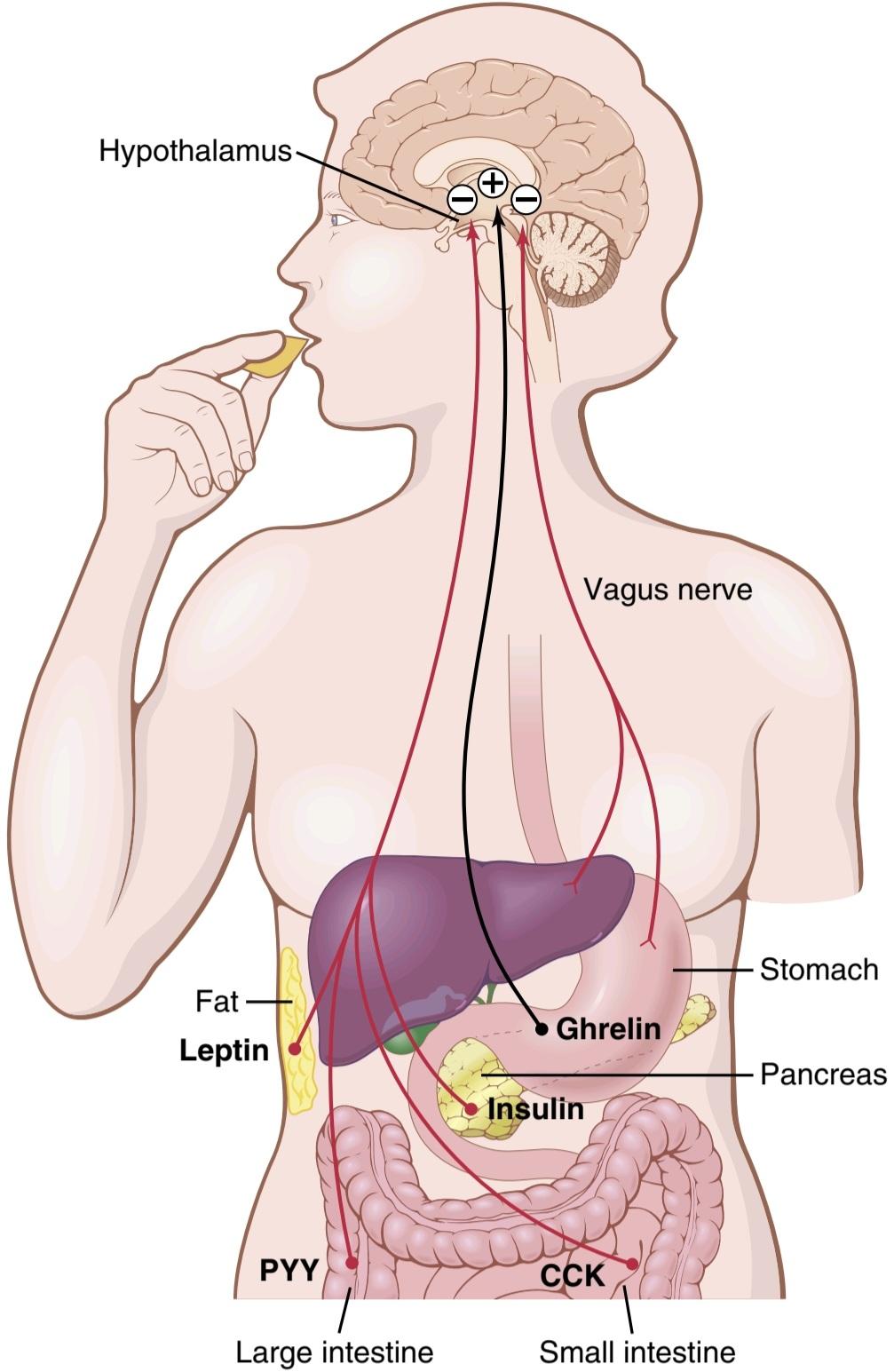
Figure 72-1. Feedback mechanisms for control of food intake. Stretch receptors in the stomach activate sensory afferent pathways in the vagus nerve and inhibit food intake. Peptide YY (PYY), cholecystokinin (CCK), and insulin are gastrointestinal hormones that are released by the ingestion of food and suppress further feeding. Ghrelin is released by the stomach, especially during fasting, and stimulates appetite. Leptin is a hormone produced in increasing amounts by fat cells as they increase in size. It inhibits food intake.
شکل 72-1. مکانیسم های بازخورد برای کنترل مصرف غذا گیرنده های کششی در معده مسیرهای آوران حسی در عصب واگ را فعال کرده و از دریافت غذا جلوگیری می کنند. پپتید YY (PYY)، کوله سیستوکینین (CCK) و انسولین هورمون های گوارشی هستند که با خوردن غذا آزاد می شوند و تغذیه بیشتر را سرکوب می کنند. گرلین به خصوص در زمان روزه داری توسط معده ترشح می شود و اشتها را تحریک می کند. لپتین هورمونی است که با افزایش اندازه سلولهای چربی به مقدار فزایندهای تولید میشود. مصرف غذا را مهار می کند.
Table 72-2 Neurotransmitters and Hormones That Influence Feeding and Satiety Centers in the Hypothalamus
جدول 72-2 انتقال دهنده های عصبی و هورمون ها که بر مراکز تغذیه و سیری در هیپوتالاموس تأثیر می گذارد

Neurons and Neurotransmitters in the Hypothalamus That Stimulate or Inhibit Feeding. Two distinct types of neurons in the arcuate nuclei of the hypothalamus are especially important as controllers of both appetite and energy expenditure (Figure 72-2): (1) pro-opiomelanocortin (POMC) neurons that produce a- melanocyte-stimulating hormone (a-MSH) together with cocaineand amphetamine-related transcript (CART); and (2) neurons that produce the orexigenic substances neuropeptide Y (NPY) and agouti-related protein (AGRP). Activation of the POMC neurons decreases food intake and increases energy expenditure, whereas activation of the NPY-AGRP neurons has the opposite effects, increasing food intake and reducing energy expenditure. Considerable cross talk occurs among these neurons and, as discussed later, POMC/CART and AGRP/NPY neurons appear to be the major targets for several hormones that regulate appetite, including leptin, insulin, cholecystokinin (CCK), and ghrelin. In fact, the neurons of the arcuate nuclei appear to be a site of convergence of many of the nervous and peripheral signals that regulate energy stores.
نورون ها و انتقال دهنده های عصبی در هیپوتالاموس که باعث تحریک یا مهار تغذیه می شوند. دو نوع متمایز نورون در هستههای کمانی هیپوتالاموس بهعنوان کنترلکننده اشتها و مصرف انرژی اهمیت ویژهای دارند (شکل 72-2): (1) نورونهای pro-opiomelanocortin (POMC) که هورمون محرک ملانوسیت (a-MSH) را همراه با کوکائین و ART مرتبط با کوکائین و آمپهتآمپهتام تولید میکنند. و (2) نورون هایی که مواد اورکسیوژنیک نوروپپتید Y (NPY) و پروتئین مرتبط با آگوتی (AGRP) را تولید می کنند. فعالسازی نورونهای POMC باعث کاهش مصرف غذا و افزایش مصرف انرژی میشود، در حالی که فعال شدن نورونهای NPY-AGRP اثرات معکوس دارد و باعث افزایش مصرف غذا و کاهش مصرف انرژی میشود. گفتگوی متقابل قابل توجهی در میان این نورون ها رخ می دهد و همانطور که بعداً بحث شد، به نظر می رسد نورون های POMC/CART و AGRP/NPY اهداف اصلی چندین هورمون تنظیم کننده اشتها از جمله لپتین، انسولین، کوله سیستوکینین (CCK) و گرلین هستند. در واقع، به نظر میرسد که نورونهای هستههای قوسی محل همگرایی بسیاری از سیگنالهای عصبی و محیطی هستند که ذخیره انرژی را تنظیم میکنند.
The POMC neurons release a-MSH, which then acts on melanocortin receptors found especially in neurons of the paraventricular nuclei. Although at least five sub- types of melanocortin receptors (MCR) exist, MCR-3 and MCR-4 are especially important in regulating food intake and energy balance. Activation of these receptors reduces food intake while increasing energy expenditure. Conversely, inhibition of MCR-3 and MCR-4 greatly increases food intake and decreases energy expenditure. The effect of MCR-4 activation to increase energy expenditure appears to be mediated, at least in part, by activation of neuronal pathways that project from the paraventricular nuclei to the nucleus tractus solitarius (NTS) and stimulate sympathetic nervous system activity. However, POMC neurons and MCR-4 are also found in brainstem neurons, including the NTS, where they also regulate food intake and energy expenditure.
نورونهای POMC a-MSH را آزاد میکنند، که سپس بر روی گیرندههای ملانوکورتین که بهویژه در نورونهای هستههای پارا بطنی یافت میشوند، عمل میکند. اگرچه حداقل پنج نوع فرعی گیرنده ملانوکورتین (MCR) وجود دارد، MCR-3 و MCR-4 به ویژه در تنظیم مصرف غذا و تعادل انرژی مهم هستند. فعال شدن این گیرنده ها باعث کاهش مصرف غذا و افزایش مصرف انرژی می شود. برعکس، مهار MCR-3 و MCR-4 مصرف غذا را تا حد زیادی افزایش می دهد و مصرف انرژی را کاهش می دهد. به نظر میرسد که اثر فعالسازی MCR-4 برای افزایش مصرف انرژی، حداقل تا حدی، با فعالسازی مسیرهای عصبی است که از هستههای پارا بطنی به سمت هسته تراکتوس سولیتریوس (NTS) پیش میروند و فعالیت سیستم عصبی سمپاتیک را تحریک میکنند. با این حال، نورونهای POMC و MCR-4 نیز در نورونهای ساقه مغز، از جمله NTS، یافت میشوند، جایی که آنها مصرف غذا و مصرف انرژی را نیز تنظیم میکنند.
The hypothalamic-brainstem melanocortin system plays a powerful role in regulating energy stores of the body, and defective signaling of this pathway is associated with extreme obesity. In fact, mutations of POMC and MCR-4 represent the most common known monogenic (single-gene) causes of human obesity, and some studies suggest that POMC and MCR-4 mutations may account for as much as 5% to 6% of early-onset severe obesity in children. In contrast, excessive activation of the melanocortin system reduces appetite. Some studies suggest that this activation may play a role in causing the loss of appetite for food (anorexia) associated with severe infections, cancer tumors, or uremia.
سیستم ملانوکورتین هیپوتالاموس ساقه مغز نقش قدرتمندی در تنظیم ذخایر انرژی بدن دارد و سیگنال دهی معیوب این مسیر با چاقی شدید همراه است. در واقع، جهشهای POMC و MCR-4 شایعترین علل شناخته شده تک ژنی (تک ژنی) چاقی انسان هستند و برخی مطالعات نشان میدهند که جهشهای POMC و MCR-4 ممکن است حدود 5 تا 6 درصد از چاقی شدید زودرس در کودکان را تشکیل دهند. در مقابل، فعال شدن بیش از حد سیستم ملانوکورتین باعث کاهش اشتها می شود. برخی از مطالعات نشان میدهند که این فعالسازی ممکن است در از دست دادن اشتها برای غذا (بیاشتهایی) مرتبط با عفونتهای شدید، تومورهای سرطانی یا اورمی نقش داشته باشد.
AGRP released from the orexigenic neurons of the hypothalamus is a natural antagonist of MCR-3 and MCR-4 and probably increases feeding by inhibiting the effects of a-MSH to stimulate melanocortin receptors (see Figure 72-2). Although the role of AGRP in normal physiological control of food intake is unclear, excessive formation of AGRP in mice and humans due to gene mutations is associated with increased food intake and obesity.
AGRP آزاد شده از نورون های اورکسیوژنیک هیپوتالاموس یک آنتاگونیست طبیعی MCR-3 و MCR-4 است و احتمالاً تغذیه را با مهار اثرات a-MSH برای تحریک گیرنده های ملانوکورتین افزایش می دهد (شکل 72-2 را ببینید). اگرچه نقش AGRP در کنترل فیزیولوژیکی طبیعی مصرف غذا مشخص نیست، تشکیل بیش از حد AGRP در موش و انسان به دلیل جهش ژنی با افزایش مصرف غذا و چاقی همراه است.
NPY is also released from orexigenic neurons of the arcuate nuclei. When energy stores of the body are low, orexigenic neurons are activated to release NPY, which stimulates appetite. At the same time, firing of the POMC neurons is reduced, thereby decreasing the activity of the melanocortin pathway and further stimulating appetite.
NPY همچنین از نورون های اورکسیوژنیک هسته های کمانی آزاد می شود. هنگامی که ذخایر انرژی بدن کم است، نورون های اورکسیژنیک فعال می شوند تا NPY آزاد کنند که اشتها را تحریک می کند. در همان زمان، شلیک نورونهای POMC کاهش مییابد، در نتیجه فعالیت مسیر ملانوکورتین را کاهش میدهد و اشتها را بیشتر تحریک میکند.

Figure 72-2. Control of energy balance by two types of neurons of the arcuate nuclei: (1) pro-opiomelanocortin (POMC) neurons that release a melanocyte-stimulating hormone (a-MSH) and cocaine- and amphetamine-regulated transcript (CART), decreasing food intake and increasing energy expenditure; and (2) neurons that produce agouti-related protein (AGRP) and neuropeptide Y (NPY), increasing food intake and reducing energy expenditure. a-MSH released by POMC neurons stimulates melanocortin receptors (MCR-3 and MCR-4) in the paraventricular nuclei (PVN), which then activate neuronal pathways that project to the nucleus tractus solitarius and increase sympathetic activity and energy expenditure. AGRP acts as an antagonist of MCR-4. Insulin, leptin, and cholecystokinin (CCK) are hormones that inhibit AGRP-NPY neurons and stimulate adjacent POMC-CART neurons, thereby reducing food intake. Ghrelin, a hormone secreted from the stomach, activates AGRP-NPY neurons and stimulates food intake. LepR, Leptin receptor; Y1r, neuropeptide Y, receptor. (Modified from Barsh GS, Schwartz MW: Genetic approaches to studying energy balance: perception and integration. Nat Rev Genet 3:589, 2002.)
شکل 72-2. کنترل تعادل انرژی توسط دو نوع نورون هسته کمانی: (1) نورون های پرو-اپیوملانوکورتین (POMC) که هورمون محرک ملانوسیت (a-MSH) و رونوشت تنظیم شده با کوکائین و آمفتامین (CART) را آزاد می کنند، مصرف غذا را کاهش می دهند و مصرف انرژی را افزایش می دهند. و (2) نورون هایی که پروتئین مرتبط با آگوتی (AGRP) و نوروپپتید Y (NPY) تولید می کنند، مصرف غذا را افزایش می دهند و مصرف انرژی را کاهش می دهند. a-MSH آزاد شده توسط نورون های POMC گیرنده های ملانوکورتین (MCR-3 و MCR-4) را در هسته های پارا بطنی (PVN) تحریک می کند، که سپس مسیرهای عصبی را فعال می کند که به سمت هسته تراکتوس سولیتریوس پیش می رود و فعالیت سمپاتیک و مصرف انرژی را افزایش می دهد. AGRP به عنوان آنتاگونیست MCR-4 عمل می کند. انسولین، لپتین و کوله سیستوکینین (CCK) هورمون هایی هستند که نورون های AGRP-NPY را مهار می کنند و نورون های POMC-CART مجاور را تحریک می کنند و در نتیجه مصرف غذا را کاهش می دهند. گرلین، هورمونی که از معده ترشح می شود، نورون های AGRP-NPY را فعال می کند و مصرف غذا را تحریک می کند. LepR، گیرنده لپتین؛ Y1r، نوروپپتید Y، گیرنده. (اصلاح شده از Barsh GS، Schwartz MW: رویکردهای ژنتیکی برای مطالعه تعادل انرژی: ادراک و ادغام. Nat Rev Genet 3:589، 2002.)
Neural Centers That Influence the Mechanical Process of Feeding. Another aspect of feeding is the mechanical act of the feeding process. If the brain is sectioned be- low the hypothalamus but above the mesencephalon, the animal can still perform the basic mechanical features of the feeding process. It can salivate, lick its lips, chew food, and swallow. Therefore, the actual mechanics of feeding are controlled by centers in the brainstem. The function of the other centers in feeding, then, is to control the quantity of food intake and to excite these centers of feeding mechanics to activity.
مراکز عصبی که بر فرآیند مکانیکی تغذیه تأثیر می گذارند. جنبه دیگر تغذیه، عمل مکانیکی فرآیند تغذیه است. اگر مغز در زیر هیپوتالاموس اما بالای مزانسفالون برش داده شود، حیوان همچنان میتواند ویژگیهای مکانیکی اولیه فرآیند تغذیه را انجام دهد. می تواند بزاق ترشح کند، لب هایش را لیس بزند، غذا بجود و قورت دهد. بنابراین، مکانیک واقعی تغذیه توسط مراکزی در ساقه مغز کنترل می شود. بنابراین، عملکرد سایر مراکز در تغذیه، کنترل مقدار غذای دریافتی و تحریک این مراکز مکانیک تغذیه به فعالیت است.
Neural centers higher than the hypothalamus also play important roles in the control of feeding, particularly in the control of appetite. These centers include the amygdala and the prefrontal cortex, which are closely coupled with the hypothalamus. Recall from the discussion of the sense of smell in Chapter 54 that portions of the amygdala are a major part of the olfactory nervous system. Destructive lesions in the amygdala have demonstrated that some of its areas increase feeding, whereas others inhibit feeding. In addition, stimulation of some areas of the amygdala elicits the mechanical act of feeding. An important effect of destruction of the amygdala on both sides of the brain is a “psychic blindness” in the choice of foods. In other words, the animal (and presumably the human being as well) at least partially loses the appetite control that determines the type and quality of food it eats.
مراکز عصبی بالاتر از هیپوتالاموس نیز نقش مهمی در کنترل تغذیه، به ویژه در کنترل اشتها دارند. این مراکز شامل آمیگدال و قشر جلوی مغز است که با هیپوتالاموس پیوند نزدیکی دارند. از بحث در مورد حس بویایی در فصل 54 به یاد بیاورید که بخش هایی از آمیگدال بخش عمده ای از سیستم عصبی بویایی است. ضایعات مخرب در آمیگدال نشان داده است که برخی از نواحی آن تغذیه را افزایش می دهند، در حالی که برخی دیگر از تغذیه جلوگیری می کنند. علاوه بر این، تحریک برخی از نواحی آمیگدال، عمل مکانیکی تغذیه را برمی انگیزد. یکی از تأثیرات مهم تخریب آمیگدال در دو طرف مغز، «کوری روانی» در انتخاب غذاها است. به عبارت دیگر، حیوان (و احتمالاً انسان نیز) حداقل تا حدی کنترل اشتهایی را که نوع و کیفیت غذایی که می خورد را تعیین می کند، از دست می دهد.
FACTORS THAT REGULATE QUANTITY OF FOOD INTAKE
Regulation of the quantity of food intake can be divided into short-term regulation, which is concerned primarily with preventing overeating at each meal, and long-term regulation, which is concerned primarily with maintenance of normal quantities of energy stores in the body.
عواملی که میزان مصرف غذا را تنظیم می کنند
تنظیم مقدار غذای دریافتی را می توان به تنظیم کوتاه مدت، که در درجه اول مربوط به جلوگیری از پرخوری در هر وعده غذایی است، و تنظیم طولانی مدت، که در درجه اول به حفظ مقادیر طبیعی ذخایر انرژی در بدن مربوط می شود، تقسیم کرد.
Short-Term Regulation of Food Intake
When a person is driven by hunger to eat voraciously and rapidly, what turns off the desire to eat when he or she has eaten enough? There has not been enough time for changes in the body’s energy stores to occur, and it takes hours for enough nutritional factors to be absorbed into the blood to cause the necessary inhibition of eating. Yet, it is important that the person not overeat and that he or she eat an amount of food that approximates nutritional needs. Several types of rapid feedback signals are important for these purposes, as described in the following sections.
تنظیم کوتاه مدت مصرف غذا
هنگامی که گرسنگی فرد را به خوردن با ولع و سریع سوق می دهد، وقتی به اندازه کافی غذا خورده باشد، چه چیزی میل به خوردن را خاموش می کند؟ زمان کافی برای ایجاد تغییرات در ذخایر انرژی بدن وجود نداشته است و ساعت ها طول می کشد تا عوامل تغذیه ای کافی در خون جذب شود تا باعث مهار لازم در خوردن شود. با این حال، مهم است که فرد پرخوری نکند و مقداری غذا بخورد که تقریباً نیازهای تغذیه ای دارد. انواع مختلفی از سیگنال های بازخورد سریع برای این اهداف مهم هستند، همانطور که در بخش های زیر توضیح داده شده است.
Gastrointestinal Filling Inhibits Feeding. When the gastrointestinal tract becomes distended, especially the stomach and the duodenum, stretch inhibitory signals are transmitted mainly by way of the vagi to suppress the feeding centers, thereby reducing the desire for food and providing a negative feedback mechanism to help limit meal size (see Figure 72-1).
پر شدن دستگاه گوارش مانع از تغذیه می شود. هنگامی که دستگاه گوارش متسع می شود، به ویژه معده و دوازدهه، سیگنال های بازدارنده کشش عمدتاً از طریق واگی برای سرکوب مراکز تغذیه منتقل می شود، در نتیجه میل به غذا کاهش می یابد و مکانیسم بازخورد منفی برای کمک به محدود کردن اندازه وعده غذایی ارائه می شود (شکل 72-1 را ببینید).
Astro intestinal Hormonal Factors Suppress Feeding. CCK, which is released mainly in response to fat and proteins entering the duodenum, enters the blood and acts as a hormone to influence several gastrointestinal functions such as gallbladder contraction, gastric emptying, gut motility, and gastric acid secretion as discussed in Chapters 63, 64, and 65. However, CCK also activates receptors on local sensory nerves in the duodenum, sending messages to the brain via the vagus nerve that contribute to satiation and meal cessation. The effect of CCK is short- lived, and chronic administration of CCK by itself has no major effect on body weight. Therefore, CCK functions mainly to prevent overeating during meals but may not play a major role in the frequency of meals or the total energy consumed.
عوامل هورمونی روده ای Astro باعث سرکوب تغذیه می شود. CCK که عمدتاً در پاسخ به ورود چربی و پروتئین به دوازدهه ترشح میشود، وارد خون میشود و بهعنوان هورمونی برای تأثیرگذاری بر عملکردهای دستگاه گوارش مانند انقباض کیسه صفرا، تخلیه معده، حرکت روده و ترشح اسید معده همانطور که در فصلهای 63، 64 و 65 بحث شد، عمل میکند. پیام هایی به مغز از طریق عصب واگ که به سیری و قطع غذا کمک می کند. اثر CCK کوتاه مدت است و تجویز مزمن CCK به خودی خود تأثیر عمده ای بر وزن بدن ندارد. بنابراین، CCK عمدتاً برای جلوگیری از پرخوری در طول وعدههای غذایی عمل میکند، اما ممکن است نقش مهمی در دفعات وعدههای غذایی یا کل انرژی مصرفشده نداشته باشد.
Peptide YY (PYY) is secreted from the entire gastrointestinal tract, but especially from the ileum and colon. Food intake stimulates release of PYY, with blood concentrations rising to peak levels 1 to 2 hours after ingesting a meal. These peak levels of PYY are influenced by the amount and composition of the food, with higher levels of PYY observed after meals with a high fat content. Although injections of PYY into mice have been shown to decrease food intake for 12 hours or more, the importance of this gastrointestinal hormone in regulating appetite in humans is still unclear.
پپتید YY (PYY) از کل دستگاه گوارش، به ویژه از ایلئوم و روده بزرگ ترشح می شود. مصرف غذا باعث تحریک ترشح PYY می شود و غلظت خون 1 تا 2 ساعت پس از خوردن غذا به اوج می رسد. این سطوح اوج PYY تحت تأثیر مقدار و ترکیب غذا قرار می گیرد و سطوح بالاتر PYY بعد از وعده های غذایی با محتوای چربی بالا مشاهده می شود. اگرچه نشان داده شده است که تزریق PYY به موش باعث کاهش مصرف غذا برای 12 ساعت یا بیشتر می شود، اهمیت این هورمون گوارشی در تنظیم اشتها در انسان هنوز مشخص نیست.
For reasons that are not entirely understood, the presence of food in the intestines stimulates them to secrete glucagon-like peptide (GLP), which in turn enhances glucose-dependent insulin production and secretion from the pancreas. GLP and insulin both tend to suppress appetite. Thus, eating a meal stimulates the release of several gastrointestinal hormones that may induce satiety and limit further intake of food (see Figure 72-1).
به دلایلی که کاملاً مشخص نیست، وجود غذا در روده ها آنها را تحریک می کند تا پپتید شبه گلوکاگون (GLP) ترشح کنند که به نوبه خود تولید انسولین وابسته به گلوکز و ترشح از پانکراس را افزایش می دهد. GLP و انسولین هر دو تمایل به سرکوب اشتها دارند. بنابراین، خوردن یک وعده غذایی، ترشح چندین هورمون گوارشی را تحریک می کند که ممکن است باعث سیری شود و مصرف بیشتر غذا را محدود کند (شکل 72-1 را ببینید).
Ghrelin, a Gastrointestinal Hormone, Increases Feeding. Ghrelin is a hormone released mainly by the oxyntic cells of the stomach but also, to much less of an extent, by the intestine. Blood levels of ghrelin rise during fasting, peak just before eating, and then fall rapidly after a meal, suggesting a possible role in stimulating feeding. Also, administration of ghrelin increases food intake in experimental animals, further supporting the possibility that it may be an orexigenic hormone.
گرلین، یک هورمون گوارشی، تغذیه را افزایش می دهد. گرلین هورمونی است که عمدتاً توسط سلول های اکسینتیک معده و همچنین به میزان بسیار کمتری از روده ترشح می شود. سطح گرلین خون در طول روزه داری افزایش می یابد، درست قبل از خوردن غذا به اوج خود می رسد و سپس به سرعت بعد از غذا کاهش می یابد، که نشان دهنده نقش احتمالی در تحریک تغذیه است. همچنین، تجویز گرلین مصرف غذا را در حیوانات آزمایشی افزایش می دهد و این احتمال را که ممکن است یک هورمون اورکسیژنیک باشد، حمایت می کند.
Oral Receptors Meter Food Intake. When an animal with an esophageal fistula is fed large quantities of food, even though this food is immediately lost to the exterior, the degree of hunger is decreased after a reasonable quantity of food has passed through the mouth. This effect occurs despite the fact that the gastrointestinal tract does not become the least bit filled. Therefore, various “oral factors” related to feeding, such as chewing, salivation,swallowing, and tasting, have been postulated to “meter” the food as it passes through the mouth, and after a certain amount has passed, the hypothalamic feeding center becomes inhibited. The inhibition caused by this metering mechanism, however, is considerably less intense and of shorter duration-usually lasting for only 20 to 40 minutes-than is the inhibition caused by gastrointestinal filling.
گیرنده های دهان اندازه گیری دریافت غذا. هنگامی که به حیوان مبتلا به فیستول مری مقادیر زیادی غذا داده می شود، حتی اگر این غذا بلافاصله به بیرون از دست می رود، پس از عبور مقدار معقول غذا از دهان، میزان گرسنگی کاهش می یابد. این اثر با وجود این واقعیت رخ می دهد که دستگاه گوارش کمترین مقداری پر نمی شود. بنابراین، «عوامل دهانی» مختلف مربوط به تغذیه، مانند جویدن، ترشح بزاق، بلع و چشیدن، فرض شده است که غذا را هنگام عبور از دهان «سنجش» میکند و پس از گذشت مقدار معینی، مرکز تغذیه هیپوتالاموس مهار میشود. با این حال، مهار ناشی از این مکانیسم اندازهگیری، بهطور قابلتوجهی شدیدتر و کوتاهتر است – معمولاً فقط 20 تا 40 دقیقه طول میکشد – نسبت به بازداری ناشی از پر شدن دستگاه گوارش.
Intermediateand Long-Term Regulation of Food Intake
An animal that has been starved for a long time and is then presented with unlimited food eats a far greater quantity than does an animal that has been on a regular diet. Conversely, an animal that has been force-fed for several weeks eats very little when allowed to eat according to its own desires. Thus, the biological feeding control mechanisms of the body are geared to the nutritional status of the body although multiple behavioral, social and environmental factors also influence food intake in humans.
تنظیم میان مدت و بلند مدت مصرف غذا
حیوانی که برای مدت طولانی گرسنه مانده است و سپس غذای نامحدودی به او ارائه می شود، نسبت به حیوانی که رژیم غذایی معمولی داشته است، مقدار بسیار بیشتری غذا می خورد. برعکس، حیوانی که برای چندین هفته به اجبار تغذیه شده است، زمانی که اجازه داده شود مطابق میل خود غذا بخورد، بسیار کم غذا می خورد. بنابراین، مکانیسمهای کنترل تغذیه بیولوژیکی بدن با وضعیت تغذیهای بدن تنظیم میشوند، اگرچه عوامل متعدد رفتاری، اجتماعی و محیطی نیز بر دریافت غذا در انسان تأثیر میگذارند.
Effect of Blood Concentrations of Glucose, Amino Acids, and Lipids on Hunger and Feeding. A decrease in blood glucose concentration has been shown in experimental studies to cause hunger, which has led to the so- called glucostatic theory of hunger and feeding regulation. Similar studies have demonstrated the same effect for blood amino acid concentration and blood concentration of breakdown products of lipids such as the keto acids and some fatty acids, leading to the aminostatic and lipostatic theories of regulation. That is, when the availability of any of the three major types of food decreases, the desire for feeding is increased, eventually returning the blood metabolite concentrations back toward normal if the appropriate foods are available.
تأثیر غلظت گلوکز، اسیدهای آمینه و لیپیدهای خون بر گرسنگی و تغذیه. در مطالعات تجربی نشان داده شده است که کاهش غلظت گلوکز خون باعث گرسنگی می شود، که منجر به به اصطلاح نظریه گلوکوستاتیک گرسنگی و تنظیم تغذیه شده است. مطالعات مشابه همین اثر را برای غلظت اسید آمینه خون و غلظت خون محصولات تجزیه لیپیدها مانند اسیدهای کتو و برخی اسیدهای چرب نشان داده است که منجر به تئوری آمینواستاتیک و لیپوستاتیک تنظیم می شود. به این معنا که وقتی دسترسی به هر یک از سه نوع اصلی غذا کاهش مییابد، میل به تغذیه افزایش مییابد و در نهایت غلظت متابولیتهای خون را به حالت عادی باز میگرداند، اگر غذای مناسب در دسترس باشد.
The following observations from neurophysiological studies of function in specific areas of the brain also sup- port the glucostatic, aminostatic, and lipostatic theories:
مشاهدات زیر از مطالعات نوروفیزیولوژیکی عملکرد در مناطق خاصی از مغز نیز از نظریه های گلوکوستاتیک، آمینواستاتیک و لیپوستاتیک حمایت می کند:
(1) a rise in blood glucose level increases the rate of firing of glucoreceptor neurons in the satiety center in the ventromedial and paraventricular nuclei of the hypothalamus, and (2) the same increase in blood glucose level simultaneously decreases the firing of glucosensitive neurons in the hunger center of the lateral hypothalamus. In addition, some amino acids and lipid substances affect the rates of firing of these same neurons or other closely associated neurons.
(1) افزایش سطح گلوکز خون باعث افزایش سرعت شلیک نورونهای گیرنده گلوکز در مرکز سیری در هستههای بطنی و پارا بطنی هیپوتالاموس میشود و (2) افزایش یکسان در سطح گلوکز خون به طور همزمان باعث کاهش شلیک نورونهای حساس به گلوکز در مرکز گرسنگی جانبی عضلانی میشود. علاوه بر این، برخی از اسیدهای آمینه و مواد لیپیدی بر سرعت شلیک همین نورون ها یا سایر نورون های نزدیک به هم تأثیر می گذارند.
Temperature Regulation and Food Intake. When an animal is exposed to cold, it tends to increase feeding; when it is exposed to heat, it tends to decrease its caloric intake. This phenomenon is caused by interaction within the hypothalamus between the temperature-regulating system (see Chapter 74) and the food intake-regulating system. This is important because increased food intake in a cold animal (1) increases its metabolic rate and (2) provides increased fat for insulation, both of which tend to protect against the cold.
تنظیم دما و مصرف غذا. هنگامی که حیوان در معرض سرما قرار می گیرد، تمایل به افزایش تغذیه دارد. هنگامی که در معرض گرما قرار می گیرد، تمایل به کاهش کالری دریافتی خود دارد. این پدیده در اثر تعامل در هیپوتالاموس بین سیستم تنظیم کننده دما (به فصل 74 مراجعه کنید) و سیستم تنظیم کننده مصرف غذا ایجاد می شود. این مهم است زیرا افزایش مصرف غذا در یک حیوان سرد (1) سرعت متابولیسم آن را افزایش می دهد و (2) افزایش چربی را برای عایق فراهم می کند، که هر دو تمایل به محافظت در برابر سرما دارند.
Feedback Signals From Adipose Tissue Regulate Food Intake. Most of the stored energy in the body consists of fat, the amount of which can vary considerably in different persons. What regulates this energy reserve, and why is there so much variability among individuals?
سیگنال های بازخورد از بافت چربی، مصرف غذا را تنظیم می کند. بیشتر انرژی ذخیره شده در بدن از چربی تشکیل شده است که مقدار آن در افراد مختلف می تواند به طور قابل توجهی متفاوت باشد. چه چیزی این ذخیره انرژی را تنظیم می کند و چرا تنوع زیادی در بین افراد وجود دارد؟
Studies in humans and in experimental animals indicate that the hypothalamus senses energy storage through the actions of leptin, a peptide hormone released from adipocytes. When the amount of adipose tissue increases (signaling excess energy storage), the adipocytes produce increased amounts of leptin, which is released into the blood. Leptin then circulates to the brain, where it moves across the blood-brain barrier by facilitated diffusion and occupies leptin receptors at multiple sites in the hypo- thalamus, especially the POMC and AGRP/NPY neurons of the arcuate nuclei and neurons of the paraventricular nuclei, as well as neurons in other areas of the brain including the brainstem.
مطالعات بر روی انسان و حیوانات آزمایشی نشان می دهد که هیپوتالاموس ذخیره انرژی را از طریق اعمال لپتین، یک هورمون پپتیدی آزاد شده از سلول های چربی، حس می کند. هنگامی که مقدار بافت چربی افزایش می یابد (که نشان دهنده ذخیره انرژی اضافی است)، سلول های چربی مقادیر بیشتری لپتین تولید می کنند که در خون آزاد می شود. لپتین سپس به مغز گردش می کند، جایی که با انتشار تسهیل شده در سراسر سد خونی مغزی حرکت می کند و گیرنده های لپتین را در چندین مکان در هیپوتالاموس اشغال می کند، به ویژه نورون های POMC و AGRP/NPY هسته های کمانی و نورون های هسته های پارا بطنی، و همچنین نورون های مغز از جمله سایر نواحی مغز.
Stimulation of leptin receptors in these central nervous system nuclei initiates multiple actions that decrease fat storage, including (1) decreased production in the hypothalamus of appetite stimulators, such as NPY and AGRP; (2) activation of POMC neurons, causing release of a-MSH and activation of melanocortin receptors; (3) increased production in the hypothalamus of substances, such as corticotropin-releasing hormone, that decrease food intake; (4) increased sympathetic nerve activity (through neural projections from the hypothalamus to the vasomotor centers), which increases metabolic rate and energy expenditure; and (5) decreased insulin secretion by the pancreatic beta cells, which decreases energy storage. Thus, leptin is an important means by which the adipose tissue signals the brain that enough energy has been stored and that intake of food is no longer necessary.
تحریک گیرنده های لپتین در این هسته های سیستم عصبی مرکزی، اقدامات متعددی را آغاز می کند که ذخیره چربی را کاهش می دهد، از جمله (1) کاهش تولید در هیپوتالاموس محرک های اشتها، مانند NPY و AGRP. (2) فعال شدن نورون های POMC، باعث آزاد شدن a-MSH و فعال شدن گیرنده های ملانوکورتین می شود. (3) افزایش تولید در هیپوتالاموس موادی مانند هورمون آزاد کننده کورتیکوتروپین که مصرف غذا را کاهش می دهد. (4) افزایش فعالیت عصب سمپاتیک (از طریق پیش بینی های عصبی از هیپوتالاموس به مراکز وازوموتور)، که باعث افزایش سرعت متابولیسم و مصرف انرژی می شود. و (5) کاهش ترشح انسولین توسط سلولهای بتای پانکراس، که ذخیره انرژی را کاهش می دهد. بنابراین، لپتین وسیله مهمی است که به وسیله آن بافت چربی به مغز سیگنال می دهد که انرژی کافی ذخیره شده است و دیگر نیازی به دریافت غذا نیست.
In mice or humans with mutations that render their fat cells unable to produce leptin or mutations that cause defective leptin receptors in the hypothalamus, marked hyperphagia and morbid obesity occur. In most obese humans, however, there does not appear to be a deficiency of leptin production because plasma leptin levels increase in proportion with increasing adiposity. Therefore, some physiologists believe that obesity may be associated with leptin resistance; that is, leptin receptors or postreceptor signaling pathways normally activated by leptin may be resistant to activation by leptin in obese people, who continue to overeat despite having very high levels of leptin.
در موشها یا انسانهای مبتلا به جهشهایی که سلولهای چربیشان را قادر به تولید لپتین نمیکنند یا جهشهایی که باعث گیرندههای لپتین معیوب در هیپوتالاموس میشوند، هیپرفاژی مشخص و چاقی مرضی رخ میدهد. با این حال، در بیشتر انسان های چاق، به نظر نمی رسد کمبود تولید لپتین وجود داشته باشد، زیرا سطح لپتین پلاسما متناسب با افزایش چربی افزایش می یابد. بنابراین، برخی از فیزیولوژیست ها معتقدند که چاقی ممکن است با مقاومت به لپتین مرتبط باشد. یعنی گیرنده های لپتین یا مسیرهای سیگنال دهی پس گیرنده که معمولاً توسط لپتین فعال می شوند ممکن است در برابر فعال شدن توسط لپتین در افراد چاق مقاوم باشند، افرادی که با وجود داشتن سطوح بسیار بالای لپتین به پرخوری ادامه می دهند.
Another explanation for the failure of leptin to prevent increasing adiposity in obese individuals is that there are many redundant systems that control feeding behavior, as well as social and cultural factors that can cause continued excess food intake even in the presence of high levels of leptin.
توضیح دیگر برای عدم موفقیت لپتین در جلوگیری از افزایش چاقی در افراد چاق این است که بسیاری از سیستمهای اضافی وجود دارند که رفتار تغذیه را کنترل میکنند و همچنین عوامل اجتماعی و فرهنگی میتوانند باعث ادامه مصرف بیش از حد غذا حتی در حضور سطوح بالای لپتین شوند.
Summary of Long-Term Regulation. Even though our information on the different feedback factors in long- term feeding regulation is imprecise, we can make the following general statement: When the energy stores of the body fall below normal, the feeding centers of the hypothalamus and other areas of the brain become highly active, and the person exhibits increased hunger, as well as the behavior of searching for food. Conversely, when the energy stores (mainly the fat stores) are already abundant, the person usually loses the sensation of hunger and develops a state of satiety. Although the precise feedback systems that regulate food intake and energy expenditure are not fully understood, rapid advances have been made in this field of research in recent years, with the discovery of many new orexigenic and anorexigenic factors.
خلاصه مقررات بلند مدت. حتی اگر اطلاعات ما در مورد فاکتورهای مختلف بازخورد در تنظیم طولانی مدت تغذیه نادرست است، میتوانیم عبارت کلی زیر را بیان کنیم: زمانی که ذخایر انرژی بدن به زیر نرمال میرسد، مراکز تغذیه هیپوتالاموس و سایر نواحی مغز بسیار فعال میشوند و فرد گرسنگی بیشتری نشان میدهد و همچنین رفتار جستجوی غذا را نشان میدهد. برعکس، زمانی که ذخایر انرژی (عمدتاً ذخایر چربی) از قبل فراوان باشد، فرد معمولاً احساس گرسنگی را از دست می دهد و حالت سیری پیدا می کند. اگرچه سیستمهای بازخورد دقیقی که مصرف غذا و مصرف انرژی را تنظیم میکنند بهطور کامل شناخته نشدهاند، پیشرفتهای سریعی در این زمینه تحقیقاتی در سالهای اخیر و با کشف بسیاری از عوامل جدید اشتها زا و بیاشتهایی ایجاد شده است.
Importance of Having Both Long- and Short-Term Regulatory Systems for Feeding
The long-term regulatory system for feeding, which includes all the nutritional energy feedback mechanisms, helps maintain constant stores of nutrients in the tissues, preventing them from becoming too low or too high. The short-term regulatory stimuli serve two other purposes. First, they tend to make the person eat smaller quantities at each eating session, thus allowing food to pass through the gastrointestinal tract at a steadier pace so that its digestive and absorptive mechanisms can work at optimal rates rather than becoming periodically overburdened. Second, they help prevent the person from eating amounts at each meal that would be too much for the metabolic storage systems once all the food has been absorbed.
اهمیت داشتن سیستم های تنظیمی بلند مدت و کوتاه مدت برای تغذیه
سیستم تنظیمی طولانی مدت برای تغذیه، که شامل تمام مکانیسم های بازخورد انرژی تغذیه ای است، به حفظ ذخایر ثابت مواد مغذی در بافت ها کمک می کند و از کم یا زیاد شدن آنها جلوگیری می کند. محرک های تنظیمی کوتاه مدت دو هدف دیگر را دنبال می کنند. اول، آنها تمایل دارند که فرد را وادار کنند در هر جلسه غذا خوردن مقادیر کمتری بخورد، بنابراین اجازه میدهند غذا با سرعت ثابتتری از دستگاه گوارش عبور کند تا مکانیسمهای گوارشی و جذبی آن بتوانند با سرعت بهینه کار کنند نه اینکه به طور دورهای بیش از حد سنگین شوند. دوم، آنها به جلوگیری از خوردن مقادیر زیادی در هر وعده غذایی به فرد کمک میکنند که پس از جذب تمام غذا برای سیستمهای ذخیرهسازی متابولیک بسیار زیاد است.
Obesity
Obesity can be defined as an excess of body fat. A surrogate marker for body fat content is the body mass index (BMI), which is calculated as:
چاقی
چاقی را می توان به عنوان چربی اضافی بدن تعریف کرد. یک نشانگر جایگزین برای محتوای چربی بدن، شاخص توده بدنی (BMI) است که به صورت زیر محاسبه می شود:

In clinical terms, a person with a BMI between 25 and 29.9 kg/m2 is deemed overweight, and a person with a BMI greater than or equal to 30 kg/m2 is considered obese. BMI is not a direct estimate of adiposity and does not take into account the fact that some individuals have a high BMI as a result of a large muscle mass. A better way to define obesity is to actually measure the percentage of total body fat. Obesity is usually defined as 25% or greater total body fat in men and 35% or greater total body fat in women. Although percentage of body fat can be estimated with various methods, such as measuring skin-fold thickness, bioelectrical impedance, or underwater weighing, these methods are not routinely used in clinical practice, where BMI is commonly used to assess obesity.
از نظر بالینی، فردی با BMI بین 25 تا 29.9 کیلوگرم بر متر مربع دارای اضافه وزن و فردی با BMI بیشتر یا مساوی 30 کیلوگرم بر متر مربع چاق در نظر گرفته می شود. BMI تخمین مستقیمی از چاقی نیست و این واقعیت را در نظر نمی گیرد که برخی افراد در نتیجه توده عضلانی بزرگ BMI بالایی دارند. یک راه بهتر برای تعریف چاقی، اندازه گیری درصد کل چربی بدن است. چاقی معمولاً به صورت ۲۵ درصد یا بیشتر چربی کل بدن در مردان و ۳۵ درصد یا بیشتر چربی کل بدن در زنان تعریف میشود. اگرچه درصد چربی بدن را میتوان با روشهای مختلفی تخمین زد، مانند اندازهگیری ضخامت چینهای پوست، امپدانس بیوالکتریکی یا توزین زیر آب، این روشها معمولاً در عمل بالینی، جایی که BMI معمولاً برای ارزیابی چاقی استفاده میشود، استفاده نمیشوند.
The adverse impact of obesity on the risk for various disorders such as cirrhosis, hypertension, heart attack, stroke, and kidney disease appears to be more closely associated with increased visceral (abdominal) adiposity than with increased subcutaneous fat storage, or storage of fat in the lower parts of the body such as the hips. Therefore, many clinicians measure waist circumference or sagittal abdominal diameter as indicators of abdominal obesity. In the United States a waist circumference greater than 102 centimeters in men and 88 centimeters in women or a waist/hip ratio of greater than 0.9 in men and 0.85 in women is often considered to indicate abdominal obesity in adults.
به نظر می رسد تاثیر نامطلوب چاقی بر خطر ابتلا به اختلالات مختلف مانند سیروز، فشار خون بالا، حمله قلبی، سکته مغزی و بیماری کلیوی با افزایش چاقی احشایی (شکمی) بیشتر از افزایش ذخیره چربی زیر جلدی یا ذخیره چربی در قسمت های تحتانی بدن مانند باسن مرتبط است. بنابراین، بسیاری از پزشکان دور کمر یا قطر ساژیتال شکم را به عنوان شاخص چاقی شکمی اندازه گیری می کنند. در ایالات متحده، دور کمر بیشتر از 102 سانتیمتر در مردان و 88 سانتیمتر در زنان یا نسبت دور کمر به باسن بیش از 0.9 در مردان و 0.85 در زنان اغلب نشاندهنده چاقی شکمی در بزرگسالان در نظر گرفته میشود.
The prevalence of obesity in children and adults in the United States and in many other industrialized countries is rapidly increasing, rising by more than 30% during the past decade. Approximately 70% of adults aged 20 and over in the United States are overweight or obese, and more than 35% of those adults are obese.
شیوع چاقی در کودکان و بزرگسالان در ایالات متحده و بسیاری از کشورهای صنعتی دیگر به سرعت در حال افزایش است و طی دهه گذشته بیش از 30 درصد افزایش یافته است. تقریباً 70 درصد از بزرگسالان 20 ساله و بالاتر در ایالات متحده دارای اضافه وزن یا چاق هستند و بیش از 35 درصد از این بزرگسالان چاق هستند.
Obesity Results From Greater Intake Than Expenditure of Energy
When greater quantities of energy (in the form of food) enter the body than are expended, the body weight increases, and most of the excess energy is stored as fat. Therefore, excessive adiposity (obesity) is caused by energy intake in excess of energy output. For each 9.3 calories of excess energy that remains in the body, approximately 1 gram of fat is stored.
چاقی ناشی از دریافت بیشتر از مصرف انرژی است
وقتی انرژی (به شکل غذا) بیشتر از مقدار مصرف شده وارد بدن می شود، وزن بدن افزایش می یابد و بیشتر انرژی اضافی به صورت چربی ذخیره می شود. بنابراین، چاقی بیش از حد (چاقی) به دلیل دریافت انرژی بیش از انرژی خروجی ایجاد می شود. به ازای هر 9.3 کالری انرژی اضافی که در بدن باقی می ماند، تقریباً 1 گرم چربی ذخیره می شود.
Fat is stored mainly in adipocytes in subcutaneous tissue and in the intraperitoneal cavity, although the liver and other tissues of the body often accumulate significant amounts of lipids in obese persons. The metabolic process- es involved in fat storage were discussed in Chapter 69.
چربی عمدتاً در سلولهای چربی در بافت زیر جلدی و در حفره داخل صفاقی ذخیره میشود، اگرچه کبد و سایر بافتهای بدن اغلب مقادیر قابلتوجهی از لیپیدها را در افراد چاق انباشته میکنند. فرآیندهای متابولیک درگیر در ذخیره چربی در فصل 69 مورد بحث قرار گرفت.
It was previously believed that the number of adipocytes could increase substantially only during infancy and childhood and that excess energy intake in children led to hyperplastic obesity, associated with increased numbers of adipocytes and only small increases in adipocyte size. In contrast, the development of obesity in adults was thought to increase only adipocyte size, resulting in hypertrophic obesity. However, research studies have shown that new adipocytes can differentiate from fibroblast-like preadipocytes at any period of life and that the development of obesity in adults is accompanied by increased numbers, as well as increased size, of adipocytes. An extremely obese person may have as many as four times as many adipocytes, each containing twice as much lipid, as a lean person.
قبلاً اعتقاد بر این بود که تعداد سلولهای چربی فقط در دوران نوزادی و کودکی میتواند به طور قابل ملاحظهای افزایش یابد و دریافت انرژی اضافی در کودکان منجر به چاقی هیپرپلاستیک میشود که با افزایش تعداد سلولهای چربی و تنها افزایش اندک در اندازه سلولهای چربی همراه است. در مقابل، تصور میشد که ایجاد چاقی در بزرگسالان فقط اندازه سلولهای چربی را افزایش میدهد و در نتیجه چاقی هیپرتروفیک ایجاد میکند. با این حال، مطالعات تحقیقاتی نشان دادهاند که سلولهای چربی جدید میتوانند در هر دوره از زندگی از پرهآدیپوسیتهای فیبروبلاستمانند متمایز شوند و ایجاد چاقی در بزرگسالان با افزایش تعداد و همچنین افزایش اندازه سلولهای چربی همراه است. یک فرد بسیار چاق ممکن است چهار برابر بیشتر از یک فرد لاغر سلول چربی داشته باشد که هرکدام دوبرابر لیپید دارند.
Once a person has become obese and a stable weight is obtained, energy intake once again equals energy output. For a person to lose weight, energy intake must be less than energy expenditure.
هنگامی که فرد چاق شد و وزن ثابتی به دست آورد، انرژی دریافتی بار دیگر با انرژی خروجی برابری می کند. برای اینکه یک فرد وزن کم کند، انرژی دریافتی باید کمتر از انرژی مصرفی باشد.
Decreased Physical Activity and Abnormal Feeding Regulation as Causes of Obesity
The causes of obesity are complex. Although genes play an important role in programming the powerful physiological mechanisms that regulate food intake and energy metabolism, lifestyle and environmental factors may play a major role in many obese people. The rapid increase in the prevalence of obesity in the past 20 to 30 years emphasizes the important role of lifestyle and environmental factors because genetic changes could not have occurred so rapidly. Still, genetic factors may predispose many people to the environmental influences that are driving the rising prevalence of obesity in most industrialized and developing countries.
کاهش فعالیت بدنی و تنظیم غیر طبیعی تغذیه به عنوان علل چاقی
دلایل چاقی پیچیده است. اگرچه ژنها نقش مهمی در برنامهریزی مکانیسمهای فیزیولوژیکی قوی ایفا میکنند که مصرف غذا و متابولیسم انرژی را تنظیم میکنند، شیوه زندگی و عوامل محیطی ممکن است نقش مهمی در بسیاری از افراد چاق داشته باشند. افزایش سریع شیوع چاقی در 20 تا 30 سال گذشته بر نقش مهم سبک زندگی و عوامل محیطی تاکید دارد زیرا تغییرات ژنتیکی نمیتوانست به این سرعت رخ دهد. با این حال، عوامل ژنتیکی ممکن است بسیاری از افراد را مستعد تأثیرات محیطی کند که باعث افزایش شیوع چاقی در اکثر کشورهای صنعتی و در حال توسعه می شود.
Sedentary Lifestyle Is a Major Cause of Obesity. Regular physical activity and physical training are known to in- crease muscle mass and decrease body fat mass, whereas inadequate physical activity is typically associated with de- creased muscle mass and increased adiposity. For example, studies have shown a close association between sedentary behaviors, such as excessive screen time (e.g., television watching), and obesity.
سبک زندگی بی تحرک یکی از دلایل اصلی چاقی است. فعالیت بدنی منظم و تمرین بدنی باعث افزایش توده عضلانی و کاهش توده چربی بدن می شود، در حالی که فعالیت بدنی ناکافی معمولاً با کاهش توده عضلانی و افزایش چاقی همراه است. به عنوان مثال، مطالعات ارتباط نزدیکی را بین رفتارهای بی تحرک، مانند زمان زیاد صفحه نمایش (مانند تماشای تلویزیون) و چاقی نشان داده است.
About 25% to 30% of the energy used each day by the average person goes into muscular activity, and in a laborer, as much as 60% to 70% is used in this way. In obese people, increased physical activity often increases energy expenditure more than food intake, resulting in significant weight loss. Even a single episode of strenuous exercise may increase basal energy expenditure for several hours after the physical activity is stopped. Because muscular activity is by far the most important means by which energy is expended in the body, increased physical activity is often an effective means of reducing fat stores.
حدود 25 تا 30 درصد انرژی مصرف شده در هر روز توسط یک فرد معمولی صرف فعالیت عضلانی می شود و در یک کارگر 60 تا 70 درصد به این روش مصرف می شود. در افراد چاق، افزایش فعالیت بدنی اغلب مصرف انرژی را بیشتر از مصرف غذا افزایش می دهد و در نتیجه باعث کاهش وزن قابل توجهی می شود. حتی یک دوره ورزش شدید ممکن است مصرف انرژی پایه را برای چند ساعت پس از توقف فعالیت بدنی افزایش دهد. از آنجایی که فعالیت عضلانی مهم ترین وسیله ای است که به وسیله آن انرژی در بدن صرف می شود، افزایش فعالیت بدنی اغلب وسیله ای موثر برای کاهش ذخایر چربی است.
Abnormal Feeding Behavior Is an Important Cause of Obesity. Although powerful physiological mechanisms regulate food intake, important environmental, social, and psychological factors also can cause abnormal feeding behavior, excessive energy intake, and obesity.
رفتار غیرعادی تغذیه یکی از دلایل مهم چاقی است. اگرچه مکانیسمهای فیزیولوژیکی قوی مصرف غذا را تنظیم میکنند، اما عوامل محیطی، اجتماعی و روانی مهم نیز میتوانند باعث رفتار غیرعادی تغذیه، دریافت بیش از حد انرژی و چاقی شوند.
As discussed previously, the importance of environmental factors is evident from the rapid increase in the prevalence of obesity in most industrialized countries, which has coincided with an abundance of high-energy foods and sedentary lifestyles.
همانطور که قبلاً بحث شد، اهمیت عوامل محیطی از افزایش سریع شیوع چاقی در اکثر کشورهای صنعتی مشهود است که با فراوانی غذاهای پرانرژی و سبک زندگی کم تحرک همزمان شده است.
Psychological and social factors may contribute to obesity in some people. For example, people often gain large amounts of weight during or after stressful situations, such as the death of a parent, a severe illness, or even mental depression. It seems that eating can be a means of relieving stress.
عوامل روانی و اجتماعی ممکن است در ایجاد چاقی در برخی افراد نقش داشته باشد. به عنوان مثال، افراد اغلب در طول یا پس از موقعیت های استرس زا، مانند مرگ والدین، یک بیماری شدید یا حتی افسردگی روانی، وزن زیادی اضافه می کنند. به نظر می رسد که خوردن می تواند وسیله ای برای کاهش استرس باشد.
Childhood Overnutrition May Contribute to Adult Obesity. One factor that may contribute to obesity is the prevalent idea that healthy eating habits require three meals a day and that each meal must be filling. Many young children are forced into this habit by overly solicitous parents, and the children continue to practice it throughout life.
تغذیه بیش از حد در دوران کودکی ممکن است به چاقی بزرگسالان کمک کند. یکی از عواملی که ممکن است به چاقی کمک کند، این ایده رایج است که عادات غذایی سالم نیاز به سه وعده غذایی در روز دارد و هر وعده غذایی باید سیر کننده باشد. بسیاری از کودکان خردسال توسط والدین بیش از حد سخت کوش مجبور به این عادت می شوند و کودکان در طول زندگی به آن ادامه می دهند.
The rate of formation of new fat cells is especially rapid in the first few years of life, and the greater the rate of fat storage, the greater the number of fat cells. The number of fat cells in obese children is often as much as three times that in normal children. Therefore, it has been suggested that overnutrition of children-especially in infancy and, to a lesser extent, during the later years of childhood-can lead to a lifetime of obesity. In fact, studies have shown that approximately 80% of obese children become obese adults.
سرعت تشکیل سلول های چربی جدید به ویژه در چند سال اول زندگی سریع است و هر چه میزان ذخیره چربی بیشتر باشد، تعداد سلول های چربی نیز بیشتر می شود. تعداد سلول های چربی در کودکان چاق اغلب سه برابر کودکان عادی است. بنابراین، پیشنهاد شده است که تغذیه بیش از حد کودکان – به ویژه در دوران شیرخوارگی و به میزان کمتر در سال های آخر کودکی – می تواند منجر به چاقی مادام العمر شود. در واقع، مطالعات نشان داده است که تقریبا 80 درصد از کودکان چاق بزرگسالان چاق می شوند.
Genetic Factors as a Cause of Obesity. Obesity definitely runs in families. However, it has been difficult to determine the precise role of genetics in contributing to obesity because family members generally share many of the same eating habits and physical activity patterns. Current evidence suggests that 20% to 25% of cases of obesity may be caused by genetic factors.
عوامل ژنتیکی به عنوان عامل چاقی. چاقی قطعا در خانواده ها وجود دارد. با این حال، تعیین نقش دقیق ژنتیک در کمک به چاقی دشوار بوده است، زیرا اعضای خانواده به طور کلی بسیاری از عادات غذایی و الگوهای فعالیت بدنی مشابهی دارند. شواهد موجود نشان می دهد که 20 تا 25 درصد موارد چاقی ممکن است ناشی از عوامل ژنتیکی باشد.
Genes can contribute to obesity by causing abnormalities of (1) one or more of the pathways that regulate the feeding centers and (2) energy expenditure and fat storage. Three of the monogenic (single-gene) causes of obesity are (1) mutations of POMC and MCR-4, the most common monogenic forms of obesity discovered thus far; (2) congenital leptin deficiency caused by mutations of the leptin gene, which are very rare; and (3) mutations of the leptin receptor, which are also rare. All these monogenic forms of obesity account for only a very small percentage of obesity.
ژن ها می توانند با ایجاد ناهنجاری در (1) یک یا چند مسیری که مراکز تغذیه را تنظیم می کنند و (2) مصرف انرژی و ذخیره چربی، به چاقی کمک کنند. سه مورد از علل تک ژنی (تک ژنی) چاقی عبارتند از (1) جهش POMC و MCR-4، رایج ترین اشکال تک ژنی چاقی که تاکنون کشف شده است. (2) کمبود مادرزادی لپتین ناشی از جهش ژن لپتین، که بسیار نادر است. و (3) جهش گیرنده لپتین که آنها نیز نادر هستند. همه این اشکال تک ژنی چاقی تنها درصد بسیار کمی از چاقی را تشکیل می دهند.
It is likely that many gene variations interact with environmental factors to influence the amount and distribution of body fat.
این احتمال وجود دارد که بسیاری از تغییرات ژنی با عوامل محیطی بر میزان و توزیع چربی بدن تأثیر بگذارند.
Parents May Contribute to Obesity in Their Offspring Through Epigenetic Mechanisms. Parents who are overweight or obese may also increase the risk for obesity and associated disorders in their offspring through “epigenetic” mechanisms that alter gene expression in the absence of a change in the DNA sequence (Figure 72-3). Epigenetic effects of parental obesity have been proposed to occur through alterations in DNA methylation, histone modifications, and expression of microRNAs that may influence gene expression in the offspring. Based mainly on studies in experimental animals, it has been suggested that epigenetic changes in the parents’ gametes (sperm from the father and oocytes from the mother) as well as an adverse intrauterine environment and epigenetic changes in the developing fetus’ germ cells (embryonic cells that give rise to the gametes) may contribute to obesity in the immediate offspring as well as in subsequent generations. However, additional research is needed to further assess the impact of germ cell epigenetics in predisposing human beings to become obese.
والدین ممکن است از طریق مکانیسم های اپی ژنتیک در ایجاد چاقی در فرزندان خود نقش داشته باشند. والدینی که دارای اضافه وزن یا چاق هستند نیز ممکن است خطر چاقی و اختلالات مرتبط با آن را در فرزندان خود از طریق مکانیسم های “اپی ژنتیک” افزایش دهند که بیان ژن را در غیاب تغییر در توالی DNA تغییر می دهد (شکل 72-3). اثرات اپی ژنتیکی چاقی والدین از طریق تغییر در متیلاسیون DNA، تغییرات هیستون و بیان microRNA ها که ممکن است بر بیان ژن در فرزندان تأثیر بگذارد، پیشنهاد شده است. بر اساس مطالعات انجام شده بر روی حیوانات آزمایشگاهی، پیشنهاد شده است که تغییرات اپی ژنتیکی در گامتهای والدین (اسپرم از پدر و تخمک از مادر) و همچنین محیط نامطلوب داخل رحمی و تغییرات اپی ژنتیکی در سلولهای زایای جنینی در حال رشد (سلولهای جنینی که گامتها را ایجاد میکنند) ممکن است به چاقی در نسل فرعی و نسل بعدی کمک کند. با این حال، تحقیقات بیشتری برای ارزیابی بیشتر تأثیر اپی ژنتیک سلولهای زایا در مستعد چاق شدن انسانها مورد نیاز است.
Regardless of the precise contribution of genetic, epigenetic and environmental factors, it is clear that the percent- age of children affected by obesity has increased steadily in many countries, including the United States, where obesity prevalence in children has more than tripled since the 1970s.
صرف نظر از سهم دقیق عوامل ژنتیکی، اپی ژنتیکی و محیطی، واضح است که درصد کودکان مبتلا به چاقی به طور پیوسته در بسیاری از کشورها از جمله ایالات متحده افزایش یافته است، جایی که شیوع چاقی در کودکان از دهه 1970 بیش از سه برابر شده است.

Figure 72-3. Possible interactions of adverse intrauterine environment, epigenetic, and development factors in programming increased risk for obesity in multiple generations. The intrauterine environment of an obese mother may influence development of the embryo, including epigenetic effects on germ cells that become sperm or oocytes. The sperm and oocytes of male and female parents may also be altered by epigenetic changes that occur secondary to obesity, thus predisposing subsequent generations to increased risk for developing obesity.
شکل 72-3. فعل و انفعالات احتمالی محیط نامطلوب داخل رحمی، عوامل اپی ژنتیکی و رشدی در برنامه ریزی، خطر چاقی را در چندین نسل افزایش می دهد. محیط داخل رحمی یک مادر چاق ممکن است بر رشد جنین تأثیر بگذارد، از جمله اثرات اپی ژنتیکی بر روی سلول های زایا که تبدیل به اسپرم یا تخمک می شوند. اسپرم و تخمک والدین زن و مرد نیز ممکن است در اثر تغییرات اپی ژنتیکی که ثانویه به چاقی رخ می دهد تغییر یابد، بنابراین نسل های بعدی مستعد افزایش خطر ابتلا به چاقی هستند.
Neurogenic Abnormalities as a Cause of Obesity. We previously pointed out that lesions in the ventromedial nuclei of the hypothalamus cause an animal to eat excessively and become obese. Progressive obesity often develops in people with hypophysial tumors that encroach on the hypothalamus, demonstrating that obesity in human beings, too, can result from damage to the hypothalamus.
ناهنجاری های نوروژنیک به عنوان علت چاقی. قبلاً اشاره کردیم که ضایعات در هسته های شکمی هیپوتالاموس باعث می شود حیوان بیش از حد غذا بخورد و چاق شود. چاقی پیشرونده اغلب در افراد مبتلا به تومورهای هیپوفیزیال که به هیپوتالاموس نفوذ می کنند، ایجاد می شود و نشان می دهد که چاقی در انسان نیز می تواند ناشی از آسیب به هیپوتالاموس باشد.
Although hypothalamic damage is almost never found in obese people, it is possible that the functional organization of the hypothalamic or other neurogenic feeding centers in obese individuals is different from that in persons who are not obese. Also, abnormalities of neurotransmitters or receptor mechanisms may be present in the neural pathways of the hypothalamus that control feeding. In sup- port of this theory, an obese person who has reduced to normal weight by strict dietary measures usually develops intense hunger that is demonstrably far greater than that of a normal person. Also, diet-induced weight loss in obese people is associated with substantial “metabolic adaptation,” which refers to slowing of metabolic rate and energy expenditure by greater amounts than predicted for the change in body composition caused by loss of fat and muscle mass. This phenomenon suggests that the “set points” of an obese person’s feeding and metabolic control systems are at a much higher level of nutrient storage than that of a nonobese person.
اگرچه آسیب هیپوتالاموس تقریباً هرگز در افراد چاق دیده نمی شود، اما ممکن است سازماندهی عملکردی مراکز تغذیه هیپوتالاموس یا سایر مراکز تغذیه عصبی در افراد چاق با افراد غیر چاق متفاوت باشد. همچنین، ناهنجاری های انتقال دهنده های عصبی یا مکانیسم های گیرنده ممکن است در مسیرهای عصبی هیپوتالاموس که تغذیه را کنترل می کنند، وجود داشته باشد. در حمایت از این نظریه، یک فرد چاق که با اقدامات رژیم غذایی سخت به وزن طبیعی کاهش یافته است، معمولاً گرسنگی شدیدی را تجربه می کند که به وضوح بسیار بیشتر از یک فرد عادی است. همچنین، کاهش وزن ناشی از رژیم غذایی در افراد چاق با “انطباق متابولیک” قابل توجهی مرتبط است، که به کاهش سرعت متابولیک و مصرف انرژی به میزان بیشتر از آنچه برای تغییر ترکیب بدن ناشی از از دست دادن چربی و توده عضلانی پیش بینی شده بود، اشاره دارد. این پدیده نشان می دهد که “نقاط تنظیم” سیستم های تغذیه و کنترل متابولیک افراد چاق در سطح بسیار بالاتری از ذخیره مواد مغذی نسبت به افراد غیر چاق قرار دارد.
Studies in experimental animals also indicate that when food intake is restricted in obese animals, marked neuro- transmitter changes occur in the hypothalamus that greatly increase hunger and oppose weight loss. Some of these changes include increased formation of orexigenic neurotransmitters such as NPY and decreased formation of anorexigenic substances such as leptin and a-MSH. Studies in humans have confirmed that diet-induced weight loss is accompanied by increased levels of hunger-stimulating hormones (e.g., ghrelin) and decreased levels of hormones (e.g., leptin) that reduce hunger. These hormonal changes persist for at least 1 year after the weight loss, perhaps explaining, in part, why it is so difficult for most people to achieve sustained weight loss by dieting alone.
مطالعات روی حیوانات آزمایشی همچنین نشان میدهد که وقتی مصرف غذا در حیوانات چاق محدود میشود، تغییرات انتقالدهنده عصبی مشخص در هیپوتالاموس رخ میدهد که گرسنگی را تا حد زیادی افزایش میدهد و با کاهش وزن مخالفت میکند. برخی از این تغییرات شامل افزایش تشکیل انتقال دهنده های عصبی اورکسیوژنیک مانند NPY و کاهش تشکیل مواد بی اشتها مانند لپتین و a-MSH می باشد. مطالعات روی انسان ها تایید کرده اند که کاهش وزن ناشی از رژیم غذایی با افزایش سطح هورمون های محرک گرسنگی (مانند گرلین) و کاهش سطح هورمون ها (مثلا لپتین) که گرسنگی را کاهش می دهند، همراه است. این تغییرات هورمونی حداقل تا یک سال پس از کاهش وزن ادامه مییابد، شاید تا حدی توضیح دهد که چرا دستیابی به کاهش وزن پایدار تنها با رژیم غذایی برای اکثر افراد دشوار است.
Treatment of Obesity
Treatment of obesity depends on decreasing energy input below energy expenditure and creating a sustained negative energy balance until the desired weight loss is achieved. The current National Institutes of Health (NIH) guidelines recommend a decrease in caloric intake of 500 Calories (kilocalories) per day for overweight and moderately obese persons (BMI >25 but <35 kg/m2) to achieve a weight loss of approximately 1 pound each week. A more aggressive energy deficit of 500 to 1000 Calories per day is recommended for persons with BMIs greater than 35 kg/m2. Typically, such an energy deficit, if it can be achieved and sustained, will cause a weight loss of about 1 to 2 pounds per week, or about a 10% weight loss after 6 months. However, it is important to prevent vitamin deficiencies during the dieting period.
درمان چاقی
درمان چاقی به کاهش انرژی ورودی کمتر از مصرف انرژی و ایجاد تعادل انرژی منفی پایدار تا رسیدن به کاهش وزن مورد نظر بستگی دارد. دستورالعملهای فعلی مؤسسه ملی بهداشت (NIH) کاهش دریافت کالری 500 کالری (کیلو کالری) در روز را برای افراد دارای اضافه وزن و چاق متوسط (BMI> 25 اما کمتر از 35 کیلوگرم بر متر مربع) توصیه میکند تا به کاهش وزن تقریباً 1 پوند در هفته دست یابند. کسری انرژی تهاجمی 500 تا 1000 کالری در روز برای افراد با BMI بیشتر از 35 کیلوگرم بر متر مربع توصیه می شود. به طور معمول، چنین کمبود انرژی، اگر بتوان آن را به دست آورد و ادامه داد، باعث کاهش وزن حدود 1 تا 2 پوند در هفته یا حدود 10 درصد کاهش وزن پس از 6 ماه می شود. با این حال، جلوگیری از کمبود ویتامین در طول دوره رژیم بسیار مهم است.
For most people, increasing physical activity is also an important component of successful long-term reductions in adiposity. Almost all current guidelines for treatment of obesity therefore recommend lifestyle modifications that include increased physical activity combined with a reduction in caloric intake.
برای اکثر افراد، افزایش فعالیت بدنی نیز جزء مهمی از کاهش موفقیت آمیز طولانی مدت چاقی است. بنابراین، تقریباً تمام دستورالعملهای فعلی برای درمان چاقی، اصلاح سبک زندگی را توصیه میکنند که شامل افزایش فعالیت بدنی همراه با کاهش کالری دریافتی است.
Pharmacological Management of Obesity. Various drugs for decreasing the degree of hunger have been used in obesity treatment. The most widely used drugs are amphetamines (or amphetamine derivatives), which directly inhibit the feeding centers in the brain. One drug for treating obesity combines phentermine, a sympathomimetic that reduces food intake and increases energy expenditure, with topiramate, which has been used as an anticonvulsant drug. The danger in using sympathomimetic drugs is that they simultaneously overexcite the sympathetic nervous system and raise blood pressure. A commonly used sympathomimetic drug, sibutramine, was removed from the United States market in 2010 for obesity treatment because clinical studies demonstrated that it increased the risk for myocardial infarction and stroke. Another drug developed for treatment of obesity is lorcaserin, which activates serotonin receptors in the brain and promotes increased POMC ex- pression. However, the FDA requested the withdrawal of lorcaserin from the market in 2020 due to safety concerns. Bupropion, a dopamine and norepinephrine reuptake inhibitor, stimulates POMC neurons and is used in combination with naltrexone, an opioid receptor antagonist, to treat obesity. Synthetic Glucagon-like peptide-1 (GLP-1) agonists, a class of drugs used to treat type 2 diabetes mellitus, also stimulates POMC neurons to cause satiety and modest weight loss.
مدیریت فارماکولوژیک چاقی. داروهای مختلفی برای کاهش گرسنگی در درمان چاقی استفاده شده است. پرمصرف ترین داروها آمفتامین ها (یا مشتقات آمفتامین) هستند که مستقیماً مراکز تغذیه در مغز را مهار می کنند. یکی از داروها برای درمان چاقی، فنترمین، یک داروی سمپاتومیمیک که مصرف غذا را کاهش می دهد و مصرف انرژی را افزایش می دهد، با توپیرامات که به عنوان یک داروی ضد تشنج استفاده می شود، ترکیب می کند. خطر استفاده از داروهای سمپاتومیمتیک این است که به طور همزمان سیستم عصبی سمپاتیک را بیش از حد تحریک کرده و فشار خون را بالا می برند. یک داروی سمپاتومیمتیک رایج، سیبوترامین، در سال 2010 برای درمان چاقی از بازار ایالات متحده حذف شد، زیرا مطالعات بالینی نشان داد که خطر انفارکتوس میوکارد و سکته مغزی را افزایش می دهد. داروی دیگری که برای درمان چاقی ساخته شده است، لورکاسرین است که گیرنده های سروتونین را در مغز فعال می کند و باعث افزایش بیان POMC می شود. با این حال، FDA به دلیل نگرانی های ایمنی درخواست خروج لورکاسرین از بازار را در سال 2020 کرد. بوپروپیون، یک مهارکننده بازجذب دوپامین و نوراپی نفرین، نورون های POMC را تحریک می کند و در ترکیب با نالترکسون، آنتاگونیست گیرنده های مواد افیونی، برای درمان چاقی استفاده می شود. آگونیستهای پپتید-1 شبیه گلوکاگون مصنوعی (GLP-1)، دستهای از داروهای مورد استفاده برای درمان دیابت نوع 2، همچنین نورونهای POMC را تحریک میکنند تا باعث سیری و کاهش وزن متوسط شوند.
Another class of medications, the sodium-glucose trans- porter 2 (SGLT2) inhibitors, promote mild weight loss by preventing reabsorption of glucose as well as water in the renal tubules and are also used in treating type 2 diabetes mellitus.
دسته دیگری از داروها، مهارکننده های انتقال دهنده سدیم-گلوکز 2 (SGLT2) با جلوگیری از جذب مجدد گلوکز و همچنین آب در لوله های کلیوی باعث کاهش وزن خفیف می شوند و همچنین در درمان دیابت نوع 2 استفاده می شوند.
Still another group of drugs works by altering lipid absorption by the gut. For example, orlistat, a lipase inhibitor, reduces the intestinal digestion of fat, causing a portion of the ingested fat to be lost in the feces and therefore reducing energy absorption. However, fecal fat loss may cause unpleasant gastrointestinal side effects, as well as loss of fat-soluble vitamins in the feces.
گروه دیگری از داروها با تغییر در جذب چربی توسط روده عمل می کنند. به عنوان مثال، اورلیستات، یک مهارکننده لیپاز، هضم روده چربی را کاهش می دهد و باعث می شود بخشی از چربی جذب شده در مدفوع از بین برود و در نتیجه جذب انرژی را کاهش دهد. با این حال، از دست دادن چربی مدفوع ممکن است عوارض جانبی ناخوشایند گوارشی و همچنین از دست دادن ویتامین های محلول در چربی در مدفوع ایجاد کند.
All of the drugs that are currently approved for long-term treatment of obesity produce modest weight loss, usually only 5% to 10%, or less in some cases, and are most effective when used in combination with lifestyle modifications aimed at increased physical activity and a healthier diet.
همه داروهایی که در حال حاضر برای درمان طولانیمدت چاقی تایید شدهاند، کاهش وزن متوسطی را ایجاد میکنند، معمولاً فقط 5 تا 10 درصد یا در برخی موارد کمتر، و زمانی مؤثرتر هستند که در ترکیب با اصلاح سبک زندگی با هدف افزایش فعالیت بدنی و رژیم غذایی سالمتر استفاده شوند.
Surgical Treatment of Obesity. For morbidly obese patients with BMIs greater than 40 kg/m2, or for patients with BMIs greater than 35 kg/m2 and conditions such as hypertension or type 2 diabetes that predispose them to other serious diseases, various surgical procedures can be used to decrease the fat mass of the body or to decrease the amount of food that can be eaten at each meal.
درمان جراحی چاقی. برای بیماران چاق مرضی با BMI بیشتر از 40 کیلوگرم بر متر مربع، یا برای بیماران با BMI بیشتر از 35 کیلوگرم بر متر مربع و شرایطی مانند فشار خون یا دیابت نوع 2 که آنها را مستعد ابتلا به سایر بیماری های جدی می کند، می توان از روش های جراحی مختلف برای کاهش توده چربی بدن یا کاهش مقدار غذایی که می توان در هر وعده خورده استفاده کرد.
Gastric bypass surgery involves construction of a small pouch in the proximal part of the stomach that is then connected to the jejunum with a section of small bowel of varying lengths; the pouch is separated from the remaining part of the stomach with staples. Gastric banding surgery involves placing an adjustable band around the stomach near its upper end; this procedure also creates a small stomach pouch that restricts the amount of food that can be eaten at each meal. A third procedure that is now becoming more widely used is vertical sleeve gastrectomy, which removes a large part of the stomach with the remaining part stapled back together. These surgical procedures generally produce substantial weight loss in obese patients. The gastric bypass and vertical sleeve procedures often lead to rapid remission of type 2 diabetes mellitus and hypertension, important complications of obesity, even before substantial weight loss has occurred. These procedures are major operations, however, and their long-term effects on overall health and mortality are still uncertain.
جراحی بای پس معده شامل ساخت یک کیسه کوچک در قسمت پروگزیمال معده است که سپس با قسمتی از روده کوچک با طول های مختلف به ژژنوم متصل می شود. کیسه با منگنه از قسمت باقی مانده معده جدا می شود. جراحی بستن معده شامل قرار دادن یک نوار قابل تنظیم در اطراف معده در نزدیکی انتهای بالایی آن است. این روش همچنین یک کیسه معده کوچک ایجاد می کند که مقدار غذایی را که می توان در هر وعده خورده محدود می کند. سومین روشی که امروزه به طور گستردهتری مورد استفاده قرار میگیرد، گاسترکتومی با آستین عمودی است که بخش بزرگی از معده را با منگنهکردن قسمتهای باقیمانده برمیدارد. این روش های جراحی به طور کلی باعث کاهش وزن قابل توجهی در بیماران چاق می شود. روش های بای پس معده و اسلیو عمودی اغلب منجر به بهبودی سریع دیابت نوع 2 و فشار خون بالا می شود که از عوارض مهم چاقی است، حتی قبل از کاهش وزن قابل توجه. با این حال، این روشها عملیاتهای عمدهای هستند و اثرات بلندمدت آنها بر سلامت کلی و مرگومیر هنوز نامشخص است.
Inanition, Anorexia, and Cachexia
Inanition is the opposite of obesity and is characterized by extreme weight loss. It can be caused by inadequate availability of food or by pathophysiological conditions that greatly decrease the desire for food, including psychogenic disturbances, hypothalamic abnormalities, and factors released from peripheral tissues. In many cases, especially in persons with serious diseases such as cancer, the reduced desire for food may be associated with increased energy expenditure, resulting in serious weight loss.
بی اشتهایی، بی اشتهایی و کاشکسی
بیانگیختگی برعکس چاقی است و با کاهش شدید وزن مشخص میشود. این می تواند ناشی از عدم دسترسی کافی غذا یا شرایط پاتوفیزیولوژیکی باشد که میل به غذا را به شدت کاهش می دهد، از جمله اختلالات روانی، ناهنجاری های هیپوتالاموس، و عوامل آزاد شده از بافت های محیطی. در بسیاری از موارد، به خصوص در افراد مبتلا به بیماری های جدی مانند سرطان، کاهش میل به غذا ممکن است با افزایش مصرف انرژی همراه باشد و در نتیجه کاهش وزن جدی را به همراه داشته باشد.
Anorexia can be defined as a reduction in food intake caused primarily by diminished appetite, as opposed to the literal definition of “not eating.” This definition emphasizes the important role of central neural mechanisms in the pathophysiology of anorexia in diseases such as cancer, when other common problems, such as pain and nausea, may also cause a person to consume less food. Anorexia nervosa is an abnormal psychic state in which a person loses all desire for food and even becomes nauseated by food; as a result, severe inanition occurs.
بی اشتهایی را می توان به عنوان کاهش در مصرف غذا که عمدتاً به دلیل کاهش اشتها ایجاد می شود، در مقابل تعریف تحت اللفظی «نخوردن» تعریف کرد. این تعریف بر نقش مهم مکانیسمهای عصبی مرکزی در پاتوفیزیولوژی بیاشتهایی در بیماریهایی مانند سرطان تاکید میکند، زمانی که سایر مشکلات رایج مانند درد و تهوع نیز ممکن است باعث شود فرد غذای کمتری مصرف کند. بی اشتهایی عصبی یک حالت روانی غیرطبیعی است که در آن فرد میل خود را به غذا از دست می دهد و حتی در اثر غذا دچار حالت تهوع می شود. در نتیجه، بیسوزی شدید رخ میدهد.
Cachexia is a metabolic disorder of increased energy expenditure leading to weight loss greater than that caused by reduced food intake alone. Anorexia and cachexia often occur together in many types of cancer or in the “wasting syndrome” observed in patients with acquired immunodeficiency syndrome (AIDS) and chronic inflammatory disorders. Almost all types of cancer cause both anorexia and cachexia, and anorexia-cachexia syndrome develops in more than half of persons with cancer during the course of their disease.
کاشکسی یک اختلال متابولیکی است که در آن افزایش مصرف انرژی منجر به کاهش وزن بیشتر از کاهش وزن ناشی از کاهش مصرف غذا به تنهایی می شود. بی اشتهایی و کاشکسی اغلب با هم در بسیاری از انواع سرطان یا در “سندرم هدر رفتن” مشاهده شده در بیماران مبتلا به سندرم نقص ایمنی اکتسابی (ایدز) و اختلالات التهابی مزمن رخ می دهند. تقریباً همه انواع سرطان باعث بی اشتهایی و کاشکسی می شوند و سندرم بی اشتهایی – کاشکسی در بیش از نیمی از افراد مبتلا به سرطان در طول دوره بیماری آنها ایجاد می شود.
Central neural and peripheral factors are believed to contribute to cancer-induced anorexia and cachexia. Several inflammatory cytokines, including tumor necrosis factor-a, interleukin-6, interleukin-1ẞ, and a proteolysis- inducing factor, have been shown to cause anorexia and cachexia. Most of these inflammatory cytokines appear to mediate anorexia by activation of the melanocortin system in the hypothalamus. The precise mechanisms by which cytokines or tumor products interact with the melanocortin pathway to decrease food intake are still unclear, but blockade of hypothalamic melanocortin receptors greatly attenuates their anorexic and cachectic effects in experimental animals. Additional research, however, is necessary to better understand the pathophysiological mechanisms of anorexia and cachexia in persons with cancer and to develop therapeutic agents to improve the nutritional status and survival of these individuals.
اعتقاد بر این است که عوامل عصبی مرکزی و محیطی به بی اشتهایی و کاشکسی ناشی از سرطان کمک می کنند. چندین سایتوکین التهابی، از جمله فاکتور نکروز تومور-a، اینترلوکین-6، اینترلوکین-1ẞ، و یک عامل القاء کننده پروتئولیز، نشان داده شده است که باعث بی اشتهایی و کاشکسی می شوند. به نظر می رسد که بیشتر این سایتوکین های التهابی با فعال شدن سیستم ملانوکورتین در هیپوتالاموس، بی اشتهایی را واسطه می کنند. مکانیسمهای دقیق تعامل سیتوکینها یا محصولات تومور با مسیر ملانوکورتین برای کاهش دریافت غذا هنوز نامشخص است، اما مسدود شدن گیرندههای ملانوکورتین هیپوتالاموس تا حد زیادی اثرات بیاشتهایی و کاهکتیک آنها را در حیوانات آزمایشگاهی کاهش میدهد. با این حال، تحقیقات بیشتری برای درک بهتر مکانیسم های پاتوفیزیولوژیک بی اشتهایی و کاشکسی در افراد مبتلا به سرطان و توسعه عوامل درمانی برای بهبود وضعیت تغذیه و بقای این افراد ضروری است.
Starvation
Depletion of Food Stores in the Body Tissues During Starvation. Even though the tissues prefer to use carbohydrate rather than fat or protein for energy, the quantity of carbohydrate normally stored in the entire body is only a few hundred grams (mainly glycogen in the liver and muscles), and it can supply the energy required for body functions for only perhaps half a day. Therefore, except for the first few hours, the major effects of starvation are progressive depletion of tissue fat and protein. Because fat is the prime source of energy (100 times as much fat energy as carbohydrate energy is stored in the average person), the rate of fat depletion continues unabated, as shown in Figure 72-4, until most of the fat stores in the body are gone.
گرسنگی
کاهش ذخایر مواد غذایی در بافت های بدن در هنگام گرسنگی. اگرچه بافتها ترجیح میدهند از کربوهیدرات به جای چربی یا پروتئین برای انرژی استفاده کنند، مقدار کربوهیدراتی که معمولاً در کل بدن ذخیره میشود تنها چند صد گرم است (عمدتاً گلیکوژن در کبد و ماهیچهها) و میتواند انرژی مورد نیاز برای عملکردهای بدن را تنها برای نیمی از روز تأمین کند. بنابراین، به جز چند ساعت اول، اثرات عمده گرسنگی کاهش تدریجی چربی و پروتئین بافتی است. از آنجایی که چربی منبع اصلی انرژی است (100 برابر انرژی چربی ذخیره شده در یک فرد معمولی انرژی کربوهیدراتی است)، سرعت تخلیه چربی بدون کاهش ادامه می یابد، همانطور که در شکل 72-4 نشان داده شده است، تا زمانی که بیشتر ذخایر چربی در بدن از بین برود.
Protein undergoes three phases of depletion: rapid depletion at first, followed by greatly slowed depletion, and finally rapid depletion again shortly before death. The initial rapid depletion is caused by the use of easily mobilized protein for direct metabolism or for conversion to glucose and then metabolism of glucose mainly by the brain. After the readily mobilized protein stores have been depleted during the early phase of starvation, the remaining protein is not so easily removed. At this time, the rate of gluconeogenesis decreases to 30% to 50% of its previous rate, and the rate of depletion of protein becomes greatly decreased. The lessened availability of glucose then initiates a series of events that leads to excessive fat utilization and conversion of some of the fat breakdown products to ketone bodies, producing ketosis, which is discussed in Chapter 69. The ketone bodies, like glucose, can cross the blood-brain barrier and can be used by the brain cells for energy. Therefore, about two-thirds of the brain’s energy is now derived from these ketone bodies, principally from ẞ-hydroxybutyrate. This sequence of events leads to at least partial preservation of the protein stores of the body.
پروتئین سه مرحله کاهش می یابد: در ابتدا تخلیه سریع، به دنبال آن کاهش بسیار آهسته، و در نهایت تخلیه سریع دوباره کمی قبل از مرگ. تخلیه سریع اولیه با استفاده از پروتئینی که به راحتی بسیج می شود برای متابولیسم مستقیم یا برای تبدیل به گلوکز و سپس متابولیسم گلوکز عمدتاً توسط مغز ایجاد می شود. پس از اینکه ذخایر پروتئینی که به راحتی بسیج می شوند در مرحله اولیه گرسنگی تخلیه شدند، پروتئین باقی مانده به راحتی حذف نمی شود. در این زمان، سرعت گلوکونئوژنز به 30 تا 50 درصد سرعت قبلی کاهش می یابد و سرعت تخلیه پروتئین به شدت کاهش می یابد. سپس کاهش در دسترس بودن گلوکز باعث شروع یک سری رویدادها می شود که منجر به استفاده بیش از حد از چربی و تبدیل برخی از محصولات تجزیه چربی به اجسام کتون می شود و باعث تولید کتوز می شود که در فصل 69 مورد بحث قرار می گیرد. بنابراین، در حال حاضر حدود دو سوم انرژی مغز از این اجسام کتون، عمدتاً از ẞ-hydroxybutyrate مشتق می شود. این توالی وقایع منجر به حفظ حداقل بخشی از ذخایر پروتئین بدن می شود.
There finally comes a time when the fat stores are almost depleted, and the only remaining source of energy is protein. At that time, the protein stores once again enter a stage of rapid depletion. Because proteins are also essential for the maintenance of cellular function, death ordinarily ensues when the body proteins have been depleted to about half their normal level.
بالاخره زمانی فرا می رسد که ذخایر چربی تقریباً تخلیه می شوند و تنها منبع انرژی باقی مانده پروتئین است. در آن زمان، ذخایر پروتئین بار دیگر وارد مرحله تخلیه سریع می شوند. از آنجایی که پروتئین ها برای حفظ عملکرد سلولی نیز ضروری هستند، معمولاً زمانی که پروتئین های بدن به حدود نصف سطح طبیعی خود کاهش یافته باشد، مرگ اتفاق می افتد.
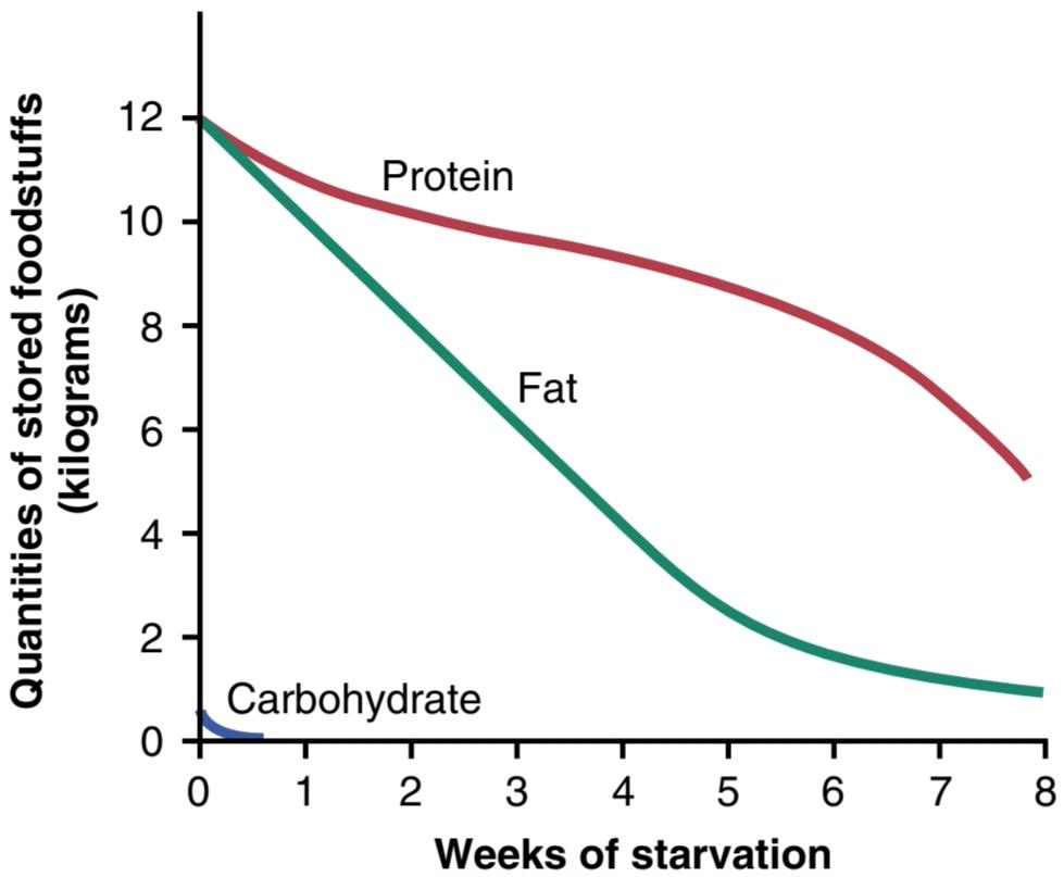
Figure 72-4. Effect of starvation on the food stores of the body.
شکل 72-4. تاثیر گرسنگی بر ذخایر غذایی بدن
Vitamin Deficiencies in Starvation. The stores of some vitamins, especially the water-soluble vitamins-the vitamin B group and vitamin C-do not last long during starvation. Consequently, after a week or more of starvation, mild vitamin deficiencies usually begin to appear, and after several weeks, severe vitamin deficiencies can occur. These deficiencies can add to the debility that leads to death.
کمبود ویتامین در گرسنگی. ذخایر برخی از ویتامینها، بهویژه ویتامینهای محلول در آب – ویتامینهای گروه B و ویتامین C – در زمان گرسنگی دوام زیادی ندارند. در نتیجه، پس از یک هفته یا بیشتر از گرسنگی، معمولاً کمبودهای خفیف ویتامین ظاهر می شود و پس از چند هفته، کمبود شدید ویتامین ممکن است رخ دهد. این کمبودها می تواند به ناتوانی منجر شود که منجر به مرگ می شود.
Vitamins
Daily Requirements of Vitamins. A vitamin is an organic compound needed in small quantities for normal metabolism that cannot be manufactured in the cells of the body. Lack of vitamins in the diet can cause important metabolic deficits. Table 72-3 lists the amounts of important vitamins required daily by the average adult man and woman. These requirements vary considerably, depending on such factors as body size, rate of growth, amount of exercise, pregnancy, and lactation.
ویتامین ها
نیاز روزانه ویتامین ها. ویتامین یک ترکیب آلی است که به مقدار کم برای متابولیسم طبیعی مورد نیاز است که در سلول های بدن ساخته نمی شود. کمبود ویتامین ها در رژیم غذایی می تواند باعث کمبودهای مهم متابولیک شود. جدول 72-3 مقادیر ویتامین های مهم مورد نیاز روزانه را برای یک زن و مرد بالغ فهرست می کند. این نیازها بسته به عواملی مانند اندازه بدن، سرعت رشد، میزان ورزش، بارداری و شیردهی بسیار متفاوت است.
Storage of Vitamins in the Body. Vitamins are stored to a slight extent in all cells. Some vitamins are stored to a major extent in the liver. For example, the quantity of vitamin A stored in the liver may be sufficient to maintain a person for 5 to 10 months without any intake of vitamin A. The quantity of vitamin D stored in the liver is usually sufficient to maintain a person for 2 to 4 months without any additional intake of vitamin D.
ذخیره ویتامین ها در بدن. ویتامین ها به مقدار کمی در تمام سلول ها ذخیره می شوند. برخی از ویتامین ها تا حد زیادی در کبد ذخیره می شوند. به عنوان مثال، مقدار ویتامین A ذخیره شده در کبد ممکن است برای حفظ یک فرد به مدت 5 تا 10 ماه بدون دریافت ویتامین A کافی باشد. مقدار ویتامین D ذخیره شده در کبد معمولاً برای حفظ یک فرد برای 2 تا 4 ماه بدون دریافت ویتامین D کافی است.
The storage of most water-soluble vitamins, especially vitamin C and most vitamin B compounds, is relatively slight. Absence of vitamin C in the diet can cause symptoms within a few weeks and can cause death from scurvy in 20 to 30 weeks. When a person’s diet is deficient in vitamin B compounds, clinical symptoms of the deficiency can sometimes be recognized within a few days (except for vitamin B12, which can last in the liver in a bound form for a year or longer).
ذخیره اکثر ویتامین های محلول در آب، به ویژه ویتامین C و بیشتر ترکیبات ویتامین B، نسبتاً اندک است. فقدان ویتامین C در رژیم غذایی می تواند در عرض چند هفته علائمی ایجاد کند و در عرض 20 تا 30 هفته باعث مرگ بر اثر اسکوربوت شود. هنگامی که رژیم غذایی فرد دچار کمبود ترکیبات ویتامین B باشد، علائم بالینی کمبود گاهی اوقات می تواند در عرض چند روز تشخیص داده شود (به جز ویتامین B12، که می تواند در کبد به صورت محدود برای یک سال یا بیشتر باقی بماند).
Table 72-3 Recommended Daily Dietary Intakes of Vitamins for Adult Men and Women
جدول 72-3 میزان مصرف روزانه ویتامین ها در رژیم غذایی برای مردان و زنان بالغ

Vitamin A
Vitamin A occurs in animal tissues as retinol. This vitamin does not occur in foods of vegetable origin, but provitamins for the formation of vitamin A do occur in abundance in many vegetable foods. These provitamins are the yellow and red carotenoid pigments, which, because their chemical structures are similar to that of vitamin A, can be changed into vitamin A in the liver.
ویتامین A
ویتامین A در بافت های حیوانی به صورت رتینول وجود دارد. این ویتامین در غذاهای با منشاء گیاهی وجود ندارد، اما پروویتامین هایی برای تشکیل ویتامین A در بسیاری از غذاهای گیاهی به وفور یافت می شود. این پروویتامینها رنگدانههای کاروتنوئیدی زرد و قرمز هستند که چون ساختار شیمیایی آنها شبیه ویتامین A است، میتوانند در کبد به ویتامین A تبدیل شوند.
Vitamin A Deficiency Causes “Night Blindness” and Abnormal Epithelial Cell Growth. One basic function of vitamin A is its use in forming retinal pigments of the eye, which is discussed in Chapter 51. Vitamin A is needed to form the visual pigments and, therefore, to prevent night blindness.
کمبود ویتامین A باعث “شب کوری” و رشد غیر طبیعی سلول های اپیتلیال می شود. یکی از عملکردهای اساسی ویتامین A استفاده از آن در تشکیل رنگدانه های شبکیه چشم است که در فصل 51 مورد بحث قرار گرفته است. ویتامین A برای تشکیل رنگدانه های بینایی و در نتیجه برای جلوگیری از شب کوری مورد نیاز است.
Vitamin A is also necessary for normal growth of most cells of the body and especially for normal growth and proliferation of the different types of epithelial cells. When vitamin A is lacking, the epithelial structures of the body tend to become stratified and keratinized. Vitamin A deficiency manifests itself by (1) scaliness of the skin and sometimes acne; (2) failure of growth of young animals, including cessation of skeletal growth; (3) failure of reproduction, associated especially with atrophy of the germinal epithelium of the testes and sometimes with interruption of the female sexual cycle; and (4) keratinization of the cornea, with resultant corneal opacity and blindness.
ویتامین A همچنین برای رشد طبیعی اکثر سلول های بدن و به ویژه برای رشد طبیعی و تکثیر انواع مختلف سلول های اپیتلیال ضروری است. هنگامی که ویتامین A کمبود داشته باشد، ساختارهای اپیتلیال بدن تمایل به لایه بندی و کراتینه شدن دارند. کمبود ویتامین A خود را با (1) پوسته پوسته شدن پوست و گاهی اوقات آکنه نشان می دهد. (2) شکست در رشد حیوانات جوان، از جمله توقف رشد اسکلتی. (3) نارسایی تولید مثل، به ویژه با آتروفی اپیتلیوم ژرمینال بیضه ها و گاهی اوقات با قطع چرخه جنسی زنان. و (4) کراتینه شدن قرنیه، با تیرگی و کوری قرنیه در نتیجه.
In vitamin A deficiency, the damaged epithelial structures often become infected (e.g., conjunctivae of the eyes, linings of the urinary tract, and respiratory passages). Vitamin A has been called an “anti-infection” vitamin.
در کمبود ویتامین A، ساختارهای اپیتلیال آسیب دیده اغلب عفونی می شوند (مانند ملتحمه چشم ها، پوشش های مجاری ادراری و مجاری تنفسی). ویتامین A را یک ویتامین “ضد عفونت” می نامند.
Thiamine (Vitamin B1)
Thiamine operates in the metabolic systems of the body principally as thiamine pyrophosphate; this compound functions as a cocarboxylase, operating mainly in conjunction with a protein decarboxylase for decarboxylation of pyruvic acid and other a-keto acids, as discussed in Chapter 68.
تیامین (ویتامین B1)
تیامین در سیستم های متابولیک بدن عمدتاً به صورت تیامین پیروفسفات عمل می کند. این ترکیب به عنوان یک کوکربوکسیلاز عمل می کند، که عمدتاً همراه با یک پروتئین دکربوکسیلاز برای دکربوکسیلاسیون اسید پیروویک و سایر اسیدهای a-keto عمل می کند، همانطور که در فصل 68 بحث شد.
Thiamine deficiency (beriberi) causes decreased utilization of pyruvic acid and some amino acids by the tissues but increased utilization of fats. Thus, thiamine is specifically needed for final metabolism of carbohydrates and many amino acids. Decreased utilization of these nutrients is responsible for many debilities associated with thiamine deficiency.
کمبود تیامین (بری بری) باعث کاهش استفاده از پیروویک اسید و برخی اسیدهای آمینه توسط بافت ها و افزایش استفاده از چربی ها می شود. بنابراین، تیامین به طور خاص برای متابولیسم نهایی کربوهیدرات ها و بسیاری از اسیدهای آمینه مورد نیاز است. کاهش استفاده از این مواد مغذی مسئول بسیاری از ناتوانی های مرتبط با کمبود تیامین است.
Thiamine Deficiency Causes Lesions of the Central and Peripheral Nervous Systems. The central nervous system normally depends almost entirely on the metabolism of carbohydrates for its energy. In thiamine deficiency, utilization of glucose by nervous tissue may be decreased by 50% to 60% and is replaced by utilization of ketone bodies derived from fat metabolism. The neuronal cells of the central nervous system frequently show chromatolysis and swelling during thiamine deficiency, changes that are characteristic of neuronal cells with poor nutrition. These changes can disrupt communication in many portions of the central nervous system.
کمبود تیامین باعث ضایعات سیستم عصبی مرکزی و محیطی می شود. سیستم عصبی مرکزی معمولاً برای انرژی خود تقریباً به طور کامل به متابولیسم کربوهیدرات ها بستگی دارد. در کمبود تیامین، استفاده از گلوکز توسط بافت عصبی ممکن است 50% تا 60% کاهش یابد و با استفاده از اجسام کتون حاصل از متابولیسم چربی جایگزین شود. سلول های عصبی سیستم عصبی مرکزی اغلب در طول کمبود تیامین، کروماتولیز و تورم را نشان می دهند، تغییراتی که مشخصه سلول های عصبی با تغذیه نامناسب است. این تغییرات می تواند ارتباط را در بسیاری از بخش های سیستم عصبی مرکزی مختل کند.
Thiamine deficiency can cause degeneration of myelin sheaths of nerve fibers in both the peripheral nerves and the central nervous system. Lesions in peripheral nerves frequently cause them to become extremely irritable, resulting in “polyneuritis,” characterized by pain radiating along the course of one or many peripheral nerves. Also, fiber tracts in the cord can degenerate to such an extent that paralysis occasionally results; even in the absence of paralysis, the muscles atrophy, resulting in severe weakness.
کمبود تیامین می تواند باعث تخریب غلاف میلین رشته های عصبی در اعصاب محیطی و سیستم عصبی مرکزی شود. ضایعات در اعصاب محیطی اغلب باعث تحریک شدید آنها می شود که منجر به “پلی نوریت” می شود که با درد در طول مسیر یک یا چند اعصاب محیطی تابش می شود. همچنین، مجاری فیبر در بند ناف می تواند به حدی تخریب شود که گهگاه فلج شود. حتی در غیاب فلج، عضلات آتروفی می شوند و در نتیجه ضعف شدید ایجاد می شود.
Thiamine Deficiency Weakens the Heart and Causes Peripheral Vasodilation. Cardiac failure eventually develops in a person with severe thiamine deficiency because of weakened cardiac muscle. Furthermore, the venous return of blood to the heart may be increased to as much as two times normal, because thiamine deficiency causes peripheral vasodilation throughout the circulatory system, presumably as a result of decreased release of metabolic energy in the tissues, leading to local vascular dilation. The cardiac effects of thiamine deficiency are due partly to high blood flow into the heart and partly to primary weakness of the cardiac muscle. Peripheral edema and ascites also occur to a major extent in some people with thiamine deficiency, mainly because of cardiac failure.
کمبود تیامین قلب را ضعیف می کند و باعث اتساع عروق محیطی می شود. نارسایی قلبی در نهایت در فردی با کمبود شدید تیامین به دلیل ضعیف شدن عضله قلب ایجاد می شود. علاوه بر این، بازگشت وریدی خون به قلب ممکن است تا دو برابر طبیعی افزایش یابد، زیرا کمبود تیامین باعث اتساع عروق محیطی در سراسر سیستم گردش خون می شود، احتمالاً در نتیجه کاهش آزاد شدن انرژی متابولیک در بافت ها، که منجر به اتساع موضعی عروق می شود. اثرات قلبی کمبود تیامین تا حدی به دلیل جریان خون بالا به قلب و تا حدی به دلیل ضعف اولیه عضله قلب است. ادم محیطی و آسیت نیز تا حد زیادی در برخی از افراد مبتلا به کمبود تیامین رخ می دهد که عمدتاً به دلیل نارسایی قلبی است.
Thiamine Deficiency Causes Gastrointestinal Tract Disturbances. Among the gastrointestinal symptoms of thiamine deficiency are indigestion, severe constipation, anorexia, gastric atony, and hypochlorhydria. All these effects presumably result from failure of the smooth muscle and glands of the gastrointestinal tract to derive sufficient energy from carbohydrate metabolism.
کمبود تیامین باعث اختلالات دستگاه گوارش می شود. از علائم گوارشی کمبود تیامین می توان به سوء هاضمه، یبوست شدید، بی اشتهایی، آتونی معده و هیپوکلریدری اشاره کرد. همه این اثرات احتمالاً ناشی از ناتوانی عضلات صاف و غدد دستگاه گوارش در به دست آوردن انرژی کافی از متابولیسم کربوهیدرات است.
The overall picture of thiamine deficiency, including polyneuritis, cardiovascular symptoms, and gastrointestinal disorders, is frequently referred to as beriberi-especially when the cardiovascular symptoms predominate.
تصویر کلی کمبود تیامین، از جمله پلی نوریت، علائم قلبی عروقی، و اختلالات گوارشی، اغلب به عنوان بری بری شناخته می شود، به خصوص زمانی که علائم قلبی عروقی غالب باشد.
Riboflavin (Vitamin B2)
Riboflavin normally combines in the tissues with phosphoric acid to form two coenzymes, flavin mononucleotide (FMN) and flavin adenine dinucleotide (FAD). They operate as hydrogen carriers in important oxidative systems of the mitochondria. NAD, operating in association with specific dehydrogenases, usually accepts hydrogen removed from various food substrates and then passes the hydrogen to FMN or FAD. Finally, the hydrogen is released as an ion into the mitochondrial matrix to become oxidized by oxygen (described in Chapter 68).
ریبوفلاوین (ویتامین B2)
ریبوفلاوین به طور معمول در بافت ها با اسید فسفریک ترکیب می شود و دو کوآنزیم فلاوین مونوکلئوتید (FMN) و فلاوین آدنین دی نوکلئوتید (FAD) را تشکیل می دهد. آنها به عنوان حامل هیدروژن در سیستم های اکسیداتیو مهم میتوکندری عمل می کنند. NAD که در ارتباط با دهیدروژنازهای خاص عمل می کند، معمولاً هیدروژن حذف شده از بسترهای مختلف غذایی را می پذیرد و سپس هیدروژن را به FMN یا FAD می دهد. در نهایت، هیدروژن به عنوان یک یون در ماتریکس میتوکندری آزاد می شود تا توسط اکسیژن اکسید شود (توضیح داده شده در فصل 68).
Deficiency of riboflavin in experimental animals causes severe dermatitis, vomiting, diarrhea, muscle spasticity that finally becomes muscle weakness, coma, and decline in body temperature, followed by death. Thus, severe riboflavin deficiency can cause many of the same effects as a lack of niacin in the diet; presumably, the debilities that result in each case are due to generally depressed oxidative processes within the cells.
کمبود ریبوفلاوین در حیوانات آزمایشگاهی باعث درماتیت شدید، استفراغ، اسهال، اسپاسم عضلانی می شود که در نهایت به ضعف عضلانی، کما و کاهش دمای بدن و به دنبال آن مرگ تبدیل می شود. بنابراین، کمبود شدید ریبوفلاوین می تواند بسیاری از اثرات مشابه کمبود نیاسین در رژیم غذایی را ایجاد کند. احتمالاً، ناتوانیهایی که در هر مورد به وجود میآیند ناشی از فرآیندهای اکسیداتیو عموماً افسرده درون سلولها است.
In the human being, there are no known cases of riboflavin deficiency severe enough to cause the marked debilities noted in animal studies, but mild riboflavin deficiency is probably common. Such deficiency causes digestive disturbances, burning sensations of the skin and eyes, cracking at the corners of the mouth, headaches, mental depression, forgetfulness, and so on.
در انسان، هیچ مورد شناخته شده ای از کمبود ریبوفلاوین وجود ندارد که به اندازه کافی شدید باشد که باعث ناتوانی های مشخص شده در مطالعات حیوانی شود، اما کمبود خفیف ریبوفلاوین احتمالاً شایع است. چنین کمبودی باعث اختلالات گوارشی، سوزش پوست و چشم، ترک خوردن گوشه های دهان، سردرد، افسردگی روانی، فراموشی و … می شود.
Although the manifestations of riboflavin deficiency are usually relatively mild, this deficiency frequently occurs in association with deficiency of thiamine, niacin, or both. Many deficiency syndromes, including pellagra, beriberi, sprue, and kwashiorkor, are probably due to a combined deficiency of a number of vitamins, as well as other aspects of malnutrition.
اگرچه تظاهرات کمبود ریبوفلاوین معمولاً نسبتاً خفیف است، این کمبود اغلب در ارتباط با کمبود تیامین، نیاسین یا هر دو رخ میدهد. بسیاری از سندرم های کمبود، از جمله پلاگرا، بری بری، اسپرو و کواشیورکور احتمالاً به دلیل کمبود ترکیبی تعدادی از ویتامین ها و همچنین سایر جنبه های سوء تغذیه است.
Niacin (Vitamin B3)
Niacin, also called nicotinic acid, functions in the body as coenzymes in the form of nicotinamide adenine dinucleotide (NAD) and NAD phosphate. These coenzymes are hydrogen acceptors and combine with hydrogen atoms as they are removed from food substrates by many types of dehydrogenases. The typical operation of both these coenzymes is presented in Chapter 68. When niacin deficiency exists, the normal rate of dehydrogenation cannot be maintained; therefore, oxidative delivery of energy from the foodstuffs to the functioning elements of all cells cannot occur at normal rates.
نیاسین (ویتامین B3)
نیاسین که اسید نیکوتینیک نیز نامیده می شود، به عنوان کوآنزیم در بدن به شکل نیکوتین آمید آدنین دی نوکلئوتید (NAD) و NAD فسفات عمل می کند. این کوآنزیمها پذیرنده هیدروژن هستند و با اتمهای هیدروژن ترکیب میشوند، زیرا توسط بسیاری از انواع دهیدروژنازها از بسترهای غذایی حذف میشوند. عملکرد معمول هر دو این کوآنزیم ها در فصل 68 ارائه شده است. هنگامی که کمبود نیاسین وجود داشته باشد، سرعت طبیعی دهیدروژناسیون نمی تواند حفظ شود. بنابراین، تحویل اکسیداتیو انرژی از مواد غذایی به عناصر عملکردی همه سلول ها نمی تواند با سرعت طبیعی انجام شود.
In the early stages of niacin deficiency, simple physiological changes such as muscle weakness and poor glandular secretion may occur, but in the case of severe niacin deficiency, actual tissue death ensues. Pathological lesions appear in many parts of the central nervous system, and permanent dementia or many types of psychoses may result. In addition, the skin develops a cracked, pigmented scaliness in areas that are exposed to mechanical irritation or sun irradiation; thus, in persons with niacin deficiency, the skin is unable to repair irritative damage.
در مراحل اولیه کمبود نیاسین، تغییرات فیزیولوژیکی ساده مانند ضعف عضلانی و ترشح ضعیف غدد ممکن است رخ دهد، اما در صورت کمبود شدید نیاسین، مرگ واقعی بافت رخ می دهد. ضایعات پاتولوژیک در بسیاری از قسمت های سیستم عصبی مرکزی ظاهر می شوند و ممکن است زوال عقل دائمی یا بسیاری از انواع روان پریشی ایجاد شود. علاوه بر این، پوست در مناطقی که در معرض تحریک مکانیکی یا تابش نور خورشید قرار دارند، پوسته پوسته شدن ترک خورده و رنگدانه ای ایجاد می کند. بنابراین، در افراد مبتلا به کمبود نیاسین، پوست قادر به ترمیم آسیب های تحریکی نیست.
Niacin deficiency causes intense irritation and inflammation of the mucous membranes of the mouth and other portions of the gastrointestinal tract, resulting in many digestive abnormalities that can lead to widespread gastrointestinal hemorrhage in severe cases. It is possible that this condition results from generalized depression of metabolism in the gastrointestinal epithelium and failure of appropriate epithelial repair.
کمبود نیاسین باعث تحریک و التهاب شدید غشاهای مخاطی دهان و سایر بخشهای دستگاه گوارش میشود و در نتیجه بسیاری از ناهنجاریهای گوارشی ایجاد میشود که در موارد شدید میتواند منجر به خونریزی گسترده دستگاه گوارش شود. ممکن است این وضعیت ناشی از افسردگی عمومی متابولیسم در اپیتلیوم دستگاه گوارش و شکست در ترمیم مناسب اپیتلیال باشد.
The clinical entity called pellagra and the canine disease called black tongue are caused mainly by niacin deficiency. Pellagra is greatly exacerbated in people on a corn diet be- cause corn is deficient in the amino acid tryptophan, which can be converted in limited quantities to niacin in the body.
موجودیت بالینی به نام پلاگر و بیماری سگ به نام زبان سیاه عمدتاً ناشی از کمبود نیاسین است. پلاگر در افرادی که از رژیم غذایی ذرت استفاده می کنند به شدت تشدید می شود زیرا ذرت دارای کمبود اسید آمینه تریپتوفان است که می تواند در مقادیر محدودی به نیاسین در بدن تبدیل شود.
Pantothenic Acid (Vitamin B5)
Pantothenic acid is mainly incorporated in the body into coenzyme A (COA), which has many metabolic roles in the cells. Two of these roles discussed at length in Chapters 68 and 69 are (1) conversion of decarboxylated pyruvic acid into acetyl-CoA before its entry into the citric acid cycle and (2) degradation of fatty acid molecules into multiple molecules of acetyl-CoA. Thus, lack of pantothenic acid can lead to depressed metabolism of carbohydrates and fats.
اسید پانتوتنیک (ویتامین B5)
اسید پانتوتنیک عمدتاً در بدن به کوآنزیم A (COA) وارد می شود که نقش های متابولیکی زیادی در سلول ها دارد. دو مورد از این نقشهایی که در فصلهای 68 و 69 به طور مفصل مورد بحث قرار گرفتند عبارتند از (1) تبدیل اسید پیروویک دکربوکسیله به استیل-CoA قبل از ورود آن به چرخه اسید سیتریک و (2) تجزیه مولکولهای اسید چرب به مولکولهای متعدد استیل-CoA. بنابراین، کمبود پانتوتنیک اسید می تواند منجر به کاهش متابولیسم کربوهیدرات ها و چربی ها شود.
Deficiency of pantothenic acid in experimental animals can cause retarded growth, failure of reproduction, graying of the hair, dermatitis, fatty liver, and hemorrhagic adrenocortical necrosis. In humans, no definite deficiency syndrome has been proved, presumably because of the wide occurrence of this vitamin in almost all foods and because small amounts can probably be synthesized in the body. This situation does not mean that pantothenic acid is not of value in the metabolic systems of the body; indeed, it is perhaps as necessary as any other vitamin.
کمبود پانتوتنیک اسید در حیوانات آزمایشگاهی می تواند باعث تاخیر در رشد، نارسایی تولید مثل، سفید شدن موها، درماتیت، کبد چرب و نکروز هموراژیک قشر آدرنال شود. در انسان، هیچ سندرم کمبود قطعی ثابت نشده است، احتمالاً به دلیل وجود گسترده این ویتامین در تقریباً همه غذاها و به دلیل اینکه احتمالاً مقادیر کمی از آن در بدن سنتز می شود. این وضعیت به این معنی نیست که اسید پانتوتنیک در سیستم های متابولیک بدن ارزشی ندارد. در واقع، شاید به اندازه هر ویتامین دیگری ضروری باشد.
Pyridoxine (Vitamin B6)
Pyridoxine exists in the form of pyridoxal phosphate in the cells and functions as a coenzyme for many chemical reactions related to amino acid and protein metabolism. Its most important role is that of coenzyme in the transamination process for the synthesis of amino acids. As a result, pyridoxine plays many key roles in metabolism, especially protein metabolism. Also, it is believed to act in the transport of some amino acids across cell membranes.
پیریدوکسین (ویتامین B6)
پیریدوکسین به شکل پیریدوکسال فسفات در سلول ها وجود دارد و به عنوان کوآنزیم برای بسیاری از واکنش های شیمیایی مربوط به متابولیسم اسید آمینه و پروتئین عمل می کند. مهمترین نقش آن کوآنزیم در فرآیند ترانس آمینواسیون برای سنتز اسیدهای آمینه است. در نتیجه، پیریدوکسین نقش های کلیدی بسیاری در متابولیسم، به ویژه متابولیسم پروتئین ایفا می کند. همچنین، اعتقاد بر این است که در انتقال برخی از اسیدهای آمینه از طریق غشای سلولی عمل می کند.
Dietary lack of pyridoxine in experimental animals can cause dermatitis, decreased rate of growth, development of fatty liver, anemia, and evidence of mental deterioration. Rarely, in children, pyridoxine deficiency has been known to cause seizures, dermatitis, and gastrointestinal disturbances such as nausea and vomiting.
کمبود پیریدوکسین در رژیم غذایی در حیوانات آزمایشگاهی می تواند باعث درماتیت، کاهش سرعت رشد، ایجاد کبد چرب، کم خونی و شواهد زوال ذهنی شود. به ندرت، کمبود پیریدوکسین در کودکان باعث تشنج، درماتیت و اختلالات گوارشی مانند تهوع و استفراغ می شود.
Vitamin B12
Several cobalamin compounds that possess a common prosthetic group exhibit so-called vitamin B12 activity. This prosthetic group contains cobalt, which has bonds similar to those of iron in the hemoglobin molecule. It is likely that the cobalt atom functions in much the same way that the iron atom functions to combine reversibly with other substances.
ویتامین B12
چندین ترکیب کوبالامین که دارای یک گروه مصنوعی مشترک هستند به اصطلاح فعالیت ویتامین B12 را نشان می دهند. این گروه مصنوعی حاوی کبالت است که پیوندهایی شبیه پیوندهای آهن در مولکول هموگلوبین دارد. این احتمال وجود دارد که اتم کبالت به همان شیوه ای عمل کند که عملکرد اتم آهن به صورت برگشت پذیر با سایر مواد ترکیب می شود.
Vitamin B12 Deficiency Causes Pernicious Anemia. Vitamin B12 performs several metabolic functions in its role as a hydrogen acceptor coenzyme. Its most important function is to act as a coenzyme for reducing ribonucleotides to deoxyribonucleotides, a step that is necessary in the replication of genes and could explain the major functions of vitamin B12: (1) promotion of growth and (2) promotion of red blood cell formation and maturation. This red blood cell function is described in detail in Chapter 33 in relation to pernicious anemia, a type of anemia caused by failure of red blood cell maturation when vitamin B12 is deficient.
کمبود ویتامین B12 باعث کم خونی خطرناک می شود. ویتامین B12 در نقش خود به عنوان کوآنزیم گیرنده هیدروژن چندین عملکرد متابولیک را انجام می دهد. مهمترین عملکرد آن این است که به عنوان یک کوآنزیم برای احیای ریبونوکلئوتیدها به دئوکسی ریبونوکلئوتیدها عمل کند، مرحله ای که در تکثیر ژن ها ضروری است و می تواند وظایف اصلی ویتامین B12 را توضیح دهد: (1) ارتقاء رشد و (2) ارتقاء تشکیل و بلوغ گلبول های قرمز. این عملکرد گلبول قرمز در فصل 33 به تفصیل در رابطه با کم خونی پرنیشیوز توضیح داده شده است، یک نوع کم خونی ناشی از عدم بلوغ گلبول های قرمز خون در صورت کمبود ویتامین B12.
Vitamin B12 Deficiency Causes Demyelination of the Large Nerve Fibers of the Spinal Cord. The demyelination of nerve fibers in people with vitamin B12 deficiency occurs especially in the posterior columns, and occasionally the lateral columns, of the spinal cord. As a result, many people with pernicious anemia have loss of peripheral sensation and, in severe cases, even become paralyzed.
کمبود ویتامین B12 باعث دمیلینه شدن رشته های عصبی بزرگ نخاع می شود. دمیلینه شدن رشته های عصبی در افراد مبتلا به کمبود ویتامین B12 به ویژه در ستون های خلفی و گاهی اوقات ستون های جانبی نخاع رخ می دهد. در نتیجه، بسیاری از افراد مبتلا به کم خونی خطرناک، حس محیطی را از دست می دهند و در موارد شدید، حتی فلج می شوند.
The usual cause of vitamin B12 deficiency is not lack of this vitamin in the food but deficiency of formation of intrinsic factor, which is normally secreted by the parietal cells of the gastric glands and is essential for absorption of vitamin B12 by the ileal mucosa. This topic is discussed in Chapters 33 and 67.
علت معمول کمبود ویتامین B12 کمبود این ویتامین در غذا نیست، بلکه کمبود تشکیل فاکتور ذاتی است که معمولاً توسط سلول های جداری غدد معده ترشح می شود و برای جذب ویتامین B12 توسط مخاط روده ضروری است. این موضوع در فصل های 33 و 67 مورد بحث قرار گرفته است.
Folic Acid (Pteroylglutamic Acid, Vitamin B,)
Several pteroylglutamic acids exhibit the “folic acid effect.” Folic acid functions as a carrier of hydroxymethyl and formyl groups. Perhaps its most important use in the body is in the synthesis of purines and thymine, which are required for DNA formation. Therefore, folic acid, like vitamin B12, is required for replication of the cellular genes and may explain one of the most important functions of folic acid-to promote growth. Indeed, when it is absent from the diet, an animal grows very little.
اسید فولیک (پتروئیل گلوتامیک اسید، ویتامین B،)
چندین اسید پتروئیل گلوتامیک “اثر اسید فولیک” را نشان می دهند. اسید فولیک به عنوان حامل گروه های هیدروکسی متیل و فرمیل عمل می کند. شاید مهمترین کاربرد آن در بدن در سنتز پورین و تیمین باشد که برای تشکیل DNA مورد نیاز است. بنابراین، اسید فولیک، مانند ویتامین B12، برای تکثیر ژنهای سلولی مورد نیاز است و ممکن است یکی از مهمترین عملکردهای اسید فولیک را برای تقویت رشد توضیح دهد. در واقع، هنگامی که در رژیم غذایی وجود ندارد، یک حیوان رشد بسیار کمی دارد.
Folic acid is an even more potent growth promoter than vitamin B12 and, like vitamin B12, it is important for the maturation of red blood cells, as discussed in Chapter 33. However, vitamin B12 and folic acid each perform specific and different chemical functions in promoting growth and maturation of red blood cells. One of the significant effects of folic acid deficiency is the development of macrocytic anemia, almost identical to that which occurs in pernicious anemia. This often can be treated effectively with folic acid alone.
اسید فولیک حتی یک محرک رشد قویتر از ویتامین B12 است و مانند ویتامین B12 برای بلوغ گلبولهای قرمز مهم است، همانطور که در فصل 33 بحث شد. با این حال، ویتامین B12 و اسید فولیک هر کدام عملکردهای شیمیایی خاص و متفاوتی را در رشد و بلوغ گلبولهای قرمز انجام میدهند. یکی از اثرات مهم کمبود اسید فولیک، ایجاد کم خونی ماکروسیتیک است که تقریباً مشابه آنمی است که در کم خونی خطرناک رخ می دهد. این اغلب می تواند به طور موثر با اسید فولیک به تنهایی درمان شود.
Ascorbic Acid (Vitamin C)
Ascorbic Acid Deficiency Weakens Collagen Fibers Throughout the Body. Ascorbic acid is essential for activating the enzyme prolyl hydroxylase, which promotes the hydroxylation step in the formation of hydroxyproline, an integral constituent of collagen. Without ascorbic acid, collagen fibers that are formed in virtually all tissues of the body are defective and weak. Therefore, this vitamin is essential for growth and strength of the fibers in subcutaneous tissue, cartilage, bone, and teeth.
اسید اسکوربیک (ویتامین C)
کمبود اسید اسکوربیک باعث ضعیف شدن فیبرهای کلاژن در سراسر بدن می شود. اسید اسکوربیک برای فعال کردن آنزیم پرولیل هیدروکسیلاز ضروری است، که مرحله هیدروکسیلاسیون را در تشکیل هیدروکسی پرولین، یک جزء جدایی ناپذیر از کلاژن، ترویج می کند. بدون اسید آسکوربیک، رشته های کلاژنی که تقریباً در تمام بافت های بدن تشکیل می شوند، معیوب و ضعیف هستند. بنابراین، این ویتامین برای رشد و استحکام الیاف در بافت زیر جلدی، غضروف، استخوان و دندان ضروری است.
Ascorbic Acid Deficiency Causes Scurvy. Deficiency of ascorbic acid for 20 to 30 weeks causes scurvy. One of the most important effects of scurvy is failure of wounds to heal. This condition is caused by failure of the cells to deposit collagen fibrils and intercellular cement substances. As a result, healing of a wound may require several months instead of the several days ordinarily necessary.
کمبود اسید اسکوربیک باعث اسکوربوت می شود. کمبود اسید اسکوربیک به مدت 20 تا 30 هفته باعث ایجاد اسکوربوت می شود. یکی از مهمترین عوارض اسکوربوت، عدم التیام زخم است. این وضعیت به دلیل شکست سلول ها در رسوب فیبرهای کلاژن و مواد سیمان بین سلولی ایجاد می شود. در نتیجه، بهبود زخم ممکن است به چندین ماه به جای چندین روز که معمولاً ضروری است نیاز داشته باشد.
Lack of ascorbic acid also causes cessation of bone growth. The cells of the growing epiphyses continue to proliferate, but no new collagen is laid down between the cells, and the bones fracture easily at the point of growth because of failure to ossify. Also, when an already ossified bone fractures in a person with ascorbic acid deficiency, the osteoblasts cannot form new bone matrix. Consequently, the fractured bone does not heal.
کمبود اسید اسکوربیک نیز باعث توقف رشد استخوان می شود. سلولهای اپیفیزهای در حال رشد به تکثیر ادامه میدهند، اما کلاژن جدیدی بین سلولها گذاشته نمیشود و استخوانها بهدلیل عدم استخوانبندی به راحتی در نقطه رشد میشکنند. همچنین، هنگامی که یک استخوان از قبل استخوانی شده در فردی با کمبود اسید اسکوربیک شکسته می شود، استئوبلاست ها نمی توانند ماتریکس استخوان جدیدی تشکیل دهند. در نتیجه، استخوان شکسته بهبود نمی یابد.
The blood vessel walls become extremely fragile in persons with scurvy because of (1) failure of the endothelial cells to be cemented together properly and (2) failure to form the collagen fibrils normally present in vessel walls. The capillaries are especially likely to rupture, and as a result, many small petechial hemorrhages occur throughout the body. The hemorrhages beneath the skin cause purpuric blotches, sometimes over the entire body. In extreme cases of scurvy, the muscle cells sometimes fragment; lesions of the gums occur, with loosening of the teeth; infections of the mouth develop; and vomiting of blood, bloody stools, and cerebral hemorrhage can all occur. Finally, high fever often develops before death.
دیوارههای رگهای خونی در افراد مبتلا به اسکوربوت به دلیل (1) عدم اتصال صحیح سلولهای اندوتلیال به هم و (2) تشکیل نشدن فیبرهای کلاژنی که معمولاً در دیوارههای عروق وجود دارد، بسیار شکننده میشوند. مویرگ ها به ویژه احتمال پارگی دارند و در نتیجه خونریزی های کوچک پتشیال در سراسر بدن رخ می دهد. خونریزی های زیر پوست باعث ایجاد لکه های پورپوریک، گاهی اوقات در کل بدن می شود. در موارد شدید اسکوربوت، سلول های ماهیچه ای گاهی تکه تکه می شوند. ضایعات لثه با شل شدن دندان ها رخ می دهد. عفونت های دهان ایجاد می شود؛ و استفراغ خون، مدفوع خونی و خونریزی مغزی همگی ممکن است رخ دهد. در نهایت، تب بالا اغلب قبل از مرگ ایجاد می شود.
Vitamin D
Vitamin D (calciferol) increases calcium absorption from the gastrointestinal tract and helps control calcium deposition in the bone. The mechanism by which vitamin D increases calcium absorption is mainly to promote active transport of calcium through the epithelium of the ileum. In particular, it increases the formation of a calcium-binding protein in the intestinal epithelial cells that aids in calcium absorption. The specific functions of vitamin D in relation to overall body calcium metabolism and bone formation are presented in Chapter 80.
ویتامین D
ویتامین D (کلسیفرول) جذب کلسیم را از دستگاه گوارش افزایش می دهد و به کنترل رسوب کلسیم در استخوان کمک می کند. مکانیسمی که توسط آن ویتامین D جذب کلسیم را افزایش می دهد، عمدتاً ترویج انتقال فعال کلسیم از طریق اپیتلیوم ایلئوم است. به طور خاص، تشکیل پروتئین اتصال دهنده کلسیم را در سلول های اپیتلیال روده افزایش می دهد که به جذب کلسیم کمک می کند. عملکردهای خاص ویتامین D در رابطه با متابولیسم کلسیم بدن و تشکیل استخوان در فصل 80 ارائه شده است.
Vitamin E (Alpha-Tocopherol)
Several related compounds exhibit so-called vitamin E activity. Only rare cases of proved vitamin E deficiency have occurred in human beings. In animal studies, lack of vitamin E can cause degeneration of the germinal epithelium in the testis and, therefore, can cause male sterility. Lack of vitamin E can also cause resorption of a fetus after conception in the female. Because of these effects of vitamin E deficiency, vitamin E is sometimes called the “antisterility vitamin.” Deficiency of vita- min E prevents normal growth and sometimes causes degeneration of the renal tubular cells and the muscle cells.
ویتامین E (آلفا توکوفرول)
چندین ترکیب مرتبط به اصطلاح فعالیت ویتامین E را نشان می دهند. تنها موارد نادری از کمبود ویتامین E ثابت شده در انسان رخ داده است. در مطالعات حیوانی، کمبود ویتامین E می تواند باعث انحطاط اپیتلیوم ژرمینال در بیضه و در نتیجه باعث عقیمی مردان شود. کمبود ویتامین E همچنین می تواند باعث تحلیل جنین پس از لقاح در زن شود. به دلیل این اثرات ناشی از کمبود ویتامین E، ویتامین E گاهی اوقات “ویتامین ضد عقیمی” نامیده می شود. کمبود ویتامین E از رشد طبیعی جلوگیری میکند و گاهی اوقات باعث تخریب سلولهای لولهای کلیه و سلولهای ماهیچهای میشود.
Vitamin E is believed to play a protective role in the prevention of oxidation of unsaturated fats. In the absence of vitamin E, the quantity of unsaturated fats in the cells becomes diminished, causing abnormal structure and function of such cellular organelles as the mitochondria, the lysosomes, and even the cell membrane.
اعتقاد بر این است که ویتامین E نقش محافظتی در جلوگیری از اکسیداسیون چربی های غیر اشباع دارد. در غیاب ویتامین E، مقدار چربی های اشباع نشده در سلول ها کاهش می یابد و باعث ایجاد ساختار و عملکرد غیر طبیعی اندامک های سلولی مانند میتوکندری، لیزوزوم ها و حتی غشای سلولی می شود.
Vitamin K
Vitamin K is an essential co-factor to a liver enzyme that adds a carboxyl group to factors II (prothrombin), VII (pro- convertin), IX, and X, all of which are important in blood coagulation. Without this carboxylation, these coagulation factors are inactive. Therefore, when vitamin K deficiency occurs, blood clotting is retarded. The function of this vitamin and its relation to some of the anticoagulants, such as dicumarol, are presented in greater detail in Chapter 37.
ویتامین K
ویتامین K یک عامل کمکی ضروری برای آنزیم کبدی است که یک گروه کربوکسیل را به فاکتورهای II (پرترومبین)، VII (پرو کانورتین)، IX و X اضافه میکند که همگی در انعقاد خون مهم هستند. بدون این کربوکسیلاسیون، این عوامل انعقادی غیر فعال هستند. بنابراین، هنگامی که کمبود ویتامین K رخ می دهد، لخته شدن خون به تاخیر می افتد. عملکرد این ویتامین و ارتباط آن با برخی از داروهای ضد انعقاد مانند دیکومارول با جزئیات بیشتر در فصل 37 ارائه شده است.
Several natural and synthetic compounds exhibit vitamin K activity. Because vitamin K is synthesized by bacteria in the colon, it is rare for a person to have a bleeding tendency because of vitamin K deficiency in the diet. However, when the bacteria of the colon are destroyed by administration of large quantities of antibiotic drugs, vitamin K deficiency occurs rapidly because of the paucity of this compound in the normal diet.
چندین ترکیب طبیعی و مصنوعی فعالیت ویتامین K را نشان می دهند. از آنجایی که ویتامین K توسط باکتری های موجود در روده بزرگ سنتز می شود، به ندرت پیش می آید که فردی به دلیل کمبود ویتامین K در رژیم غذایی، تمایل به خونریزی داشته باشد. با این حال، هنگامی که باکتری های روده بزرگ با تجویز مقادیر زیادی از داروهای آنتی بیوتیک از بین می روند، کمبود ویتامین K به سرعت به دلیل کمبود این ترکیب در رژیم غذایی معمولی رخ می دهد.
Mineral Metabolism
The functions of many minerals, such as sodium, potassium, and chloride, are presented at appropriate points in the text. Only specific functions of minerals not covered elsewhere are mentioned here. The average recommended or adequate daily dietary intakes of these minerals for adult men and women are provided in Table 72-4.
متابولیسم مواد معدنی
عملکرد بسیاری از مواد معدنی مانند سدیم، پتاسیم و کلرید در نقاط مناسب در متن ارائه شده است. در اینجا فقط به وظایف خاصی از مواد معدنی اشاره شده است که در جای دیگری پوشش داده نشده است. متوسط مصرف روزانه توصیه شده یا کافی این مواد معدنی برای مردان و زنان بالغ در جدول 72-4 ارائه شده است.
Magnesium. Magnesium is about one sixth as plentiful in cells as potassium. Magnesium is required as a catalyst for many intracellular enzymatic reactions, particularly those related to carbohydrate metabolism.
منیزیم. منیزیم در سلول ها حدود یک ششم به اندازه پتاسیم فراوان است. منیزیم به عنوان یک کاتالیزور برای بسیاری از واکنش های آنزیمی درون سلولی، به ویژه آنهایی که مربوط به متابولیسم کربوهیدرات هستند، مورد نیاز است.
The extracellular fluid magnesium concentration is slight, only 1.8 to 2.5 mEq/L. Increased extracellular concentration of magnesium depresses nervous system activity, as well as skeletal muscle contraction. This latter effect can be blocked by the administration of calcium. Low magnesium concentration causes increased irritability of the nervous system, peripheral vasodilation, and cardiac arrhythmias, especially after acute myocardial infarction.
غلظت منیزیم مایع خارج سلولی اندک است، تنها 1.8 تا 2.5 mEq/L. افزایش غلظت خارج سلولی منیزیم باعث کاهش فعالیت سیستم عصبی و همچنین انقباض عضلات اسکلتی می شود. این اثر اخیر را می توان با تجویز کلسیم مسدود کرد. غلظت کم منیزیم باعث افزایش تحریک پذیری سیستم عصبی، اتساع عروق محیطی و آریتمی قلبی به ویژه پس از انفارکتوس حاد میوکارد می شود.
Calcium. Calcium is present in the body mainly in the form of calcium phosphate in the bone. This subject is dis- cussed in detail in Chapter 80, as is the calcium content of extracellular fluid. Excess quantities of calcium ions in extracellular fluid can cause the heart to stop in systole and can act as a mental depressant. At the other extreme, low levels of calcium can cause spontaneous discharge of nerve fibers, resulting in tetany, as discussed in Chapter 80.
کلسیم. کلسیم در بدن عمدتا به شکل فسفات کلسیم در استخوان وجود دارد. این موضوع به طور مفصل در فصل 80 مورد بحث قرار گرفته است، همانطور که محتوای کلسیم مایع خارج سلولی نیز مورد بحث قرار گرفته است. مقادیر بیش از حد یون کلسیم در مایع خارج سلولی می تواند باعث توقف قلب در سیستول شود و می تواند به عنوان یک افسردگی روانی عمل کند. از طرف دیگر، سطوح پایین کلسیم می تواند باعث تخلیه خود به خودی رشته های عصبی شود که منجر به کزاز می شود، همانطور که در فصل 80 بحث شد.
Phosphorus. Phosphate is the major anion of intracellular fluid. Phosphates have the ability to combine reversibly with many coenzyme systems and with multiple other compounds that are necessary for the operation of metabolic processes. Many important reactions of phosphates have been catalogued at other points in this text, especially in relation to the functions of adenosine triphosphate, adenosine diphosphate, phosphocreatine, and so forth. Also, bone contains a large amount of calcium phosphate, which is discussed in Chapter 80.
فسفر. فسفات آنیون اصلی مایع داخل سلولی است. فسفات ها توانایی ترکیب برگشت پذیر با بسیاری از سیستم های کوآنزیمی و چندین ترکیب دیگر را دارند که برای عملکرد فرآیندهای متابولیک ضروری هستند. بسیاری از واکنشهای مهم فسفاتها در سایر نقاط این متن فهرستبندی شدهاند، به ویژه در رابطه با عملکرد آدنوزین تری فسفات، آدنوزین دی فسفات، فسفوکراتین و غیره. همچنین استخوان حاوی مقدار زیادی فسفات کلسیم است که در فصل 80 مورد بحث قرار گرفته است.
Iron. The function of iron in the body, especially in relation to the formation of hemoglobin, is discussed in Chapter 33. Two thirds of the iron in the body is in the form of hemoglobin, although smaller quantities are present in other forms, especially in the liver and bone marrow. Electron carriers containing iron (especially the cytochromes) are present in the mitochondria of all cells of the body and are essential for most of the oxidation that occurs in the cells. Therefore, iron is absolutely essential for transport of oxygen to the tissues and operation of oxidative systems within the tissue cells, without which life would cease within a few seconds.
آهن. عملکرد آهن در بدن، به ویژه در رابطه با تشکیل هموگلوبین، در فصل 33 مورد بحث قرار گرفته است. دو سوم آهن موجود در بدن به شکل هموگلوبین است، اگرچه مقادیر کمتری در اشکال دیگر، به ویژه در کبد و مغز استخوان وجود دارد. حامل های الکترونی حاوی آهن (به ویژه سیتوکروم ها) در میتوکندری تمام سلول های بدن وجود دارند و برای بیشتر اکسیداسیونی که در سلول ها اتفاق می افتد ضروری هستند. بنابراین آهن برای انتقال اکسیژن به بافتها و عملکرد سیستمهای اکسیداتیو درون سلولهای بافت کاملاً ضروری است که بدون آن حیات در عرض چند ثانیه متوقف میشود.
Table 72-4 Recommended or Adequate Daily Dietary Intakes of Minerals for Adult Men and Women
جدول 72-4 دریافت توصیه شده یا کافی روزانه مواد معدنی برای مردان و زنان بالغ

Important Trace Elements in the Body
A few elements are present in the body in such small quantities that they are called trace elements. The amounts of these elements in foods are also usually minute. Yet, without any one of them, a specific deficiency syndrome is likely to develop. Three of the most important trace elements are iodine, zinc, and fluorine.
عناصر کمیاب مهم در بدن
تعداد کمی از عناصر در بدن به مقدار کم وجود دارد که به آنها عناصر کمیاب می گویند. مقدار این عناصر در غذاها نیز معمولاً اندک است. با این حال، بدون هیچ یک از آنها، احتمالاً یک سندرم کمبود خاص ایجاد می شود. سه مورد از مهمترین عناصر کمیاب عبارتند از ید، روی و فلوئور.
lodine. The best known of the trace elements is iodine. This element is discussed in Chapter 77 in connection with the formation and function of thyroid hormone; as shown in Table 72-4, the entire body contains an average of only 14 milligrams. Iodine is essential for the formation of thyroxine and triiodothyronine, the two thyroid hormones that are essential for maintenance of normal metabolic rates in all cells of the body.
لودین. شناخته شده ترین عناصر کمیاب ید است. این عنصر در فصل 77 در ارتباط با تشکیل و عملکرد هورمون تیروئید مورد بحث قرار گرفته است. همانطور که در جدول 72-4 نشان داده شده است، کل بدن به طور متوسط فقط 14 میلی گرم دارد. ید برای تشکیل تیروکسین و تری یدوتیرونین، دو هورمون تیروئید که برای حفظ سرعت متابولیک طبیعی در تمام سلولهای بدن ضروری هستند، ضروری است.
Zinc. Zinc is an integral part of many enzymes, one of the most important of which is carbonic anhydrase, which is present in especially high concentration in the red blood cells. This enzyme is responsible for rapid combination of carbon dioxide with water in red blood cells of peripheral capillary blood and for rapid release of carbon dioxide from pulmonary capillary blood into the alveoli. Carbonic anhydrase is also present to a major extent in the gastrointestinal mucosa, the tubules of the kidney, and the epithelial cells of many glands of the body. Consequently, zinc in small quantities is essential for the performance of many reactions related to carbon dioxide metabolism.
روی. روی جزء لاینفک بسیاری از آنزیمها است که یکی از مهمترین آنها کربنیک انیدراز است که بهویژه غلظت بالایی در گلبولهای قرمز وجود دارد. این آنزیم مسئول ترکیب سریع دی اکسید کربن با آب در گلبول های قرمز خون مویرگی محیطی و انتشار سریع دی اکسید کربن از خون مویرگی ریوی به داخل آلوئول است. کربنیک انیدراز همچنین تا حد زیادی در مخاط دستگاه گوارش، لوله های کلیه و سلول های اپیتلیال بسیاری از غدد بدن وجود دارد. در نتیجه، روی در مقادیر کم برای انجام بسیاری از واکنش های مربوط به متابولیسم دی اکسید کربن ضروری است.
Zinc is also a component of lactic dehydrogenase and is therefore important for the interconversions between pyruvic acid and lactic acid. Finally, zinc is a component of some peptidases and is important for the digestion of proteins in the gastrointestinal tract.
روی همچنین جزء لاکتیک دهیدروژناز است و بنابراین برای تبدیل بین پیروویک اسید و اسید لاکتیک مهم است. در نهایت، روی جزء برخی از پپتیدازها است و برای هضم پروتئین ها در دستگاه گوارش مهم است.
Fluorine. Fluorine does not seem to be a necessary element for metabolism, but the presence of a small quantity of fluorine in the body during the period of life when the teeth are being formed subsequently protects against caries. Fluorine does not make the teeth stronger but suppresses the cariogenic process. It has been suggested that fluorine is deposited in the hydroxyapatite crystals of the tooth enamel and combines with and therefore blocks the functions of various trace metals that are necessary for activation of the bacterial enzymes that cause caries. Therefore, when fluorine is present, the enzymes remain inactive and cause no caries.
فلوئور. فلوئور عنصر ضروری برای متابولیسم به نظر نمی رسد، اما وجود مقدار کمی فلوئور در بدن در طول دوره زندگی که دندان ها در حال شکل گیری هستند، متعاقباً از پوسیدگی محافظت می کند. فلوئور دندان ها را قوی تر نمی کند، اما فرآیند پوسیدگی را سرکوب می کند. پیشنهاد شده است که فلوئور در کریستال های هیدروکسی آپاتیت مینای دندان رسوب می کند و با فلزات کمیاب مختلف که برای فعال شدن آنزیم های باکتریایی که باعث پوسیدگی می شوند ضروری هستند ترکیب می شود و در نتیجه آن را مسدود می کند. بنابراین، هنگامی که فلوئور وجود دارد، آنزیم ها غیر فعال می مانند و باعث پوسیدگی نمی شوند.
Excessive intake of fluorine causes fluorosis, which manifests in its mild state by mottled teeth and in its more severe state by enlarged bones. It has been postulated that in this condition, fluorine combines with trace metals in some of the metabolic enzymes, including the phosphatases, so that various metabolic systems become partially inactivated. According to this theory, the mottled teeth and enlarged bones are due to abnormal enzyme systems in the odontoblasts and osteoblasts. Even though the mottled teeth are highly resistant to the development of caries, the structural strength of these teeth may be lessened by the mottling process.
مصرف زیاد فلوئور باعث فلوئوروزیس می شود که در حالت خفیف با دندان های خالدار و در حالت شدیدتر با بزرگ شدن استخوان ها ظاهر می شود. فرض بر این است که در این شرایط، فلوئور با فلزات کمیاب در برخی از آنزیمهای متابولیک از جمله فسفاتازها ترکیب میشود، به طوری که سیستمهای متابولیک مختلف تا حدی غیرفعال میشوند. بر اساس این نظریه، دندان های خالدار و استخوان های بزرگ شده به دلیل سیستم های آنزیمی غیر طبیعی در ادنتوبلاست ها و استئوبلاست ها است. حتی با وجود اینکه دندان های خالدار در برابر ایجاد پوسیدگی بسیار مقاوم هستند، استحکام ساختاری این دندان ها ممکن است با فرآیند لکه بینی کاهش یابد.
کلیک کنید «Bibliography: فهرست کتب مربوطه»
Al-Najim W, Docherty NG, le Roux CW: Food intake and eating behavior after bariatric surgery. Physiol Rev 98:1113, 2018.
Anderson EJ, Çakir I, Carrington SJ, Cone RD et al: 60 Years of POMC: Regulation of feeding and energy homeostasis by a-MSH. J Mol Endocrinol 56:T157, 2016.
Apovian CM, Aronne LJ, Bessesen DH, et al: Endocrine Society Pharmacological management of obesity: an Endocrine Society clinical practice guideline. J Clin Endocrinol Metab 100:342, 2015.
Bray GA, Heisel WE, Afshin A, et al: The science of obesity management: an endocrine society scientific statement. Endocr Rev 39:79, 2018.
Christakos S, Dhawan P, Verstuyf A, et al: Vitamin D: Metabolism, molecular mechanism of action, and pleiotropic effects. Physiol Rev 96:365, 2016.
Clemmensen C, Müller TD, Woods SC, et al: Gut-brain cross-talk in metabolic control. Cell 168:758, 2017.
Fernandez-Twinn DS, Hjort L, Novakovic B, et al: Intrauterine programming of obesity and type 2 diabetes. Diabetologia 62:1789, 2019.
Friedman J: The long road to leptin. J Clin Invest. 126:4727, 2016. Hall JE, do Carmo JM, da Silva AA, Wang Z, Hall ME: Obesity, kidney dysfunction and hypertension: mechanistic links. Nat Rev Nephrol 15:367, 2019.
Hall JE, do Carmo JM, da Silva AA, Wang Z, Hall ME: Obesity-induced hypertension: interaction of neurohumoral and renal mechanisms. Circ Res 116:991, 2015.
Hall JE, Hall ME: Cardiometabolic Surgery for Treatment of Hypertension? Hypertension 73:543, 2019.
Heymsfield SB, Wadden TA: Mechanisms, pathophysiology, and management of obesity. N Engl J Med 376:254, 2017.
Kim KS, Seeley RJ, Sandoval DA: Signalling from the periphery to the brain that regulates energy homeostasis. Nat Rev Neurosci 19:185, 2018.
National Academy of Sciences, Health and Medicine Division. http ://nationalacademies.org/hmd/ Activities/Nutrition/SummaryDRIs/D RI-Tables.aspx
National Institutes of Health, Office of Dietary Supplements. https://o ds.od.nih.gov/factsheets/list-VitaminsMinerals/
Neeland IJ, Poirier P, Després JP: Cardiovascular and metabolic heterogeneity of obesity: clinical challenges and implications for man- agement. Circulation 137:1391, 2018.
Pan WW, Myers MG Jr: Leptin and the maintenance of elevated body weight. Nat Rev Neurosci 19:95, 2018.
Pareek M, Schauer PR, Kaplan LM, et al: Metabolic surgery: weight loss, diabetes, and beyond. J Am Coll Cardiol 71:670, 2018.
Rohde K, Keller M, la Cour Poulsen L, et al: Genetics and epigenetics in obesity. Metabolism 92:37, 2019.
Rohm M, Zeigerer A, Machado J, Herzig S: Energy metabolism in cachexia. EMBO Rep 2019 Apr;20(4). pii: e47258. doi: 10.15252/ embr.201847258. Epub 2019 Mar 19
Sales VM, Ferguson-Smith AC, Patti ME: Epigenetic mechanisms of transmission of metabolic disease across generations. Cell Metab 25:559, 2017.
Schwartz MW, Seeley RJ, Zeltser LM, et al: Obesity pathogenesis: an endocrine society scientific statement. Endocr Rev 38:267, 2017.
Srivastava G, Apovian CM: Current pharmacotherapy for obesity. Nat Rev Endocrinol 14:12, 2018.
Tchernof A, Després JP: Pathophysiology of human visceral obesity: an update. Physiol Rev 93:359, 2013.
🚀 با ما همراه شوید!
تازهترین مطالب و آموزشهای مغز و اعصاب را از دست ندهید. با فالو کردن کانال تلگرام آیندهنگاران مغز، از ما حمایت کنید!



 ورود/ ثبت نام با جیمیل
