
ظاهر کلی نیمکرههای مغزی؛ شیارهای اصلی و لوبهای نیمکرههای مخ


دعای مطالعه [ نمایش ]
بِسْمِ الله الرَّحْمنِ الرَّحیمِ
اَللّهُمَّ اَخْرِجْنى مِنْ ظُلُماتِ الْوَهْمِ
خدايا مرا بيرون آور از تاريكىهاى وهم،
وَ اَكْرِمْنى بِنُورِ الْفَهْمِ
و به نور فهم گرامى ام بدار،
اَللّهُمَّ افْتَحْ عَلَيْنا اَبْوابَ رَحْمَتِكَ
خدايا درهاى رحمتت را به روى ما بگشا،
وَانْشُرْ عَلَيْنا خَزائِنَ عُلُومِكَ بِرَحْمَتِكَ يا اَرْحَمَ الرّاحِمينَ
و خزانههاى علومت را بر ما باز كن به امید رحمتت اى مهربانترين مهربانان.
آیندهنگاران مغز: «ما مغز را میشناسیم، تا آینده را بسازیم.»
کتاب «نوروآناتومی بالینی اسنل» اثر دکتر ریچارد اس. اسنل، با ویرایش جدید دکتر رایان اسپلیتگربر، همچنان بهعنوان مرجع طلایی این حوزه شناخته میشود. این اثر با تلفیق تازهترین پژوهشها و کاربردهای بالینی، پلی استوار میان دانش پایه و عمل پزشکی بنا میکند.
تلاشهای علمی و سالها تدریس ارزشمند استاد گرانقدر آقای دکتر محمدتقی جغتایی الهامبخش ترجمه و گسترش این دانش در ایران بوده است.
ترجمه دقیق این کتاب توسط برند علمی آیندهنگاران مغز به مدیریت داریوش طاهری، امکان دسترسی فارسیزبانان به مرزهای نوین علوم اعصاب را فراهم کرده و رسالتی علمی در جهت شناخت عمیقتر مغز و ترسیم آیندهای روشنتر بر عهده گرفته است.
»» فصل ۷: مخ؛ بخش دوم مبانی و اصول
»» Chapter 7: Cerebrum
GENERAL APPEARANCE OF THE CEREBRAL HEMISPHERES
The cerebral hemispheres are the largest part of the brain; they are separated by a deep midline sagittal fissure, the longitudinal cerebral fissure (Fig. 7-6; see also Atlas Plates 1 and 2). The fissure contains the sickle-shaped fold of dura mater, the falx cerebri, and the anterior cerebral arteries. In the depths of the fissure, the great commissure, the corpus callosum, connects the hemispheres across the midline. A second horizontal fold of dura mater separates the cerebral hemispheres from the cerebellum and is called the tentorium cerebelli.
ظاهر کلی نیمکرههای مغزی
نیمکرههای مغزی بزرگترین بخش مغز هستند؛ آنها توسط یک شکاف ساژیتال عمیق در خط میانی، شکاف مغزی طولی (شکل 7-6؛ همچنین به صفحات اطلس 1 و 2 مراجعه کنید) از هم جدا میشوند. این شکاف شامل چین داسی شکل سختشامه، داس مغزی و شریانهای مغزی قدامی است. در اعماق شکاف، رابط بزرگ، جسم پینهای، نیمکرهها را در خط میانی به هم متصل میکند. یک چین افقی دوم از سختشامه، نیمکرههای مغزی را از مخچه جدا میکند و چادرینه مخچه نامیده میشود.

Figure 7-6 Superior view of the cerebral hemispheres.
شکل ۷-۶ نمای فوقانی از نیمکرههای مغزی.
To increase the surface area of the cerebral cortex maximally, the surface of each cerebral hemisphere is thrown into folds or gyri, which are separated from each other by sulci or fissure. For ease of description, each hemisphere is divided into lobes, which are named according to the cranial bones under which they lie. The central and parieto-occipital sulci and the lateral and calcarine sulci are boundaries used for the division of the cerebral hemisphere into frontal, parietal, temporal, and occipital lobes (Fig. 7-7; also see Fig. 7-10).
برای افزایش حداکثری سطح قشر مغز، سطح هر نیمکره مغزی به صورت چینها یا شکنجهایی درمیآید که توسط شیارها یا شکافهایی از یکدیگر جدا میشوند. برای سهولت در توصیف، هر نیمکره به لوبهایی تقسیم میشود که بر اساس استخوانهای جمجمهای که زیر آنها قرار دارند، نامگذاری میشوند. شیارهای مرکزی و جداری-پسسری و شیارهای جانبی و کالکارین، مرزهایی هستند که برای تقسیم نیمکره مغزی به لوبهای پیشانی، جداری، گیجگاهی و پسسری استفاده میشوند (شکل 7-7؛ همچنین به شکل 7-10 مراجعه کنید).

Figure 7-7 Lateral view of the right cerebral hemisphere.
شکل ۷-۷ نمای جانبی نیمکره راست مغز.
MAIN SULCI
The central sulcus (see Fig. 7-7; see also Atlas Plate 3) is of great importance because the gyrus that lies anterior to it contains the motor cells that initiate the movements of the opposite side of the body; posterior to it lies the general sensory cortex that receives sensory information from the opposite side of the body. The central sulcus indents the superior medial border of the hemisphere about 0.4 in (1 cm) behind the midpoint (Fig. 7-8). It runs downward and forward across the lateral aspect of the hemisphere, and its lower end is separated from the posterior ramus of the lateral sulcus by a narrow bridge of cortex. The central sulcus is the only sulcus of any length on this surface of the hemisphere that indents the superomedial border and lies between two parallel gyri.
شیارهای اصلی
شیار مرکزی (شکل 7-7 را ببینید؛ همچنین به صفحه اطلس 3 مراجعه کنید) از اهمیت بالایی برخوردار است زیرا شیاری که در جلوی آن قرار دارد حاوی سلولهای حرکتی است که حرکات طرف مقابل بدن را آغاز میکنند؛ در پشت آن قشر حسی عمومی قرار دارد که اطلاعات حسی را از طرف مقابل بدن دریافت میکند. شیار مرکزی حدود 1 سانتیمتر (0.4 اینچ) پشت نقطه میانی، مرز داخلی فوقانی نیمکره را فرو میکند (شکل 7-8). این شیار به سمت پایین و جلو در سراسر جنبه جانبی نیمکره امتداد دارد و انتهای پایینی آن توسط یک پل باریک قشری از شاخه خلفی شیار جانبی جدا میشود. شیار مرکزی تنها شیار با هر طولی در این سطح نیمکره است که مرز فوقانی داخلی را فرو میکند و بین دو شیار موازی قرار دارد.

Figure 7-8 Medial view of the right cerebral hemisphere.
شکل ۷-۸ نمای داخلی نیمکره راست مغز.
The lateral sulcus (see Fig. 7-7; see also Atlas Plate 3) is a deep cleft found mainly on the inferior and lateral surfaces of the cerebral hemisphere. It consists of a short stem that divides into three rami. The stem arises on the inferior surface, and on reaching the lateral surface, it divides into the anterior horizontal ramus and the anterior ascending ramus and continues as the posterior ramus (see Fig. 7-7). An area of cortex called the insula lies at the bottom of the deep lateral sulcus and cannot be seen from the surface unless the lips of the sulcus are separated (Fig. 7-9).
شیار جانبی (شکل 7-7 را ببینید؛ همچنین به صفحه اطلس 3 مراجعه کنید) یک شکاف عمیق است که عمدتاً در سطوح تحتانی و جانبی نیمکره مغزی یافت میشود. این شیار از یک ساقه کوتاه تشکیل شده است که به سه شاخه تقسیم میشود. ساقه در سطح تحتانی ظاهر میشود و با رسیدن به سطح جانبی، به شاخه افقی قدامی و شاخه صعودی قدامی تقسیم میشود و به عنوان شاخه خلفی ادامه مییابد (شکل 7-7 را ببینید). ناحیهای از قشر مغز به نام اینسولا در انتهای شیار جانبی عمیق قرار دارد و از سطح دیده نمیشود مگر اینکه لبهای شیار از هم جدا شوند (شکل 7-9).
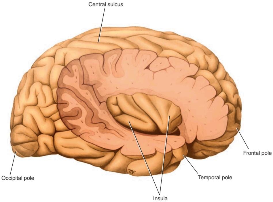
Figure 7-9 Lateral view of the right cerebral hemisphere dissected to reveal the right insula.
شکل ۷-۹ نمای جانبی نیمکره راست مغز که برای آشکار کردن اینسولای راست تشریح شده است.
The parieto-occipital sulcus begins on the superior medial margin of the hemisphere about 2 in (5 cm) anterior to the occipital pole (see Fig. 7-8; see also Atlas Plate 3). It passes downward and anteriorly on the medial surface to meet the calcarine sulcus (see Fig. 7-8).
شیار آهیانه-پسسری از لبه داخلی فوقانی نیمکره، حدود ۵ سانتیمتر (۲ اینچ) جلوتر از قطب پسسری شروع میشود (شکل ۷-۸ را ببینید؛ همچنین به صفحه اطلس ۳ مراجعه کنید). این شیار به سمت پایین و جلو روی سطح داخلی امتداد مییابد تا به شیار کالکارین برسد (شکل ۷-۸ را ببینید).
The calcarine sulcus is found on the medial surface of the hemisphere (see Fig. 7-8; see also Atlas Plate 3). It commences under the posterior end of the corpus. callosum and arches upward and backward to reach the occipital pole, where it stops. In some brains, however, it continues for a short distance onto the lateral surface of the hemisphere. The calcarine sulcus is joined at an acute angle by the parieto-occipital sulcus about half- way along its length.
شیار کالکارین در سطح داخلی نیمکره یافت میشود (شکل ۷-۸ را ببینید؛ همچنین به صفحه اطلس ۳ مراجعه کنید). این شیار از زیر انتهای خلفی جسم پینهای شروع میشود و به سمت بالا و عقب قوس میزند تا به قطب پسسری برسد، جایی که متوقف میشود. با این حال، در برخی مغزها، این شیار برای مسافت کوتاهی تا سطح جانبی نیمکره ادامه مییابد. شیار کالکارین با زاویه حاده به شیار آهیانه-پسسری تقریباً در نیمه طول خود متصل میشود.
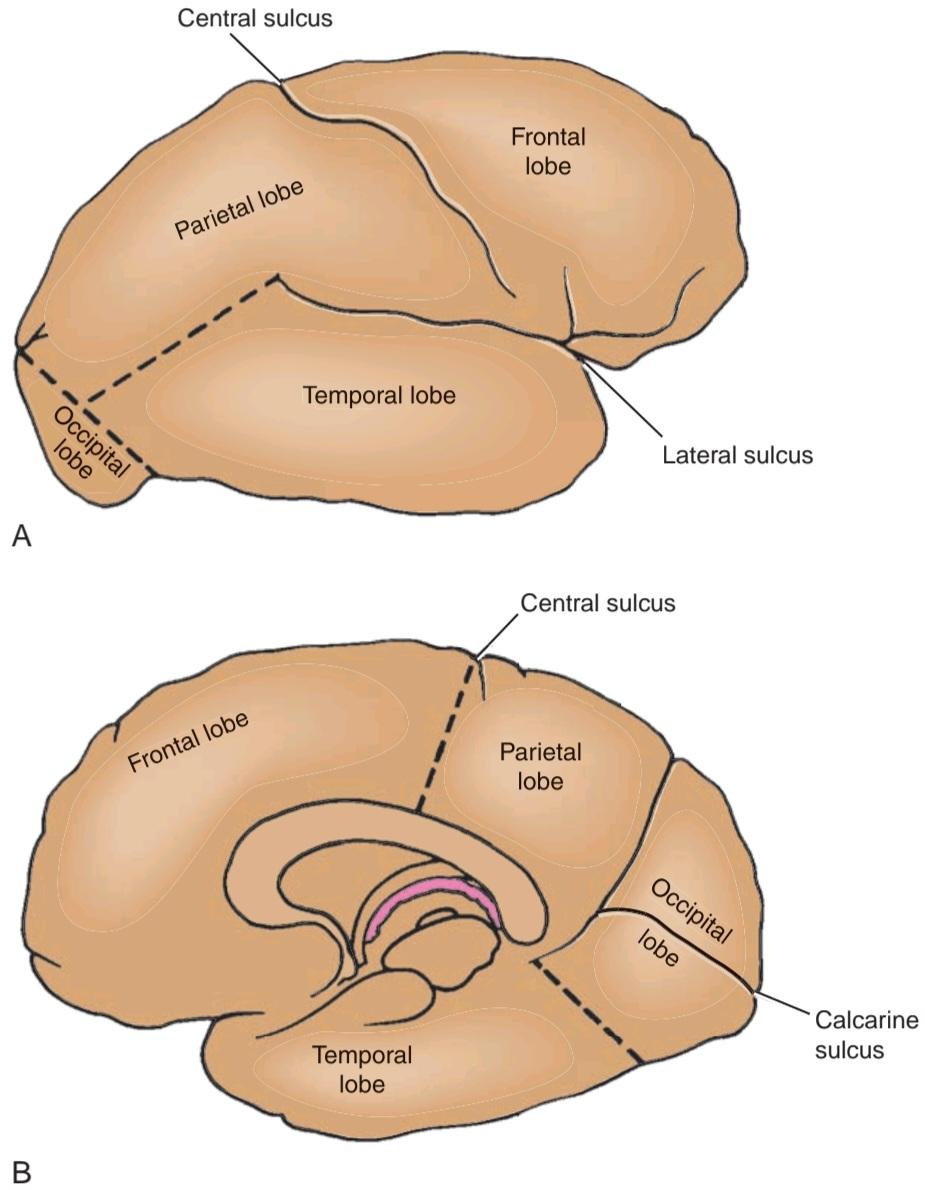
Figure 7-10 A: Lateral view of the right cerebral hemisphere showing the lobes. B: Medial view of the right cerebral hemisphere showing the lobes. Note that the dashed lines indicate the approximate position of the boundaries where no sulci are present.
شکل ۷-۱۰ الف: نمای جانبی نیمکره راست مغز که لوبها را نشان میدهد. ب: نمای داخلی نیمکره راست مغز که لوبها را نشان میدهد. توجه داشته باشید که خطوط چیندار موقعیت تقریبی مرزهایی را نشان میدهند که در آنها هیچ شیاری وجود ندارد.
CEREBRAL HEMISPHERE LOBES
Cerebral hemispheres are functionally divided into lobes, each of which are anatomically identified using specific gyri and sulci.
لوبهای نیمکره مغزی
نیمکرههای مغزی از نظر عملکردی به لوبهایی تقسیم میشوند که هر یک از آنها با استفاده از شیارها و چینهای خاص، از نظر آناتومیکی شناسایی میشوند.
Superolateral Surface (Atlas Plate 3)
The frontal lobe occupies the area anterior to the central sulcus and superior to the lateral sulcus (Fig. 7-10). The superolateral surface of the frontal lobe is divided by three sulci into four gyri. The precentral sulcus runs parallel to the central sulcus, and the precentral gyrus lies between them (see Fig. 7-7). Extending anteriorly from the precentral sulcus are the superior and inferior frontal sulci. The superior frontal gyrus lies superior to the superior frontal sulcus, the middle frontal gyrus lies between the superior and inferior frontal sulci, and the inferior frontal gyrus lies inferior to the inferior frontal sulcus. The inferior frontal gyrus is invaded by the anterior and ascending rami of the lateral sulcus.
سطح فوقانی جانبی (صفحه اطلس ۳)
لوب پیشانی ناحیهای را در جلوی شیار مرکزی و بالاتر از شیار جانبی اشغال میکند (شکل ۷-۱۰). سطح فوقانی جانبی لوب پیشانی توسط سه شیار به چهار شکنج تقسیم میشود. شیار پیشمرکزی به موازات شیار مرکزی امتداد دارد و شکنج پیشمرکزی بین آنها قرار دارد (شکل ۷-۷ را ببینید). شیارهای پیشانی فوقانی و تحتانی از شیار پیشمرکزی به سمت جلو امتداد یافتهاند. شکنج پیشانی فوقانی در بالای شیار پیشانی فوقانی، شکنج پیشانی میانی در بین شیارهای پیشانی فوقانی و تحتانی و شکنج پیشانی تحتانی در پایین شیار پیشانی تحتانی قرار دارد. شکنج پیشانی تحتانی توسط شاخههای قدامی و صعودی شیار جانبی مورد حمله قرار میگیرد.
The parietal lobe occupies the area posterior to the central sulcus and superior to the lateral sulcus; it extends posteriorly as far as the parieto-occipital sulcus (see Figs. 7-7 to 7-10). The lateral surface of the parietal lobe is divided by two sulci into three gyri. The postcentral sulcus runs parallel to the central sulcus, and the postcentral gyrus lies between them. Running posteriorly from the middle of the postcentral sulcus is the intraparietal sulcus (see Fig. 7-7). Superior to the intraparietal sulcus is the superior parietal lobule (gyrus), and inferior to the intraparietal sulcus is the inferior parietal lobule (gyrus).
لوب آهیانه ناحیهای را در خلف شیار مرکزی و بالاتر از شیار جانبی اشغال میکند؛ این ناحیه از خلف تا شیار آهیانه-پسسری امتداد مییابد (شکلهای ۷-۷ تا ۷-۱۰ را ببینید). سطح جانبی لوب آهیانه توسط دو شیار به سه شکنج تقسیم میشود. شیار پسمرکزی به موازات شیار مرکزی امتداد دارد و شکنج پسمرکزی بین آنها قرار دارد. از وسط شیار پسمرکزی به سمت خلف شیار درونجداری امتداد دارد (شکل ۷-۷ را ببینید). بالای شیار درونجداری، لوبول آهیانه فوقانی (شکنج) قرار دارد و پایینتر از شیار درونجداری، لوبول آهیانه تحتانی (شکنج) قرار دارد.
The temporal lobe occupies the area inferior to the lateral sulcus (see Figs. 7-7 to 7-10). The lateral surface of the temporal lobe is divided into three gyri by two sulci. The superior and middle temporal sulci run parallel to the posterior ramus of the lateral sulcus and divide the temporal lobe into the superior, middle, and inferior temporal gyri; the inferior temporal gyrus is continued onto the inferior surface of the hemisphere (see Fig. 7-7).
لوب گیجگاهی ناحیهای را در پایین شیار جانبی اشغال میکند (شکلهای ۷-۷ تا ۷-۱۰ را ببینید). سطح جانبی لوب گیجگاهی توسط دو شیار به سه شکنج تقسیم میشود. شیارهای گیجگاهی فوقانی و میانی به موازات شاخ خلفی شیار جانبی امتداد یافته و لوب گیجگاهی را به شکنجهای گیجگاهی فوقانی، میانی و تحتانی تقسیم میکنند؛ شکنج گیجگاهی تحتانی تا سطح تحتانی نیمکره ادامه دارد (شکل 7-7 را ببینید).
The occipital lobe occupies the small area behind the parieto-occipital sulcus (see Figs. 7-7 to 7-10).
لوب پسسری ناحیه کوچکی را در پشت شیار جداری-پسسری اشغال میکند (شکلهای 7-7 تا 7-10 را ببینید).
Medial and Inferior Surfaces (Atlas Plates 3, 6, and 8)
The lobes of the cerebral hemisphere are not clearly defined on the medial and inferior surfaces. However, many important areas should be recognized. The corpus callosum, which is the largest commissure of the brain, forms a striking feature on this surface (see Fig. 7-8). The cingulate gyrus begins beneath the anterior end of the corpus callosum and continues above the corpus callosum until it reaches its posterior end. The gyrus is separated from the corpus callosum by the callosal sulcus. The cingulate gyrus is separated from the superior frontal gyrus by the cingulate sulcus (see Fig. 7-8).
سطوح داخلی و تحتانی (صفحات اطلس ۳، ۶ و ۸)
لوبهای نیمکره مغزی به وضوح در سطوح داخلی و تحتانی مشخص نشدهاند. با این حال، بسیاری از نواحی مهم باید شناسایی شوند. جسم پینهای، که بزرگترین رابط مغز است، یک ویژگی برجسته در این سطح تشکیل میدهد (شکل ۷-۸ را ببینید). شکنج سینگولیت از زیر انتهای قدامی جسم پینهای شروع میشود و بالای جسم پینهای ادامه مییابد تا به انتهای خلفی آن برسد. شکنج توسط شیار پینهای از جسم پینهای جدا میشود. شکنج سینگولیت توسط شیار سینگولیت از شکنج پیشانی فوقانی جدا میشود (شکل ۷-۸ را ببینید).
The paracentral lobule is the area of the cerebral cortex that surrounds the indentation produced by the central sulcus on the superior border. The anterior part of this lobule is a continuation of the precentral gyrus on the superior lateral surface, and the posterior part of the lobule is a continuation of the postcentral gyrus.
لوبول پاراسنترال ناحیهای از قشر مغز است که فرورفتگی ایجاد شده توسط شیار مرکزی در مرز فوقانی را احاطه میکند. قسمت قدامی این لوبول ادامهی شکنج پیشمرکزی در سطح جانبی فوقانی است و قسمت خلفی لوبول ادامهی شکنج پسمرکزی است.
The precuneus is an area of cortex bounded anteriorly by the upturned posterior end of the cingulate sulcus and posteriorly by the parieto-occipital sulcus.
پرکونئوس ناحیهای از قشر مغز است که از جلو توسط انتهای خلفی رو به بالای شیار سینگولیت و از عقب توسط شیار جداری-پسسری محدود شده است.
The cuneus is a triangular area of cortex bounded above by the parieto-occipital sulcus, inferiorly by the calcarine sulcus, and posteriorly by the superior medial margin.
کونئوس ناحیهای مثلثی شکل از قشر مغز است که از بالا توسط شیار جداری-پسسری، از پایین توسط شیار کالکارین و از عقب توسط حاشیه میانی فوقانی محدود شده است.
The collateral sulcus is situated on the inferior surface of the hemisphere (Fig. 7-11; also see Fig. 7-8). This runs anteriorly below the calcarine sulcus. Between the collateral sulcus and the calcarine sulcus is the lingual gyrus. Anterior to the lingual gyrus is the parahippocampal gyrus; the latter terminates in front as the hooklike uncus (see Fig. 7-11).
شیار جانبی در سطح تحتانی نیمکره قرار دارد (شکل 7-11؛ همچنین به شکل 7-8 مراجعه کنید). این شیار در قسمت قدامی زیر شیار کالکارین امتداد دارد. بین شیار جانبی و شیار کالکارین، شکنج زبانی قرار دارد. در جلوی شکنج زبانی، شکنج پاراهیپوکامپ قرار دارد. دومی در جلو به صورت آنکوس قلابمانند خاتمه مییابد (شکل 7-11 را ببینید).

Figure 7-11 Inferior view of the brain; the medulla oblongata, the pons, and the cerebellum have been removed.
شکل ۷-۱۱ نمای تحتانی مغز؛ بصل النخاع، پل مغزی و مخچه برداشته شدهاند.
The medial occipitotemporal gyrus extends from the occipital pole to the temporal pole. It is bounded medially by the collateral and rhinal sulci and laterally by the occipitotemporal sulcus. The occipitotemporal gyrus lies lateral to the sulcus and is continuous with the inferior temporal gyrus.
شکنج پسسری-گیجگاهی داخلی از قطب پسسری تا قطب گیجگاهی امتداد دارد. این شکنج از سمت داخل توسط شیارهای جانبی و بینی و از سمت خارج توسط شیار پسسری-گیجگاهی محدود میشود. شکنج پسسری-گیجگاهی در سمت خارج شیار قرار دارد و با شکنج گیجگاهی تحتانی امتداد مییابد.
On the inferior surface of the frontal lobe, the olfactory bulb and tract overlie a sulcus called the olfactory sulcus. Medial to the olfactory sulcus is the gyrus rectus, and lateral to the sulcus are a number of orbital gyri.
در سطح تحتانی لوب پیشانی، پیاز بویایی و مسیر بویایی روی یک شیار به نام شیار بویایی قرار دارند. در سمت داخل شیار بویایی، شکنج رکتوس قرار دارد و در سمت خارج شیار، تعدادی شکنج مداری وجود دارد.
«فصل هفتم کتاب نورواناتومی بالینی اسنل»
🚀 با ما همراه شوید!
تازهترین مطالب و آموزشهای مغز و اعصاب را از دست ندهید. با فالو کردن کانال تلگرام آیندهنگاران مغز، از ما حمایت کنید!



