ساختار داخلی نیمکرههای مغزی: بطنهای جانبی، هستههای قاعدهای، الیاف رابط و پرتابی


دعای مطالعه [ نمایش ]
بِسْمِ الله الرَّحْمنِ الرَّحیمِ
اَللّهُمَّ اَخْرِجْنى مِنْ ظُلُماتِ الْوَهْمِ
خدايا مرا بيرون آور از تاريكىهاى وهم،
وَ اَكْرِمْنى بِنُورِ الْفَهْمِ
و به نور فهم گرامى ام بدار،
اَللّهُمَّ افْتَحْ عَلَيْنا اَبْوابَ رَحْمَتِكَ
خدايا درهاى رحمتت را به روى ما بگشا،
وَانْشُرْ عَلَيْنا خَزائِنَ عُلُومِكَ بِرَحْمَتِكَ يا اَرْحَمَ الرّاحِمينَ
و خزانههاى علومت را بر ما باز كن به امید رحمتت اى مهربانترين مهربانان.
آیندهنگاران مغز: «ما مغز را میشناسیم، تا آینده را بسازیم.»
کتاب «نوروآناتومی بالینی اسنل» اثر دکتر ریچارد اس. اسنل، با ویرایش جدید دکتر رایان اسپلیتگربر، همچنان بهعنوان مرجع طلایی این حوزه شناخته میشود. این اثر با تلفیق تازهترین پژوهشها و کاربردهای بالینی، پلی استوار میان دانش پایه و عمل پزشکی بنا میکند.
تلاشهای علمی و سالها تدریس ارزشمند استاد گرانقدر آقای دکتر محمدتقی جغتایی الهامبخش ترجمه و گسترش این دانش در ایران بوده است.
ترجمه دقیق این کتاب توسط برند علمی آیندهنگاران مغز به مدیریت داریوش طاهری، امکان دسترسی فارسیزبانان به مرزهای نوین علوم اعصاب را فراهم کرده و رسالتی علمی در جهت شناخت عمیقتر مغز و ترسیم آیندهای روشنتر بر عهده گرفته است.
»» فصل ۷: مخ؛ بخش مبانی و اصول
»» Chapter 7: Cerebrum
INTERNAL STRUCTURE OF THE CEREBRAL HEMISPHERES (ATLAS PLATES 4 AND 5)
The cerebral hemispheres are covered with a layer of gray matter, the cerebral cortex; the structure and function of the cerebral cortex are discussed in Chapter 15. Located in the interior of the cerebral hemispheres are the lateral ventricles, masses of gray matter, the basal nuclei, and nerve fibers. The nerve fibers are embedded in neuroglia and constitute the white matter (Fig. 7-12).
ساختار داخلی نیمکرههای مغزی (صفحات اطلس ۴ و ۵)
نیمکرههای مغزی با لایهای از ماده خاکستری، قشر مغز، پوشیده شدهاند؛ ساختار و عملکرد قشر مغز در فصل ۱۵ مورد بحث قرار گرفته است. بطنهای جانبی، تودههایی از ماده خاکستری، هستههای قاعدهای و رشتههای عصبی در داخل نیمکرههای مغزی قرار دارند. رشتههای عصبی در نوروگلیاها فرو رفته و ماده سفید را تشکیل میدهند (شکل ۷-۱۲).

Figure 7-12 Horizontal section of the cerebrun, as seen from above, showing the relationship between the lentiform nucleus, the caudate nucleus, the thalamus, and the internal capsule.
شکل ۷-۱۲ برش افقی مخ، از نمای بالا، که رابطه بین هسته عدسی شکل، هسته دم دار، تالاموس و کپسول داخلی را نشان میدهد.
Lateral Ventricles
The two lateral ventricles each occupy one cerebral hemisphere (Fig. 7-13; also see Fig. 7-12). Each ventricle is a roughly C-shaped cavity lined with ependyma and filled with CSF. The lateral ventricle may be divided into a body, which occupies the parietal lobe, and from which anterior, posterior, and inferior horns extend into the frontal, occipital, and temporal lobes, respectively. The lateral ventricle communicates with the cavity of the third ventricle through the interventricular foramen (see Figs. 7-8 and 7-13). This opening, which lies in the anterior part of the medial wall of the lateral ventricle, is bounded anteriorly by the anterior column of the fornix and posteriorly by the anterior end of the thalamus.
بطنهای جانبی
دو بطن جانبی هر کدام یک نیمکره مغزی را اشغال میکنند (شکل 7-13؛ همچنین به شکل 7-12 مراجعه کنید). هر بطن تقریباً یک حفره C شکل است که با اپاندیم پوشیده شده و با مایع مغزی نخاعی پر شده است. بطن جانبی را میتوان به یک بدنه تقسیم کرد که لوب جداری را اشغال میکند و از آن شاخهای قدامی، خلفی و تحتانی به ترتیب به لوبهای پیشانی، پسسری و گیجگاهی امتداد مییابند. بطن جانبی از طریق سوراخ بین بطنی با حفره بطن سوم ارتباط برقرار میکند (شکلهای 7-8 و 7-13 را ببینید). این دهانه که در قسمت قدامی دیواره داخلی بطن جانبی قرار دارد، از جلو توسط ستون قدامی فورنیکس و از عقب توسط انتهای قدامی تالاموس محدود میشود.

Figure 7-13 Ventricular cavities of the brain. A: Lateral view. B: Superior view.
شکل ۷-۱۳ حفرههای بطنی مغز. الف: نمای جانبی. ب: نمای فوقانی.
Basal Nuclei
The term basal nuclei (basal ganglia) is applied to a collection of masses of gray matter situated within each cerebral hemisphere. They are the corpus striatum, the amygdaloid nucleus, and the claustrum.
هستههای قاعدهای
اصطلاح هستههای قاعدهای (عقدههای قاعدهای) به مجموعهای از تودههای ماده خاکستری واقع در هر نیمکره مغزی اطلاق میشود. این هستهها عبارتند از جسم مخطط، هسته آمیگدالوئید و کلاستروم.
Corpus Striatum
The corpus striatum is situated lateral to the thalamus. It is almost completely divided by a band of nerve fibers, the internal capsule, into the caudate nucleus and the lentiform nucleus (see Figs. 7-12 and 7-17).
جسم مخطط
جسم مخطط در کنار تالاموس قرار دارد. تقریباً به طور کامل توسط نواری از الیاف عصبی، کپسول داخلی، به هسته دمدار و هسته عدسی شکل تقسیم میشود (شکلهای 7-12 و 7-17 را ببینید).
The caudate nucleus, a large C-shaped mass of gray matter that is closely related to the lateral ventricle, lies lateral to the thalamus (Fig. 7-14). The lateral surface of the nucleus is related to the internal capsule, which separates it from the lentiform nucleus.
هسته دمدار، یک توده بزرگ C شکل از ماده خاکستری که ارتباط نزدیکی با بطن جانبی دارد، در کنار تالاموس قرار دارد (شکل 7-14). سطح جانبی هسته به کپسول داخلی مرتبط است که آن را از هسته عدسی شکل جدا میکند.
The lentiform nucleus is a wedge-shaped mass of gray matter whose broad convex base is directed laterally and its blade medially (see Figs. 7-12 and 7-14). It is buried deep in the white matter of the cerebral hemisphere and is related medially to the internal capsule, which separates it from the caudate nucleus and the thalamus. The lentiform nucleus is related laterally to a thin sheet of white matter, the external capsule (see Fig. 7-12), that separates it from a thin sheet of gray matter, called the claustrum. The claustrum, in turn, separates the external capsule from the subcortical white matter of the insula. Inferiorly at its anterior end, the lentiform nucleus is continuous with the caudate nucleus.
هسته عدسی شکل، تودهای گوهای شکل از ماده خاکستری است که قاعده محدب پهن آن به سمت طرفین و تیغه آن به سمت داخل است (شکلهای ۷-۱۲ و ۷-۱۴ را ببینید). این هسته در عمق ماده سفید نیمکره مغزی قرار دارد و از سمت داخل به کپسول داخلی متصل است که آن را از هسته دمدار و تالاموس جدا میکند. هسته عدسی شکل از سمت خارج به یک صفحه نازک از ماده سفید، کپسول خارجی (شکل ۷-۱۲ را ببینید) متصل است که آن را از یک صفحه نازک از ماده خاکستری به نام کلاستروم جدا میکند. کلاستروم نیز به نوبه خود، کپسول خارجی را از ماده سفید زیر قشری اینسولا جدا میکند. هسته عدسی شکل در انتهای قدامی خود، در امتداد هسته دمدار قرار دارد.
The detailed structure and connections of the corpus striatum are considered in Chapter 10. Briefly, it may be stated that the corpus striatum receives afferent fibers from different areas of the cerebral cortex, the thalamus, subthalamus, and brainstem. Efferent fibers then travel back to the same areas of the nervous system. The function of the corpus striatum is concerned with muscular movement, which is accomplished by controlling the cerebral cortex rather than through direct descending pathways to the brainstem and spinal cord.
ساختار و اتصالات دقیق جسم مخطط در فصل 10 مورد بررسی قرار گرفته است. به طور خلاصه، میتوان گفت که جسم مخطط، فیبرهای آوران را از نواحی مختلف قشر مغز، تالاموس، ساب تالاموس و ساقه مغز دریافت میکند. سپس فیبرهای وابران به همان نواحی سیستم عصبی باز میگردند. عملکرد جسم مخطط مربوط به حرکت عضلانی است که با کنترل قشر مغز انجام میشود، نه از طریق مسیرهای نزولی مستقیم به ساقه مغز و نخاع.
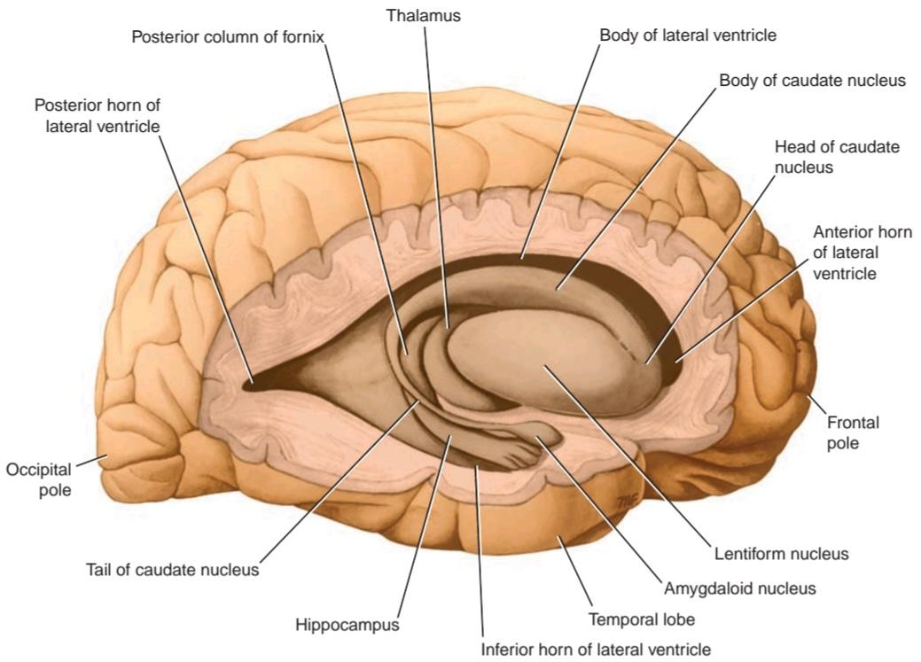
Figure 7-14 Lateral view of the right cerebral hemisphere dissected to show the position of the lentiform nucleus, the caudate nucleus, the thalamus, and the hippocampus.
شکل ۷-۱۴ نمای جانبی نیمکره راست مغز که برای نشان دادن موقعیت هسته عدسی شکل، هسته دم دار، تالاموس و هیپوکامپ تشریح شده است.
Amygdaloid Nucleus
The amygdaloid nucleus is situated in the temporal lobe close to the uncus (see Fig. 7-14). The amygdaloid nucleus is considered part of the limbic system and is described in Chapter 9 (see p. 303).
هسته آمیگدالوئید
هسته آمیگدالوئید در لوب گیجگاهی نزدیک به آنکوس قرار دارد (شکل 7-14 را ببینید). هسته آمیگدالوئید بخشی از سیستم لیمبیک محسوب میشود و در فصل 9 توضیح داده شده است (به صفحه 303 مراجعه کنید).
The claustrum is a thin sheet of gray matter that is separated from the lateral surface of the lentiform nucleus by the external capsule (see Fig. 7-12). Lateral to the claustrum is the subcortical white matter of the insula. The function of the claustrum is unknown.
کلاستروم یک صفحه نازک از ماده خاکستری است که توسط کپسول خارجی از سطح جانبی هسته عدسی شکل جدا شده است (شکل 7-12 را ببینید). در کنار کلاستروم، ماده سفید زیر قشری اینسولا قرار دارد. عملکرد کلاستروم ناشناخته است.
White Matter
The white matter is composed of myelinated nerve fibers of different diameters supported by neuroglia. The nerve fibers may be classified into three groups according to their connections: (1) commissural fibers, (2) association fibers, and (3) projection fibers.
ماده سفید
ماده سفید از فیبرهای عصبی میلیندار با قطرهای مختلف تشکیل شده است که توسط نوروگلیا پشتیبانی میشوند. فیبرهای عصبی را میتوان بر اساس اتصالاتشان به سه گروه طبقهبندی کرد: (1) فیبرهای ارتباطی، (2) فیبرهای ارتباطی و (3) فیبرهای برآمدگی.
Commissure Fibers
Commissure fibers essentially connect corresponding regions of the two hemispheres. They are as follows: the corpus callosum, the anterior commissure, the posterior commissure, the fornix, and the habenular commissure.
الیاف رابط
الیاف رابط اساساً نواحی متناظر دو نیمکره را به هم متصل میکنند. آنها به شرح زیر هستند: جسم پینهای، رابط قدامی، رابط خلفی، فورنیکس و رابط هابنولار.
The corpus callosum, the largest commissure of the brain, connects the two cerebral hemispheres (Fig. 7-15; see also Fig. 7-8 and Atlas Plate 8). It lies at the bottom of the longitudinal fissure. For purposes of description, it is divided into the rostrum, the genu, the body, and the splenium.
جسم پینهای، بزرگترین رابط مغز، دو نیمکره مغزی را به هم متصل میکند (شکل 7-15؛ همچنین به شکل 7-8 و صفحه اطلس 8 مراجعه کنید). این جسم در پایین شکاف طولی قرار دارد. برای توصیف، به روستروم، زانو، بدنه و اسپلنیوم تقسیم میشود.

Figure 7-15 A: Coronal section of the brain passing through the anterior horn of the lateral ventricle and the optic chiasma. B: Superior view of the brain dissected to show the fibers of the corpus callosum and the corona radiata.
شکل ۷-۱۵ الف: برش کرونال مغز که از شاخ قدامی بطن جانبی و کیاسمای بینایی عبور میکند. ب: نمای فوقانی مغز که برای نشان دادن الیاف جسم پینهای و تاج شعاعی تشریح شده است.
The rostrum is the thin part of the anterior end of the corpus callosum, which is prolonged posteriorly to be continuous with the upper end of the lamina terminalis (see Fig. 7-8).
روستروم قسمت نازک انتهای قدامی جسم پینهای است که به سمت عقب امتداد یافته تا در امتداد انتهای فوقانی لامینا ترمینالیس (lamina terminalis) قرار گیرد (شکل ۷-۸ را ببینید).
The genu is the curved anterior end of the corpus callosum that bends inferiorly in front of the septum pellucidum (see Figs. 7-8 and 7-15).
ژنو انتهای قدامی خمیده جسم پینهای است که در جلوی سپتوم پلوسیدوم به سمت پایین خم میشود (شکلهای ۷-۸ و ۷-۱۵ را ببینید).
The body of the corpus callosum arches posteriorly and ends as the thickened posterior portion called the splenium (see Fig. 7-15).
بدنه جسم پینهای به سمت عقب قوس برمیدارد و به قسمت خلفی ضخیمشدهای به نام اسپلنیوم (splenium) ختم میشود (شکل ۷-۱۵ را ببینید).
Traced laterally, the fibers of the genu curve forward into the frontal lobes and form the forceps minor (see Fig. 7-15B). The fibers of the body extend laterally as the radiation of the corpus callosum. They intersect with bundles of association and projection fibers as they pass to the cerebral cortex. Some of the fibers form the roof and lateral wall of the posterior horn of the lateral ventricle and the lateral wall of the inferior horn of the lateral ventricle; these fibers are referred to as the tapetum. Traced laterally, the fibers in the splenium arch backward into the occipital lobe and form the forceps major.
الیاف زانو که به صورت جانبی ردیابی میشوند، به سمت جلو به داخل لوبهای پیشانی خم میشوند و فورسپس مینور را تشکیل میدهند (شکل 7-15B را ببینید). الیاف بدنه به صورت جانبی به عنوان شعاع جسم پینهای امتداد مییابند. آنها هنگام عبور به قشر مغز با دستههایی از الیاف ارتباطی و برآمدگی تقاطع میکنند. برخی از الیاف، سقف و دیواره جانبی شاخ خلفی بطن جانبی و دیواره جانبی شاخ تحتانی بطن جانبی را تشکیل میدهند. این الیاف به عنوان تاپتوم شناخته میشوند. الیاف اسپلنیوم که به صورت جانبی ردیابی میشوند، به سمت عقب به داخل لوب پسسری قوس میخورند و فورسپس مینور را تشکیل میدهند.
The anterior commissure is a small bundle of nerve fibers that crosses the midline in the lamina terminalis (see Fig. 7-8). When traced laterally, a smaller or anterior bundle curves forward on each side toward the anterior perforated substance and the olfactory tract. A larger bundle curves posteriorly on each side and grooves the inferior surface of the lentiform nucleus to reach the temporal lobes.
رابط قدامی دسته کوچکی از الیاف عصبی است که از خط میانی در لامینا ترمینالیس عبور میکند (شکل ۷-۸ را ببینید). وقتی به صورت جانبی ردیابی شود، یک دسته کوچکتر یا قدامی در هر طرف به سمت ماده سوراخدار قدامی و مجرای بویایی به جلو خم میشود. یک دسته بزرگتر در هر طرف به عقب خم میشود و سطح تحتانی هسته عدسی شکل را شیار میدهد تا به لوبهای گیجگاهی برسد.
The posterior commissure is a bundle of nerve fibers that crosses the midline immediately above the opening of the cerebral aqueduct into the third ventricle (see Fig. 7-3); it is related to the inferior part of the stalk of the pineal gland. Various collections of nerve cells are situated along its length. The destinations and functional significance of many of the nerve fibers are not known. However, the fibers from the pretectal nuclei involved in the pupillary light reflex are believed to cross in this commissure on their way to the para- sympathetic part of the oculomotor nuclei.
رابط خلفی دستهای از الیاف عصبی است که بلافاصله بالای دهانه قنات مغزی به بطن سوم از خط میانی عبور میکند (شکل ۷-۳ را ببینید)؛ این دسته به قسمت تحتانی ساقه غده پینهآل مربوط میشود. مجموعههای مختلفی از سلولهای عصبی در طول آن قرار دارند. مقصد و اهمیت عملکردی بسیاری از الیاف عصبی مشخص نیست. با این حال، اعتقاد بر این است که الیاف هستههای پرهتکتال که در رفلکس نوری مردمک نقش دارند، در مسیر خود به سمت قسمت پاراسمپاتیک هستههای اوکولوموتور از این رابط عبور میکنند.
The fornix is composed of myelinated nerve fibers and constitutes the efferent system of the hippocampus that passes to the mammillary bodies of the hypothalamus. The nerve fibers first form the alveus (see Fig. 9-5), which is a thin layer of white matter covering the ventricular surface of the hippocampus, and then converge to form the fimbria. The fimbriae of the two sides increase in thickness and, on reaching the posterior end of the hippocampus, arch forward above the thalamus and below the corpus callosum to form the posterior columns of the fornix. The two columns then come together in the midline to form the body of the fornix (Fig. 7-16). The commissure of the fornix consists of transverse fibers that cross the midline from one column to another just before the formation of the body of the fornix. The function of the commissure of the fornix is to connect the hippocampal formations of the two sides.
فورنیکس از فیبرهای عصبی میلیندار تشکیل شده است و سیستم وابران هیپوکامپ را تشکیل میدهد که به اجسام پستانی هیپوتالاموس میرود. فیبرهای عصبی ابتدا آلوئوس را تشکیل میدهند (شکل 9-5 را ببینید)، که یک لایه نازک از ماده سفید است که سطح بطنی هیپوکامپ را میپوشاند، و سپس برای تشکیل فیمبریا همگرا میشوند. فیمبریاهای دو طرف افزایش ضخامت مییابند و با رسیدن به انتهای خلفی هیپوکامپ، به سمت جلو در بالای تالاموس و زیر جسم پینهای قوس میخورند تا ستونهای خلفی فورنیکس را تشکیل دهند. سپس دو ستون در خط میانی به هم میرسند تا بدنه فورنیکس را تشکیل دهند (شکل 7-16). رابط فورنیکس از فیبرهای عرضی تشکیل شده است که درست قبل از تشکیل بدنه فورنیکس، از یک ستون به ستون دیگر از خط میانی عبور میکنند. وظیفه رابط فورنیکس اتصال تشکیلات هیپوکامپ دو طرف است.
The habenular commissure is a small bundle of nerve fibers that crosses the midline in the superior part of the root of the pineal stalk (see Fig. 7-3). The commissure is associated with the habenular nuclei, which are situated on either side of the midline in this region. The habenular nuclei receive many afferents from the amygdaloid nuclei and the hippocampus. These afferent fibers pass to the habenular nuclei in the stria medullaris thalami. Some of the fibers cross the midline to reach the contralateral nucleus through the habenular commissure. The function of the habenular nuclei and its connections in humans is unknown.
رابط هابنولار دسته کوچکی از فیبرهای عصبی است که از خط میانی در قسمت فوقانی ریشه ساقه پینه آل عبور میکند (شکل 7-3 را ببینید). این رابط با هستههای هابنولار مرتبط است که در دو طرف خط میانی در این ناحیه قرار دارند. هستههای هابنولار آورانهای زیادی را از هستههای آمیگدالوئید و هیپوکامپ دریافت میکنند. این فیبرهای آوران به هستههای هابنولار در استریا بصلالنخاع تالامی منتقل میشوند. برخی از فیبرها از خط میانی عبور میکنند تا از طریق رابط هابنولار به هسته مقابل برسند. عملکرد هستههای هابنولار و اتصالات آن در انسان ناشناخته است.
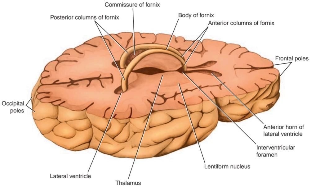
Figure 7-16 Horizontal section of the brain leaving the fornix in position.
شکل ۷-۱۶ برش افقی مغز که فورنیکس را در جای خود باقی میگذارد.
Association Fibers
Association fibers are nerve fibers that essentially connect various cortical regions within the same hemisphere and may be divided into short and long groups (Fig. 7-17). The short association fibers lie immediately beneath the cortex and connect adjacent gyri; these fibers run transversely to the long axis of the sulci. The long association fibers are collected into named bundles that can be dissected in a formalin-hardened brain. The uncinate fasciculus connects the first motor speech area and the gyri on the inferior surface of the frontal lobe with the cortex of the pole of the temporal lobe. The cingulum is a long, curved fasciculus lying within the white matter of the cingulate gyrus (see Fig. 7-8). It connects the frontal and parietal lobes with parahippocampal and adjacent temporal cortical regions. The superior longitudinal fasciculus is the largest bundle of nerve fibers. It connects the anterior part of the frontal lobe to the occipital and temporal lobes. The inferior longitudinal fasciculus runs anteriorly from the occipital lobe, passing lateral to the optic radiation, and is distributed to the temporal lobe. The fronto-occipital fasciculus connects the frontal lobe to the occipital and temporal lobes. It is situated deep within the cerebral hemisphere and is related to the lateral border of the caudate nucleus.
الیاف ارتباطی
الیاف ارتباطی، الیاف عصبی هستند که اساساً نواحی مختلف قشری را در یک نیمکره به هم متصل میکنند و میتوانند به گروههای کوتاه و بلند تقسیم شوند (شکل 7-17). الیاف ارتباطی کوتاه بلافاصله در زیر قشر مغز قرار دارند و شکنجهای مجاور را به هم متصل میکنند. این الیاف به صورت عرضی به محور طولی شیارها امتداد دارند. الیاف ارتباطی بلند در دستههای نامگذاری شدهای جمع میشوند که میتوانند در مغز سخت شده با فرمالین تشریح شوند. فاسیکولوس چنگکی، اولین ناحیه حرکتی گفتار و شکنجها را در سطح تحتانی لوب فرونتال با قشر قطب لوب گیجگاهی متصل میکند. سینگولوم یک فاسیکولوس بلند و خمیده است که در ماده سفید شکنج سینگولیت قرار دارد (شکل 7-8 را ببینید). این فاسیکولوس لوبهای فرونتال و پاریتال را با نواحی قشری پاراهیپوکامپ و مجاور متصل میکند. فاسیکولوس طولی فوقانی بزرگترین دسته الیاف عصبی است. این دسته قسمت قدامی لوب فرونتال را به لوبهای پس سری و گیجگاهی متصل میکند. دسته عصبی طولی تحتانی از لوب پسسری به سمت جلو امتداد مییابد، از کنار مجرای بینایی عبور میکند و در لوب گیجگاهی توزیع میشود. دسته عصبی پیشانی-پسسری، لوب فرونتال را به لوبهای پسسری و گیجگاهی متصل میکند. این دسته عصبی در اعماق نیمکره مغزی قرار دارد و به لبه جانبی هسته دمی مرتبط است.
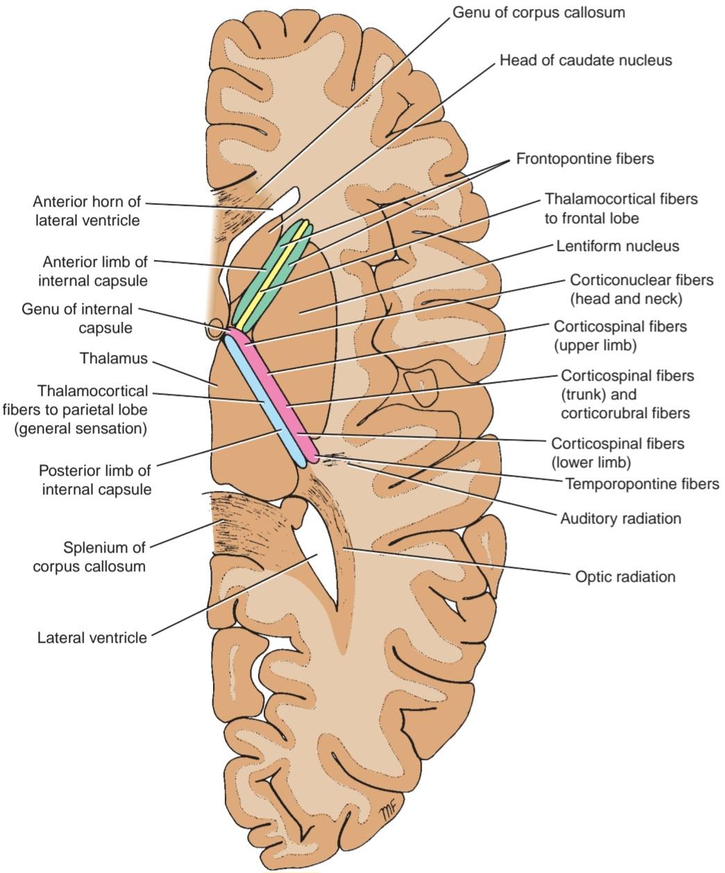
Figure 7-17 Horizontal section of the right cerebral hemisphere showing the relationships and different parts of the internal capsule.
شکل ۷-۱۷ برش افقی از نیمکره راست مغز که روابط و بخشهای مختلف کپسول داخلی را نشان میدهد.
Projection Fibers
Afferent and efferent nerve fibers passing to and from the brainstem to the entire cerebral cortex must travel between large nuclear masses of gray matter within the cerebral hemisphere. At the upper part of the brainstem, these fibers form a compact band known as the internal capsule, which is flanked medially by the caudate nucleus and the thalamus and laterally by the lentiform nucleus (see Fig. 7-12). Because of the wedge shape of the lentiform nucleus, as seen on horizontal section, the internal capsule is bent to form an anterior limb and a posterior limb, which are continuous with each other at the genu (Figs. 7-18 and 7-19). Once the nerve fibers have emerged superiorly from between the nuclear masses, they radiate in all directions to the cerebral cortex. These radiating projection fibers are known as the corona radiata (Fig. 7-19). Most of the projection fibers lie medial to the association fibers, but they intersect the commissural fibers of the corpus callosum and the anterior commissure. The nerve fibers lying within the most posterior part of the posterior limb of the internal capsule radiate toward the calcarine sulcus and are known as the optic radiation (Fig. 7-18). The detailed arrangement of the fibers within the internal capsule is shown in Figure 7-18.
الیاف پرتابی
الیاف عصبی آوران و وابران که به ساقه مغز و از آن به کل قشر مغز میروند، باید بین تودههای هستهای بزرگ ماده خاکستری در نیمکره مغزی حرکت کنند. در قسمت فوقانی ساقه مغز، این الیاف یک نوار فشرده به نام کپسول داخلی تشکیل میدهند که از طرف داخل توسط هسته دمدار و تالاموس و از طرف خارج توسط هسته عدسی شکل احاطه شده است (شکل 7-12 را ببینید). به دلیل شکل گوهای هسته عدسی شکل، همانطور که در مقطع افقی دیده میشود، کپسول داخلی خم شده و یک شاخه قدامی و یک شاخه خلفی تشکیل میدهد که در زانو به یکدیگر متصل هستند (شکلهای 7-18 و 7-19). هنگامی که الیاف عصبی از بین تودههای هستهای به سمت بالا خارج میشوند، در تمام جهات به قشر مغز منتشر میشوند. این الیاف پرتابی ساطع کننده به عنوان تاج شعاعی شناخته میشوند (شکل 7-19). بیشتر فیبرهای برآمدگی در سمت داخلی فیبرهای ارتباطی قرار دارند، اما فیبرهای رابط جسم پینهای و رابط قدامی را قطع میکنند. فیبرهای عصبی که در خلفیترین قسمت اندام خلفی کپسول داخلی قرار دارند، به سمت شیار کالکارین تابش میکنند و به عنوان تابش بینایی شناخته میشوند (شکل 7-18). ترتیب دقیق فیبرها در داخل کپسول داخلی در شکل 7-18 نشان داده شده است.
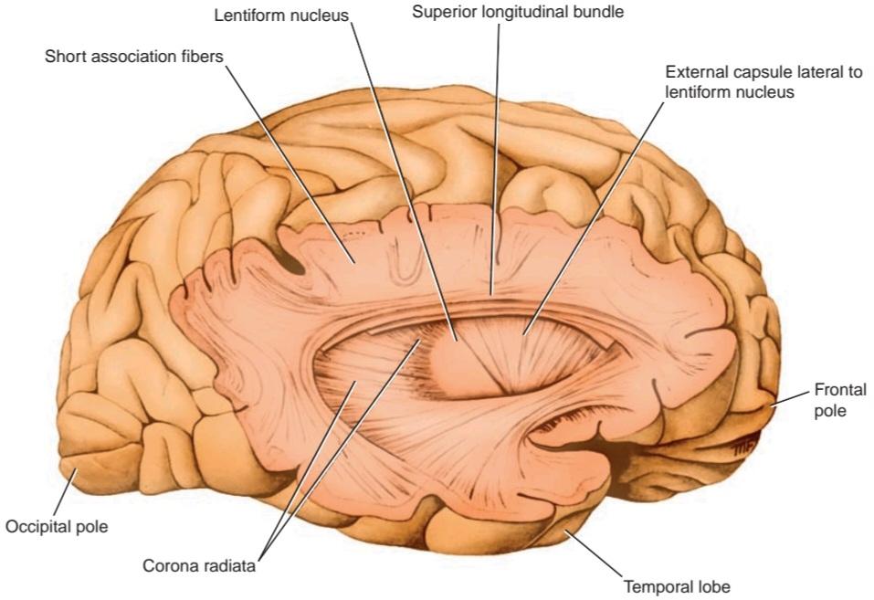
Figure 7-18 Lateral view of the right cerebral hemisphere, which has been dissected to show some of the principal association fibers.
شکل ۷-۱۸ نمای جانبی نیمکره راست مغز، که برای نشان دادن برخی از فیبرهای ارتباطی اصلی تشریح شده است.

Figure 7-19 Medial view of the right cerebral hemisphere, which has been dissected to show the internal capsule and the corona radiata. The thalamus has been removed. Note the interdigitation of the horizontally running fibers of the corpus callosum and the vertical fibers of the corona radiata.
شکل ۷-۱۹ نمای داخلی نیمکره راست مغز، که برای نشان دادن کپسول داخلی و تاج شعاعی تشریح شده است. تالاموس برداشته شده است. به تداخل الیاف افقی جسم پینهای و الیاف عمودی تاج شعاعی توجه کنید.
Septum Pellucidum
The septum pellucidum is a thin vertical sheet of nervous tissue consisting of white and gray matter covered on either side by ependyma (see Figs. 7-8 and 7-12; see also Atlas Plate 8). It stretches between the fornix and the corpus callosum. Anteriorly, it occupies the interval between the body of the corpus callosum and the rostrum. It is essentially a double membrane with a closed, slitlike cavity between the membranes. The septum pellucidum forms a partition between the anterior horns of the lateral ventricles.
سپتوم پلوسیدوم
سپتوم پلوسیدوم یک صفحه عمودی نازک از بافت عصبی است که از ماده سفید و خاکستری تشکیل شده و از دو طرف توسط اپاندیم پوشانده شده است (شکلهای ۷-۸ و ۷-۱۲ را ببینید؛ همچنین به صفحه اطلس ۸ مراجعه کنید). این صفحه بین فورنیکس و جسم پینهای امتداد دارد. در جلو، فاصله بین جسم پینهای و روستروم را اشغال میکند. اساساً یک غشای دوتایی با یک حفره بسته و شکاف مانند بین غشاها است. سپتوم پلوسیدوم یک جدار بین شاخهای قدامی بطنهای جانبی تشکیل میدهد.
Tela Choroidea
The tela choroidea is a two-layered fold of pia mater. It is situated between the fornix superiorly and the roof of the third ventricle and the upper surfaces of the two thalami inferiorly. When seen from above, the anterior end is situated at the interventricular foramina (see Fig. 16-6). Its lateral edges are irregular and project laterally into the body of the lateral ventricles. Here, they are covered by ependyma and form the choroid plexuses of the lateral ventricle. Posteriorly, the lateral edges continue into the inferior horn of the lateral ventricle and are covered with ependyma so that the choroid plexus projects through the choroidal fissure.
تلا مشیمیه
تلای مشیمیه یک چین دو لایه از نرم شامه است. این چین بین فورنیکس در بالا و سقف بطن سوم و سطوح بالایی دو تالاموس در پایین قرار دارد. وقتی از بالا دیده میشود، انتهای قدامی آن در سوراخهای بین بطنی قرار دارد (شکل 16-6 را ببینید). لبههای جانبی آن نامنظم هستند و به صورت جانبی به داخل بدنه بطنهای جانبی بیرون زدهاند. در اینجا، آنها توسط اپاندیم پوشیده شدهاند و شبکههای مشیمیه بطن جانبی را تشکیل میدهند. در عقب، لبههای جانبی به داخل شاخ تحتانی بطن جانبی ادامه مییابند و با اپاندیم پوشیده میشوند به طوری که شبکه مشیمیه از طریق شکاف مشیمیه بیرون زده است.
On either side of the midline, the tela choroidea projects down through the roof of the third ventricle to form the choroid plexuses of the third ventricle.
در دو طرف خط میانی، تلا مشیمیه از سقف بطن سوم به پایین امتداد مییابد و شبکههای مشیمیه بطن سوم را تشکیل میدهد.
The blood supply of the tela choroidea and, therefore, also of the choroid plexuses of the third and lateral ventricles is derived from the choroidal branches of the internal carotid and basilar arteries. The venous blood drains into the internal cerebral veins, which unite to form the great cerebral vein. The great cerebral vein joins the inferior sagittal sinus to form the straight sinus.
خونرسانی تلا مشیمیه و بنابراین، همچنین شبکههای مشیمیه بطنهای سوم و جانبی از شاخههای مشیمیه شریانهای کاروتید داخلی و بازیلار گرفته میشود. خون وریدی به وریدهای مغزی داخلی تخلیه میشود که با هم ادغام شده و ورید مغزی بزرگ را تشکیل میدهند. ورید مغزی بزرگ به سینوس ساژیتال تحتانی میپیوندد و سینوس مستقیم را تشکیل میدهد.
» ویرایش شده این بخش از فصل مخ که قبلاً برای آزمون از معلمان آموزش و پرورش آماده شده بود.
«فصل هفتم کتاب نورواناتومی بالینی اسنل»
🚀 با ما همراه شوید!
تازهترین مطالب و آموزشهای مغز و اعصاب را از دست ندهید. با فالو کردن کانال تلگرام آیندهنگاران مغز، از ما حمایت کنید!