بخشهای اصلی سیستم عصبی محیطی: اعصاب جمجمهای و نخاعی و گانگلیونها


دعای مطالعه [ نمایش ]
بِسْمِ الله الرَّحْمنِ الرَّحیمِ
اَللّهُمَّ اَخْرِجْنى مِنْ ظُلُماتِ الْوَهْمِ
خدايا مرا بيرون آور از تاريكىهاى وهم،
وَ اَكْرِمْنى بِنُورِ الْفَهْمِ
و به نور فهم گرامى ام بدار،
اَللّهُمَّ افْتَحْ عَلَيْنا اَبْوابَ رَحْمَتِكَ
خدايا درهاى رحمتت را به روى ما بگشا،
وَانْشُرْ عَلَيْنا خَزائِنَ عُلُومِكَ بِرَحْمَتِكَ يا اَرْحَمَ الرّاحِمينَ
و خزانههاى علومت را بر ما باز كن به امید رحمتت اى مهربانترين مهربانان.
آیندهنگاران مغز: «ما مغز را میشناسیم، تا آینده را بسازیم.»
کتاب «نوروآناتومی بالینی اسنل» اثر دکتر ریچارد اس. اسنل، با ویرایش جدید دکتر رایان اسپلیتگربر، همچنان بهعنوان مرجع طلایی این حوزه شناخته میشود. این اثر با تلفیق تازهترین پژوهشها و کاربردهای بالینی، پلی استوار میان دانش پایه و عمل پزشکی بنا میکند.
تلاشهای علمی و سالها تدریس ارزشمند استاد گرانقدر آقای دکتر محمدتقی جغتایی الهامبخش ترجمه و گسترش این دانش در ایران بوده است.
ترجمه دقیق این کتاب توسط برند علمی آیندهنگاران مغز به مدیریت داریوش طاهری، امکان دسترسی فارسیزبانان به مرزهای نوین علوم اعصاب را فراهم کرده و رسالتی علمی در جهت شناخت عمیقتر مغز و ترسیم آیندهای روشنتر بر عهده گرفته است.
»» فصل ۱: مقدمه و سازماندهی سیستم عصبی؛ قسمت مبانی و اصول
»» Chapter 1: Introduction and Organization of the Nervous System
MAJOR DIVISIONS OF THE PERIPHERAL NERVOUS SYSTEM
The PNS comprises the cranial and spinal nerves and their associated ganglia.
بخشهای اصلی سیستم عصبی محیطی
سیستم عصبی محیطی (PNS) شامل اعصاب جمجمهای و نخاعی و گانگلیونهای مرتبط با آنهاست.
Cranial and Spinal Nerves
The cranial and spinal nerves are made up of bundles of nerve fibers supported by connective tissue.
اعصاب جمجمهای و نخاعی
اعصاب جمجمهای و نخاعی از دستههایی از رشتههای عصبی تشکیل شدهاند که توسط بافت همبند پشتیبانی میشوند.
The 12 pairs of cranial nerves (see Fig. 1-8) leave the brain and pass through foramina in the skull. The 31 pairs of spinal nerves (see Fig. 1-5) leave the spinal cord and pass through intervertebral foramina in the vertebral column. The spinal nerves are associated with regions of the spinal cord: 8 cervical, 12 thoracic, 5 lumbar, 5 sacral, and 1 coccygeal. Note that there are 8 cervical nerves yet only 7 cervical vertebrae and that there is 1 coccygeal nerve but 4 coccygeal vertebrae.
۱۲ جفت عصب جمجمهای (شکل ۱-۸ را ببینید) از مغز خارج شده و از سوراخهای جمجمه عبور میکنند. ۳۱ جفت عصب نخاعی (شکل ۱-۵ را ببینید) از نخاع خارج شده و از سوراخهای بین مهرهای در ستون فقرات عبور میکنند. اعصاب نخاعی با نواحی از نخاع مرتبط هستند: ۸ عصب گردنی، ۱۲ عصب سینهای، ۵ عصب کمری، ۵ عصب خاجی و ۱ عصب دنبالچهای. توجه داشته باشید که ۸ عصب گردنی وجود دارد، اما فقط ۷ مهره گردنی وجود دارد و ۱ عصب دنبالچهای وجود دارد، اما ۴ مهره دنبالچهای وجود دارد.
Each spinal nerve is connected to the spinal cord by two roots: the anterior root and the posterior root (see Fig. 1-5B). The anterior root consists of bundles of nerve fibers carrying nerve impulses away from the CNS efferent fibers. Those that go to skeletal muscles and cause them to contract are motor fibers. Their cells of origin lie in the anterior gray horn of the spinal cord. The posterior root consists of bundles of afferent fibers that carry nervous impulses to the CNS. Because these fibers convey information about sensations of touch, pain, temperature, and vibration, they are called sensory fibers. The cell bodies of these nerve fibers are situated in a swelling on the posterior root called the posterior root ganglion.
هر عصب نخاعی توسط دو ریشه به نخاع متصل میشود: ریشه قدامی و ریشه خلفی (شکل 1-5B را ببینید). ریشه قدامی شامل دستههایی از الیاف عصبی است که تکانههای عصبی را از الیاف وابران سیستم عصبی مرکزی (CNS) دور میکنند. آنهایی که به عضلات اسکلتی میروند و باعث انقباض آنها میشوند، الیاف حرکتی هستند. سلولهای منشأ آنها در شاخ خاکستری قدامی نخاع قرار دارند. ریشه خلفی شامل دستههایی از الیاف آوران است که تکانههای عصبی را به سیستم عصبی مرکزی (CNS) حمل میکنند. از آنجایی که این الیاف اطلاعات مربوط به حس لامسه، درد، دما و ارتعاش را منتقل میکنند، به آنها الیاف حسی میگویند. اجسام سلولی این الیاف عصبی در یک برآمدگی روی ریشه خلفی به نام گانگلیون ریشه خلفی قرار دارند.
The spinal nerve roots pass from the spinal cord to the level of their respective intervertebral foramina, where they unite to form a spinal nerve (Fig. 1-14). Here, the motor and sensory fibers mix together; thus, a spinal nerve comprises both motor and sensory fibers.
ریشههای عصب نخاعی از نخاع به سطح سوراخهای بین مهرهای مربوطه خود عبور میکنند، جایی که برای تشکیل عصب نخاعی به هم میپیوندند (شکل 1-14). در اینجا، فیبرهای حرکتی و حسی با هم مخلوط میشوند؛ بنابراین، یک عصب نخاعی شامل هر دو فیبر حرکتی و حسی است.

Figure 1-14 Posterior view of the spinal cord showing the origins of the roots of the spinal nerves and their relationship to the different vertebrae. On the right, the laminae have been removed to expose the right half of the spinal cord and the nerve roots.
شکل ۱-۱۴ نمای خلفی نخاع که منشأ ریشههای اعصاب نخاعی و ارتباط آنها با مهرههای مختلف را نشان میدهد. در سمت راست، لامیناها برداشته شدهاند تا نیمه راست نخاع و ریشههای عصبی نمایان شوند.

Figure 1-15 Oblique posterior view of the lower end of the spinal cord and the cauda equina. On the right, the laminae have been removed to expose the right half of the spinal cord and the nerve roots.
شکل ۱-۱۵ نمای خلفی مایل از انتهای تحتانی نخاع و دم اسب. در سمت راست، لامیناها برداشته شدهاند تا نیمه راست نخاع و ریشههای عصبی نمایان شوند.
Because of the disproportionate growth in length of the vertebral column during development, compared with that of the spinal cord, the length of the roots increases progressively from above downward (see Fig. 1-15). In the upper cervical region, the spinal nerve roots are short and run almost horizontally, but the roots of the lumbar and sacral nerves below the level of the termination of the cord (lower border of the 1st lumbar vertebra in the adult) form a vertical leash of nerves around the filum terminale (Fig. 1-15). Together, these lower nerve roots are called the cauda equina.
به دلیل رشد نامتناسب طول ستون فقرات در طول رشد، در مقایسه با طول نخاع، طول ریشهها از بالا به پایین به تدریج افزایش مییابد (شکل 1-15 را ببینید). در ناحیه گردنی فوقانی، ریشههای عصبی نخاعی کوتاه هستند و تقریباً به صورت افقی امتداد دارند، اما ریشههای اعصاب کمری و خاجی در زیر سطح انتهای نخاع (مرز پایینی اولین مهره کمری در بزرگسالان) یک طناب عمودی از اعصاب را در اطراف فیلوم ترمینال تشکیل میدهند (شکل 1-15). این ریشههای عصبی پایینی با هم، دم اسب نامیده میشوند.
After emerging from the intervertebral foramen, each spinal nerve immediately divides into a large anterior ramus and a smaller posterior ramus, each containing both motor and sensory fibers. The posterior ramus passes posteriorly around the vertebral column to supply the muscles and skin of the back. The anterior ramus continues anteriorly to supply the muscles and skin over the anterolateral body wall and all the muscles and skin of the limbs.
پس از خروج از سوراخ بین مهرهای، هر عصب نخاعی بلافاصله به یک شاخه قدامی بزرگ و یک شاخه خلفی کوچکتر تقسیم میشود که هر کدام حاوی فیبرهای حرکتی و حسی هستند. شاخه خلفی به صورت خلفی در اطراف ستون مهرهها عبور میکند تا عضلات و پوست پشت را عصبدهی کند. شاخه قدامی به صورت قدامی ادامه مییابد تا عضلات و پوست روی دیواره قدامی-جانبی بدن و تمام عضلات و پوست اندامها را عصبدهی کند.
The anterior rami join one another at the root of the limbs to form complicated nerve plexuses (see Fig. 1-1B). The cervical and brachial plexuses are found at the root of the upper limbs, and the lumbar and sacral plexuses are found at the root of the lower limbs.
شاخههای قدامی در ریشه اندامها به یکدیگر متصل میشوند تا شبکههای عصبی پیچیدهای را تشکیل دهند (شکل 1-1B را ببینید). شبکههای گردنی و بازویی در ریشه اندامهای فوقانی و شبکههای کمری و خاجی در ریشه اندامهای تحتانی قرار دارند.
Ganglia
Ganglia can be divided into sensory ganglia of spinal nerves (posterior root ganglia) and cranial nerves and autonomic ganglia.
گانگلیونها
گانگلیونها را میتوان به گانگلیونهای حسی اعصاب نخاعی (گانگلیونهای ریشه خلفی) و اعصاب جمجمهای و گانگلیونهای اتونوم تقسیم کرد.
Sensory Ganglia
Sensory ganglia are fusiform swellings (see Fig. 1-5) on the posterior root of each spinal nerve just proximal to the root’s junction with a corresponding anterior root. They are referred to as posterior root ganglia. Similar ganglia found along the course of cranial nerves V, VII, VIII, IX, and X are the sensory ganglia of these nerves.
گانگلیونهای حسی
گانگلیونهای حسی، برآمدگیهای دوکیشکل (شکل ۱-۵ را ببینید) روی ریشه خلفی هر عصب نخاعی هستند که درست قبل از محل اتصال ریشه با ریشه قدامی مربوطه قرار دارند. به آنها گانگلیون ریشه خلفی گفته میشود. گانگلیونهای مشابهی که در امتداد اعصاب جمجمهای V، VII، VIII، IX و X یافت میشوند، گانگلیونهای حسی این اعصاب هستند.
Autonomic Ganglia
Autonomic ganglia, which are often irregular in shape, are situated along the course of efferent nerve fibers of the ANS. They are found in the paravertebral sympathetic chains (see Figs. 14-1 and 14-2) around the roots of the great visceral arteries in the abdomen and close to, or embedded within, the walls of various viscera.
گانگلیونهای اتونومیک
گانگلیونهای اتونومیک، که اغلب شکل نامنظمی دارند، در امتداد رشتههای عصبی وابران ANS قرار دارند. آنها در زنجیرههای سمپاتیک پاراورتبرال (شکلهای 14-1 و 14-2 را ببینید) در اطراف ریشههای شریانهای احشایی بزرگ در شکم و نزدیک به دیوارههای احشای مختلف یا در داخل آنها یافت میشوند.
EARLY DEVELOPMENT OF THE NERVOUS SYSTEM
Before the formation of the nervous system in the embryo, three main cell layers differentiate. The innermost layer, the entoderm, gives rise to the gastrointestinal tract, the lungs, and the liver. The mesoderm gives rise to the muscle, connective tissues, and the vascular system. The third and outermost layer, the ectoderm, formed of columnar epithelium, gives rise to the entire nervous system.
رشد اولیه سیستم عصبی
قبل از تشکیل سیستم عصبی در جنین، سه لایه سلولی اصلی تمایز مییابند. داخلیترین لایه، آنتودرم، دستگاه گوارش، ریهها و کبد را ایجاد میکند. مزودرم، عضله، بافتهای همبند و سیستم عروقی را ایجاد میکند. سومین و خارجیترین لایه، اکتودرم، که از اپیتلیوم ستونی تشکیل شده است، کل سیستم عصبی را ایجاد میکند.
During the third week of development, the ectoderm on the dorsal surface of the embryo between the primitive knot and the buccopharyngeal membrane thickens to form the neural plate. The plate, which is pear shaped and wider cranially, develops a longitudinal neural groove. The groove now deepens so that it is bounded on either side by neural folds (Fig. 1-16).
در طول هفته سوم رشد، اکتودرم روی سطح پشتی جنین بین گره اولیه و غشای دهانی-حلقی ضخیم میشود و صفحه عصبی را تشکیل میدهد. این صفحه که گلابی شکل و از نظر جمجمه پهنتر است، یک شیار عصبی طولی ایجاد میکند. این شیار اکنون عمیقتر میشود به طوری که از دو طرف توسط چینهای عصبی محدود میشود (شکل 1-16).

Figure 1-16 Formation of the neural plate, neural groove, and neural tube. The cells of the neural crest differentiate into the cells of the posterior root ganglia, the sensory ganglia of cranial nerves, autonomic ganglia, neurilemmal cells (Schwann cells), the cells of the suprarenal medulla, and melanocytes.
شکل ۱-۱۶ تشکیل صفحه عصبی، شیار عصبی و لوله عصبی. سلولهای تاج عصبی به سلولهای گانگلیون ریشه خلفی، گانگلیونهای حسی اعصاب جمجمهای، گانگلیونهای اتونوم، سلولهای نوریلمی (سلولهای شوان)، سلولهای مدولای فوق کلیوی و ملانوسیتها تمایز مییابند.

Figure 1-17 A: Expansion of the cephalic end of the neural tube to form the forebrain, midbrain, and hindbrain vesicles. B, C: Cross section of the developing neural tube in the region of the spinal cord. The cells of the neuroepithelial layer have been widely separated for clarity.
شکل ۱-۱۷ الف: انبساط انتهای سری لوله عصبی برای تشکیل وزیکولهای مغز قدامی، مغز میانی و مغز خلفی. ب، ج: برش عرضی لوله عصبی در حال رشد در ناحیه نخاع. سلولهای لایه نورواپیتلیال برای وضوح بیشتر به طور گسترده از هم جدا شدهاند.
With further development, the neural folds fuse, converting the neural groove into a neural tube. Fusion starts at about the midpoint along the groove and extends cranially and caudally so that, in the earliest stage, the cavity of the tube remains in communication with the amniotic cavity through the anterior and posterior neuropores. The anterior neuropore closes first and the posterior neuropore 2 days later. Thus, normally, the neural tube closure is complete within 28 days. Meanwhile, the neural tube has sunk beneath the surface ectoderm.
با رشد بیشتر، چینهای عصبی به هم جوش میخورند و شیار عصبی را به یک لوله عصبی تبدیل میکنند. جوش خوردن تقریباً از نقطه میانی در امتداد شیار شروع میشود و به سمت جمجمه و دم گسترش مییابد، به طوری که در مراحل اولیه، حفره لوله از طریق نوروپورهای قدامی و خلفی با حفره آمنیوتیک در ارتباط باقی میماند. نوروپور قدامی ابتدا و نوروپور خلفی ۲ روز بعد بسته میشود. بنابراین، به طور معمول، بسته شدن لوله عصبی ظرف ۲۸ روز کامل میشود. در همین حال، لوله عصبی در زیر اکتودرم سطحی فرو رفته است.
During the invagination of the neural plate to form the neural groove, the cells forming the lateral margin of the plate do not become incorporated in the neural tube but instead form a strip of ectodermal cells that lie between the neural tube and the covering ectoderm, the neural crest (Fig. 1-16). Subsequently, this group of cells will migrate ventrolaterally on each side around the neural tube. Ultimately, the neural crest cells will differentiate into the cells of the posterior root ganglia, the sensory ganglia of the cranial nerves, autonomic ganglia, the cells of the suprarenal medulla, and the melanocytes. These cells also probably give rise to mesenchymal cells in the head and neck.
در طول فرورفتگی صفحه عصبی برای تشکیل شیار عصبی، سلولهایی که حاشیه جانبی صفحه را تشکیل میدهند، در لوله عصبی ادغام نمیشوند، بلکه در عوض نواری از سلولهای اکتودرمی را تشکیل میدهند که بین لوله عصبی و اکتودرم پوشاننده، یعنی ستیغ عصبی، قرار دارند (شکل 1-16). متعاقباً، این گروه از سلولها به صورت شکمی-جانبی در هر طرف در اطراف لوله عصبی مهاجرت میکنند. در نهایت، سلولهای ستیغ عصبی به سلولهای گانگلیون ریشه خلفی، گانگلیونهای حسی اعصاب جمجمهای، گانگلیونهای اتونوم، سلولهای مدولای فوق کلیوی و ملانوسیتها تمایز مییابند. این سلولها همچنین احتمالاً باعث ایجاد سلولهای مزانشیمی در سر و گردن میشوند.
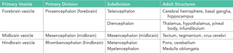
Meanwhile, the proliferation of cells at the cephalic end of the neural tube causes it to dilate and form three primary brain vesicles: the forebrain, midbrain, and hindbrain vesicles (Fig. 1-17) (Table 1-2). The rest of the tube elongates and remains smaller in diameter; it will form the spinal cord.
در همین حال، تکثیر سلولها در انتهای سری لوله عصبی باعث اتساع آن و تشکیل سه وزیکول مغزی اولیه میشود: وزیکولهای مغز قدامی، مغز میانی و مغز خلفی (شکل 1-17) (جدول 1-2). بقیه لوله کشیده میشود و قطر کوچکتری پیدا میکند؛ این لوله نخاع را تشکیل خواهد داد.
Table 1-2 The Primary Divisions of the Developing Brain
جدول ۱-۲ بخشهای اولیه مغز در حال رشد
The subsequent differentiation of cells in the neural tube is brought about by the inductive interactions of one group of cells with another. The inducing factors influence the control of the gene expression in the target cells. Ultimately, the simplest progenitor cell will differentiate into neurons and neuroglial cells. Interestingly, excessive numbers of neurons and neuroglial cells develop, and many (nearly half of the developing neurons) will be programmed to die by a process known as programmed cell death. Research into the identification of neurotrophic factors that promote the development and survival of neurons is of great importance, as the results could possibly be applied to the problem of regeneration of the spinal cord neurons following trauma or the inhibition of degenerative diseases such as Alzheimer disease.
تمایز بعدی سلولها در لوله عصبی توسط برهمکنشهای القایی یک گروه از سلولها با گروه دیگر انجام میشود. عوامل القاکننده بر کنترل بیان ژن در سلولهای هدف تأثیر میگذارند. در نهایت، سادهترین سلول پیشساز به نورونها و سلولهای نوروگلیال تمایز مییابد. جالب توجه است که تعداد زیادی نورون و سلولهای نوروگلیال ایجاد میشوند و بسیاری (تقریباً نیمی از نورونهای در حال توسعه) توسط فرآیندی که به عنوان مرگ برنامهریزیشده سلولی شناخته میشود، برنامهریزی میشوند تا بمیرند. تحقیق در مورد شناسایی عوامل نوروتروفیک که باعث رشد و بقای نورونها میشوند از اهمیت بالایی برخوردار است، زیرا نتایج آن میتواند در مسئله بازسازی نورونهای نخاع پس از تروما یا مهار بیماریهای دژنراتیو مانند بیماری آلزایمر به کار رود.
The further development of the nervous system will be fully described in Chapter 18 following the description of the different parts of the nervous system and their neuronal connections.
رشد بیشتر سیستم عصبی به طور کامل در فصل 18 و پس از شرح بخشهای مختلف سیستم عصبی و ارتباطات عصبی آنها شرح داده خواهد شد.
«فصل اول کتاب نورواناتومی بالینی اسنل»
🚀 با ما همراه شوید!
تازهترین مطالب و آموزشهای مغز و اعصاب را از دست ندهید. با فالو کردن کانال تلگرام آیندهنگاران مغز، از ما حمایت کنید!