
نوروآناتومی بالینی اسنل | مقدمه و سازمان سیستم عصبی – نکات و کاربردهای بالینی


دعای مطالعه [ نمایش ]
بِسْمِ الله الرَّحْمنِ الرَّحیمِ
اَللّهُمَّ اَخْرِجْنى مِنْ ظُلُماتِ الْوَهْمِ
خدايا مرا بيرون آور از تاريكىهاى وهم،
وَ اَكْرِمْنى بِنُورِ الْفَهْمِ
و به نور فهم گرامى ام بدار،
اَللّهُمَّ افْتَحْ عَلَيْنا اَبْوابَ رَحْمَتِكَ
خدايا درهاى رحمتت را به روى ما بگشا،
وَانْشُرْ عَلَيْنا خَزائِنَ عُلُومِكَ بِرَحْمَتِكَ يا اَرْحَمَ الرّاحِمينَ
و خزانههاى علومت را بر ما باز كن به امید رحمتت اى مهربانترين مهربانان.
آیندهنگاران مغز: «ما مغز را میشناسیم، تا آینده را بسازیم.»
کتاب «نوروآناتومی بالینی اسنل» اثر دکتر ریچارد اس. اسنل، با ویرایش جدید دکتر رایان اسپلیتگربر، همچنان بهعنوان مرجع طلایی این حوزه شناخته میشود. این اثر با تلفیق تازهترین پژوهشها و کاربردهای بالینی، پلی استوار میان دانش پایه و عمل پزشکی بنا میکند.
تلاشهای علمی و سالها تدریس ارزشمند استاد گرانقدر آقای دکتر محمدتقی جغتایی الهامبخش ترجمه و گسترش این دانش در ایران بوده است.
ترجمه دقیق این کتاب توسط برند علمی آیندهنگاران مغز به مدیریت داریوش طاهری، امکان دسترسی فارسیزبانان به مرزهای نوین علوم اعصاب را فراهم کرده و رسالتی علمی در جهت شناخت عمیقتر مغز و ترسیم آیندهای روشنتر بر عهده گرفته است.
»» فصل ۱: مقدمه و سازماندهی سیستم عصبی؛ بخش نکات و کاربردهای بالینی
»» Chapter 1: Introduction and Organization of the Nervous System
Clinical Notes
Relationship of Spinal Cord Segments to Vertebral Numbers
یادداشتهای بالینی
ارتباط بخشهای نخاع با تعداد مهرهها
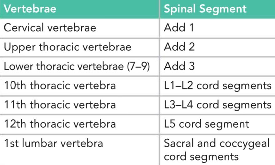
Because the spinal cord is shorter than the vertebral column, the spinal cord segments do not correspond numerically with the vertebrae that lie at the same level (see Fig. 1-14). The following table will help a clinician determine which spinal segment is related to a given vertebral body (Table 1-3).
از آنجا که نخاع کوتاهتر از ستون مهرهها است، بخشهای نخاع از نظر عددی با مهرههایی که در همان سطح قرار دارند، مطابقت ندارند (شکل 1-14 را ببینید). جدول زیر به پزشک کمک میکند تا مشخص کند کدام بخش نخاعی مربوط به یک جسم مهرهای خاص است (جدول 1-3).
Examination of a patient’s back shows that the spinous processes lie approximately at the same level as the vertebral bodies. In the lower thoracic region, however, because of the length and extreme obliquity of the spinous processes, the tips of the spines lie at the level of the vertebral body below.
معاینه کمر بیمار نشان میدهد که زوائد خاری تقریباً در همان سطح جسم مهرهها قرار دارند. با این حال، در ناحیه تحتانی قفسه سینه، به دلیل طول و شیب شدید زوائد خاری، نوک خارها در سطح جسم مهره زیرین قرار دارند.
Table 1-3 kelationship of spinal cord segments to vertebral Numbers
جدول ۱-۳ نسبت قطعات نخاع به مهرهها
Spinal Cord and Brain Injuries
The spinal cord and brain are well protected. Both are suspended in cerebrospinal fluid (CSF) and are surrounded by the bones of the vertebral column and skull (see Chapters 4 and 5). However, with enough force, these protective structures can be damaged, with consequent injury to the delicate underlying nervous tissue. Moreover, the cranial and spinal nerves and blood vessels are also likely to be injured.
آسیبهای نخاعی و مغزی
نخاع و مغز به خوبی محافظت میشوند. هر دو در مایع مغزی نخاعی (CSF) معلق هستند و توسط استخوانهای ستون فقرات و جمجمه احاطه شدهاند (به فصلهای ۴ و ۵ مراجعه کنید). با این حال، با نیروی کافی، این ساختارهای محافظ میتوانند آسیب ببینند و در نتیجه به بافت عصبی ظریف زیرین آسیب وارد شود. علاوه بر این، اعصاب جمجمهای و نخاعی و رگهای خونی نیز احتمالاً آسیب میبینند.
Spinal Cord Injuries
The degree of spinal cord injury at different vertebral levels is determined largely by anatomical factors. In the cervical region, dislocation or fracture dislocation is common, but the large size of the vertebral canal usually prevents severe injury to the spinal cord. However, with considerable displacement of the bones or bone fragments, the cord is sectioned. Respiration ceases if the cord is completely severed above the segmental origin of the phrenic nerves (C3-C5) because the intercostal muscles and the diaphragm are paralyzed, resulting in death.
آسیبهای نخاعی
درجه آسیب نخاعی در سطوح مختلف مهرهای تا حد زیادی توسط عوامل آناتومیکی تعیین میشود. در ناحیه گردنی، دررفتگی یا شکستگی شایع است، اما اندازه بزرگ کانال مهرهای معمولاً از آسیب شدید به نخاع جلوگیری میکند. با این حال، با جابجایی قابل توجه استخوانها یا قطعات استخوان، نخاع قطع میشود. اگر نخاع به طور کامل در بالای مبدا سگمنتال اعصاب فرنیک (C3-C5) قطع شود، تنفس متوقف میشود زیرا عضلات بین دندهای و دیافراگم فلج میشوند و منجر به مرگ میشود.
In fracture dislocations of the thoracic region, displacement can be considerable. The small size of the vertebral canal results in severe injury to this region of the spinal cord.
در دررفتگیهای شکستگی ناحیه سینهای، جابجایی میتواند قابل توجه باشد. اندازه کوچک کانال مهرهای منجر به آسیب شدید به این ناحیه از نخاع میشود.
In fracture dislocations of the lumbar region, two anatomic facts help prevent substantial nerve injury. First, the spinal cord in the adult extends down only as far as the level of the lower border of the 1st lumbar vertebra (see Fig. 1-15). Second, the large size of the vertebral foramen in this region gives the roots of the cauda equina ample room.
در دررفتگیهای شکستگی ناحیه کمری، دو واقعیت آناتومیکی به جلوگیری از آسیب قابل توجه عصب کمک میکند. اول، نخاع در بزرگسالان فقط تا سطح مرز پایینی اولین مهره کمری امتداد مییابد (شکل 1-15 را ببینید). دوم، اندازه بزرگ سوراخ مهرهای در این ناحیه، فضای کافی به ریشههای دم اسب میدهد.
Injury to the spinal cord may produce partial or complete loss of function at the level of the lesion and partial or complete loss of function of afferent and efferent nerve tracts below the level of the lesion. The symptoms and signs of such injuries are discussed after the detailed structure of the spinal cord, and the ascending and descending tracts are discussed in Chapter 4.
آسیب به نخاع ممکن است باعث از دست دادن جزئی یا کامل عملکرد در سطح ضایعه و از دست دادن جزئی یا کامل عملکرد مسیرهای عصبی آوران و وابران در زیر سطح ضایعه شود. علائم و نشانههای چنین آسیبهایی پس از ساختار دقیق نخاع مورد بحث قرار گرفته و مسیرهای صعودی و نزولی در فصل 4 مورد بحث قرار گرفتهاند.
Spinal Nerve Injuries
The intervertebral foramina (Fig. 1-18) transmit the spinal nerves and the small segmental arteries and veins, all of which are embedded in areolar tissue. Each foramen is bounded superiorly and inferiorly by the pedicles of adjacent vertebrae, anteriorly by the lower part of the vertebral body and the intervertebral disc, and posteriorly by the articular processes and the joint between them. In this situation, the spinal nerve is very vulnerable and may be pressed on or irritated by disease of the surrounding structures. Herniation of the intervertebral disc, fractures of the vertebral bodies, and osteoarthritis involving the joints of the articular processes or the joints between the vertebral bodies may all result in pressure, stretching, or edema of the emerging spinal nerve. Such pressure would give rise to dermatomal pain, muscle weakness, and diminished or absent reflexes.
آسیبهای عصب نخاعی
سوراخهای بین مهرهای (شکل ۱-۱۸) اعصاب نخاعی و شریانها و وریدهای کوچک سگمنتال را منتقل میکنند که همگی در بافت آرئولار قرار دارند. هر سوراخ از بالا و پایین توسط پایههای مهرههای مجاور، از جلو توسط قسمت پایینی جسم مهره و دیسک بین مهرهای و از عقب توسط زوائد مفصلی و مفصل بین آنها محدود میشود. در این وضعیت، عصب نخاعی بسیار آسیبپذیر است و ممکن است توسط بیماری ساختارهای اطراف تحت فشار قرار گیرد یا تحریک شود. فتق دیسک بین مهرهای، شکستگیهای جسم مهرهها و آرتروز که مفاصل زوائد مفصلی یا مفاصل بین جسم مهرهها را درگیر میکند، همگی ممکن است منجر به فشار، کشش یا ادم عصب نخاعی در حال ظهور شوند. چنین فشاری باعث درد پوستی، ضعف عضلانی و کاهش یا عدم وجود رفلکسها میشود.
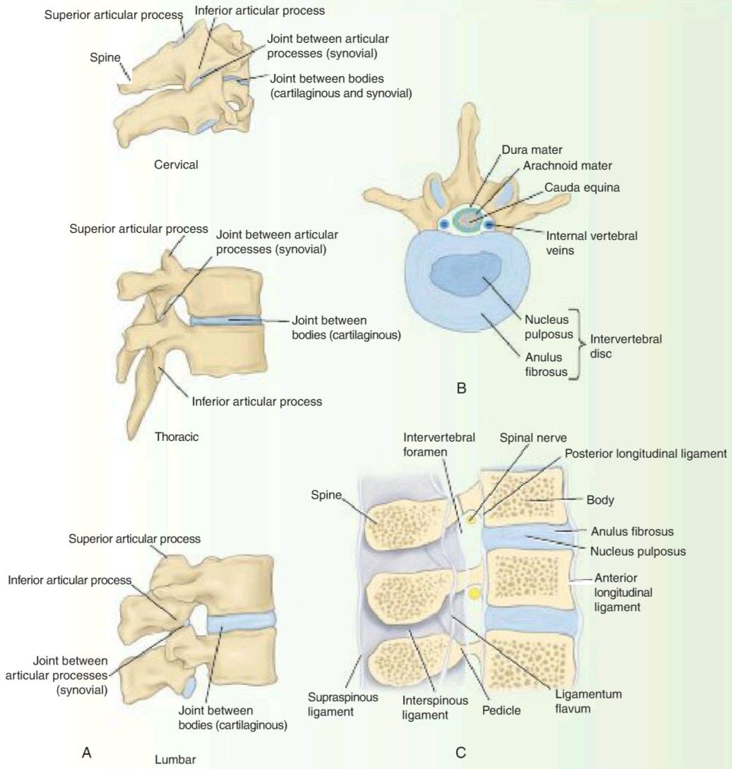
Figure 1-18 A: Joints in the cervical, thoracic, and lumbar regions of the vertebral column. B: Third lumbar vertebra seen from above showing the relationship between the intervertebral disc and the cauda equina. C: Sagittal section through three lumbar vertebrae showing the ligaments and the intervertebral discs. Note the relationship between the emerging spinal nerve in an intervertebral foramen and the intervertebral disc.
شکل ۱-۱۸ الف: مفاصل در نواحی گردنی، سینهای و کمری ستون مهرهها. ب: مهره سوم کمری که از بالا دیده میشود و رابطه بین دیسک بین مهرهای و دم اسب را نشان میدهد. ج: برش ساژیتال از طریق سه مهره کمری که رباطها و دیسکهای بین مهرهای را نشان میدهد. به رابطه بین عصب نخاعی که از سوراخ بین مهرهای بیرون میآید و دیسک بین مهرهای توجه کنید.
HERNIATED INTERVERTEBRAL DISCS
Herniation of the intervertebral discs occurs most commonly in areas of the vertebral column where a mobile part joins a relatively immobile part-for example, the cervicothoracic and lumbosacral junctions. In these areas, the posterior part of the anulus fibrosus of the disc ruptures, and the central nucleus pulposus is forced posteriorly like toothpaste out of a tube. This herniation of the nucleus pulposus may result either in a central protrusion in the midline under the posterior longitudinal ligament of the vertebrae or in a lateral protrusion at the side of the posterior ligament close to the intervertebral foramen (Fig. 1-19).
فتق دیسکهای بین مهرهای
فتق دیسکهای بین مهرهای بیشتر در مناطقی از ستون مهرهها رخ میدهد که یک قسمت متحرک به یک قسمت نسبتاً بیحرکت متصل میشود – به عنوان مثال، اتصالات گردنی-سینهای و کمری-خاجی. در این مناطق، قسمت خلفی آنولوس فیبروزوس دیسک پاره میشود و هسته مرکزی پالپوزوس مانند خمیردندان از یک لوله به سمت عقب رانده میشود. این فتق هسته پالپوزوس ممکن است منجر به یک بیرونزدگی مرکزی در خط وسط زیر رباط طولی خلفی مهرهها یا یک بیرونزدگی جانبی در کنار رباط خلفی نزدیک به سوراخ بین مهرهای شود (شکل 1-19).
Cervical disc herniations are less common than herniations in the lumbar region. The discs most susceptible are those between the 5th and 6th and the 6th and 7th cervical vertebrae. Lateral protrusions cause pressure on a spinal nerve or its roots. Each spinal nerve emerges above the corresponding vertebra; thus, the protrusion of the disc between the 5th and 6th cervical vertebrae may compress the C6 spinal nerve or its roots. Pain is felt near the lower part of the back of the neck and shoulder and along the area in the distribution of the spinal nerve involved. Central protrusions may press on the spinal cord and the anterior spinal artery and involve the various spinal tracts.
فتق دیسک گردن کمتر از فتق در ناحیه کمری شایع است. دیسکهایی که بیشتر مستعد ابتلا هستند، دیسکهای بین مهرههای پنجم و ششم و ششم و هفتم گردن هستند. بیرونزدگیهای جانبی باعث فشار بر عصب نخاعی یا ریشههای آن میشوند. هر عصب نخاعی از بالای مهره مربوطه خارج میشود؛ بنابراین، بیرونزدگی دیسک بین مهرههای پنجم و ششم گردن ممکن است عصب نخاعی C6 یا ریشههای آن را فشرده کند. درد در نزدیکی قسمت پایینی پشت گردن و شانه و در امتداد ناحیه توزیع عصب نخاعی درگیر احساس میشود. بیرونزدگیهای مرکزی ممکن است بر نخاع و شریان نخاعی قدامی فشار وارد کنند و مسیرهای مختلف نخاعی را درگیر کنند.
Lumbar disc herniations are more common than cervical disc herniations. The discs usually affected are those between the 4th and 5th lumbar vertebrae and between the 5th lumbar vertebra and the sacrum. In the lumbar region, the roots of the cauda equina run posteriorly over a number of intervertebral discs. A lateral herniation may press on one or two roots and commonly involves the nerve root going to the intervertebral foramen just below. The nucleus pulposus occasionally herniates directly backward, and if it is a large herniation, the whole cauda equina may be compressed, causing paraplegia.
فتق دیسک کمری شایعتر از فتق دیسک گردنی است. دیسکهایی که معمولاً تحت تأثیر قرار میگیرند، دیسکهای بین مهرههای چهارم و پنجم کمری و بین مهره پنجم کمری و استخوان خاجی هستند. در ناحیه کمری، ریشههای دم اسب به سمت عقب روی تعدادی از دیسکهای بین مهرهای امتداد مییابند. فتق جانبی ممکن است به یک یا دو ریشه فشار وارد کند و معمولاً ریشه عصبی را که به سوراخ بین مهرهای درست در زیر میرود، درگیر میکند. هسته پالپوس گاهی اوقات مستقیماً به سمت عقب فتق پیدا میکند و اگر فتق بزرگ باشد، ممکن است کل دم اسب فشرده شود و باعث فلج اندام تحتانی شود.
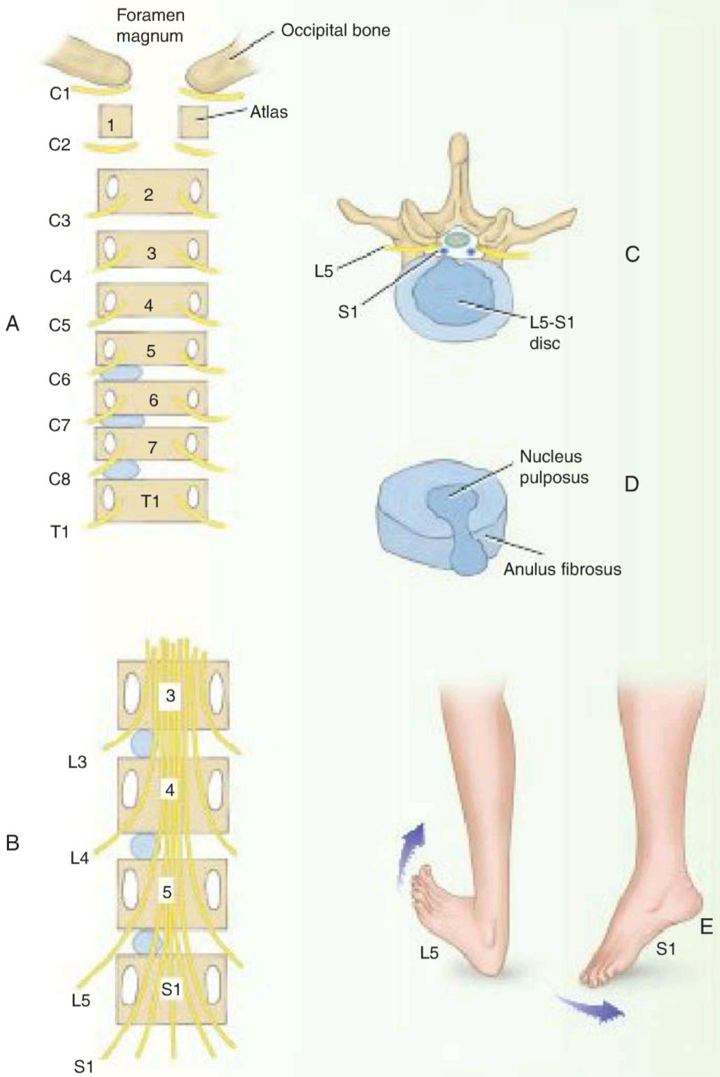
Figure 1-19 A, B: Posterior views of the vertebral bodies in the cervical and lumbar regions showing the relationship that might exist between a herniated nucleus pulposus and spinal nerve roots. Note there are eight cervical spinal nerves and only seven cervical vertebrae. In the lumbar region, for example, the emerging L4 nerve roots pass out laterally close to the pedicle of the 4th lumbar vertebra and are not related to the intervertebral disc between the 4th and 5th lumbar vertebrae. C: Posterolateral herniation of the nucleus pulposus of the intervertebral disc between the 5th lumbar vertebra and the 1st sacral vertebra showing pressure on the S1 nerve root. D: An intervertebral disc that has E herniated its nucleus pulposus posteriorly. E: Pressure on the L5 motor nerve root produces weakness of dorsiflexion of the ankle; pressure on the $1 motor nerve root produces weakness of plantar flexion of the ankle joint.
شکل 1-19 الف، ب: نماهای خلفی جسم مهرهها در نواحی گردنی و کمری که رابطهی احتمالی بین فتق هستهی پالپوزوس و ریشههای عصبی نخاعی را نشان میدهد. توجه داشته باشید که هشت عصب نخاعی گردنی و تنها هفت مهرهی گردنی وجود دارد. به عنوان مثال، در ناحیهی کمری، ریشههای عصبی L4 که در حال ظهور هستند، به صورت جانبی نزدیک به پایهی مهرهی چهارم کمری خارج میشوند و به دیسک بین مهرهای بین مهرههای چهارم و پنجم کمری مرتبط نیستند. ج: فتق خلفی-جانبی هستهی پالپوزوس دیسک بین مهرهای بین مهرهی پنجم کمری و مهرهی اول خاجی که فشار بر ریشهی عصبی S1 را نشان میدهد. د: دیسک بین مهرهای که هستهی پالپوزوس آن در خلف دچار فتق شده است. ه: فشار بر ریشهی عصب حرکتی L5 باعث ضعف خم شدن مچ پا به سمت پشت میشود؛ فشار بر ریشهی عصب حرکتی $1 باعث ضعف خم شدن کف پا در مفصل مچ پا میشود.
In lumbar disc herniations, pain is referred down the leg and foot in the distribution of the affected nerve. Because the sensory posterior roots most commonly pressed on are the 5th lumbar and 1st sacral, pain is usually felt down the back and lateral side of the leg, radiating to the sole of the foot, a condition known as sciatica. In severe cases, paresthesia or actual sensory loss may occur.
در فتق دیسک کمری، درد به سمت پایین پا و ساق پا در محل توزیع عصب آسیبدیده منتشر میشود. از آنجا که ریشههای حسی خلفی که معمولاً تحت فشار قرار میگیرند، عصب پنجم کمری و عصب اول خاجی هستند، درد معمولاً در پشت و کنارههای ساق پا احساس میشود و به کف پا انتشار مییابد، وضعیتی که به عنوان سیاتیک شناخته میشود. در موارد شدید، ممکن است پارستزی یا از دست دادن حس واقعی رخ دهد.
Pressure on the anterior motor roots causes muscle weakness. Involvement of the 5th lumbar motor root weakens dorsiflexion of the ankle, whereas pressure on the 1st sacral motor root causes plantar flexion weakness. The ankle jerk reflex may be diminished or absent (see Fig. 1-19E).
فشار بر ریشههای حرکتی قدامی باعث ضعف عضلات میشود. درگیری ریشه حرکتی پنجم کمری، خم شدن مچ پا به سمت عقب را ضعیف میکند، در حالی که فشار بر ریشه حرکتی اول خاجی باعث ضعف خم شدن کف پا میشود. رفلکس تکان دادن مچ پا ممکن است کاهش یابد یا وجود نداشته باشد (شکل 1-19E را ببینید).
A large, centrally placed protrusion may give rise to bilateral pain and muscle weakness in both legs. Acute retention of urine may also occur.
یک برآمدگی بزرگ و مرکزی ممکن است باعث درد دو طرفه و ضعف عضلات در هر دو پا شود. احتباس حاد ادرار نیز ممکن است رخ دهد.
Spinal Tap
Spinal tap (lumbar puncture) may be performed to withdraw a sample of CSF for microscopic or bacteriologic examination or to inject drugs to combat infection or induce anesthesia. Fortunately, the spinal cord terminates inferiorly at the level of the lower border of the 1st lumbar vertebra in adults (in infants, it may reach inferiorly to the 3rd lumbar vertebra). The subarachnoid space extends inferiorly as far as the lower border of the 2nd sacral vertebra. The lower lumbar part of the vertebral canal is thus occupied by the subarachnoid space, which contains the lumbar and sacral nerve roots and the filum terminale (the cauda equina). A needle inserted into the subarachnoid space in this region usually pushes the nerve roots to one side without causing damage.
ضربه نخاعی
ضربه نخاعی (پونکسیون کمری) ممکن است برای برداشتن نمونهای از CSF برای بررسی میکروسکوپی یا باکتریشناسی یا تزریق دارو برای مبارزه با عفونت یا القای بیهوشی انجام شود. خوشبختانه، نخاع در بزرگسالان در سطح لبه پایینی اولین مهره کمری به پایین ختم میشود (در نوزادان، ممکن است به پایین تا سومین مهره کمری برسد). فضای زیر عنکبوتیه در پایین تا لبه پایینی دومین مهره خاجی امتداد مییابد. بنابراین قسمت کمری پایینی کانال مهرهای توسط فضای زیر عنکبوتیه اشغال شده است که شامل ریشههای عصبی کمری و خاجی و فیلوم ترمینال (دم اسب) است. سوزنی که در این ناحیه وارد فضای زیر عنکبوتیه میشود، معمولاً ریشههای عصبی را بدون ایجاد آسیب به یک طرف هل میدهد.
With the patient lying on his or her side or in the upright sitting position, with the vertebral column well flexed, the space between adjoining laminae in the lumbar region is opened to a maximum (Fig. 1-20). An imaginary line joining the highest points on the iliac crests passes over the 4th lumbar spine. Using a careful aseptic technique and local anesthesia, the clinician passes the lumbar puncture needle fitted with a stylet into the vertebral canal above or below the 4th lumbar spine. The needle will pass through the following anatomical structures before it enters the subarachnoid space: (a) skin, (b) superficial fascia, (c) supraspinous ligament, (d) interspinous ligament, (e) ligamentum flavum, (f) areolar tissue containing the internal vertebral venous plexus, (g) dura mater, and (h) arachnoid mater. The depth to which the needle will have to pass will vary from 1 in (2.5 cm) or less in a child to as much as 4 in (10 cm) in an obese adult.
در حالتی که بیمار به پهلو دراز کشیده یا در حالت نشسته و ستون فقرات به خوبی خم شده است، فضای بین لایههای مجاور در ناحیه کمری تا حداکثر باز میشود (شکل 1-20). یک خط فرضی که بالاترین نقاط روی ستیغهای ایلیاک را به هم متصل میکند، از روی چهارمین مهره کمری عبور میکند. پزشک با استفاده از یک تکنیک آسپتیک دقیق و بیحسی موضعی، سوزن پونکسیون کمری مجهز به استیلت را از بالا یا پایین چهارمین مهره کمری وارد کانال مهرهای میکند. سوزن قبل از ورود به فضای زیر عنکبوتیه، از ساختارهای آناتومیکی زیر عبور میکند: (الف) پوست، (ب) فاسیای سطحی، (ج) رباط فوق خاری، (د) رباط بین خاری، (ه) رباط زرد، (و) بافت آرئولار حاوی شبکه وریدی مهرهای داخلی، (ز) سختشامه و (ح) ماده عنکبوتیه. عمقی که سوزن باید وارد شود از ۱ اینچ (۲.۵ سانتیمتر) یا کمتر در یک کودک تا ۴ اینچ (۱۰ سانتیمتر) در یک بزرگسال چاق متغیر است.
As the stylet is withdrawn, a few drops of blood may escape. This usually indicates that the point of the needle is in one of the veins of the internal vertebral plexus and has not yet reached the subarachnoid space. If the needle stimulates one of the nerve roots of the cauda equina, the patient will experience a fleeting discomfort in one of the dermatomes or a muscle will twitch, depending on whether a sensory or a motor root was affected.
هنگامی که استایلت بیرون کشیده میشود، ممکن است چند قطره خون خارج شود. این معمولاً نشان میدهد که نوک سوزن در یکی از رگهای شبکه مهرهای داخلی قرار دارد و هنوز به فضای زیر عنکبوتیه نرسیده است. اگر سوزن یکی از ریشههای عصبی دم اسب را تحریک کند، بیمار بسته به اینکه ریشه حسی یا حرکتی تحت تأثیر قرار گرفته باشد، احساس ناراحتی زودگذری در یکی از درماتومها خواهد داشت یا عضله دچار پرش میشود.
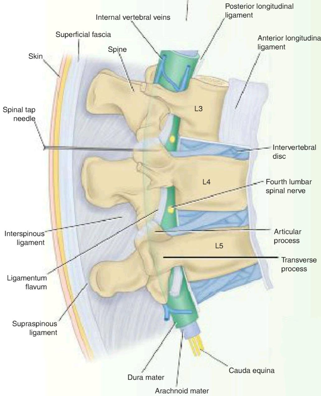
Figure 1-20 Sagittal section through the lumbar part of the vertebral column in a position of flexion. Note that the spines and laminae are Cauda equina well separated in this position allowing insertion of the spinal tap needle into the subarachnoid space.
شکل ۱-۲۰ برش ساژیتال از قسمت کمری ستون فقرات در حالت خمیدگی. توجه داشته باشید که خارها و لامیناها در این حالت به خوبی از هم جدا هستند و به این ترتیب سوزن اسپاینال تیپی به راحتی وارد فضای زیر عنکبوتیه میشود.
CSF pressure can be measured by attaching a manometer to the needle. When the patient is in the recumbent position, the normal pressure is about 60 to 150 mm of water. The pressure shows oscillations corresponding to the movements of respiration and the arterial pulse.
فشار CSF را میتوان با اتصال یک مانومتر به سوزن اندازهگیری کرد. وقتی بیمار در حالت خوابیده است، فشار طبیعی حدود ۶۰ تا ۱۵۰ میلیمتر آب است. فشار، نوساناتی را نشان میدهد که مربوط به حرکات تنفس و نبض شریانی است.
A block of the subarachnoid space in the vertebral canal, which may be caused by a tumor of the spinal cord or the meninges, can be detected by compressing the internal jugular veins in the neck. This raises the cerebral venous pressure and inhibits the absorption of CSF in the arachnoid granulations, thereby increasing the manometer reading of the CSF pressure. If this rise does not occur, the subarachnoid space is blocked, and the patient exhibits a positive Queckenstedt sign.
انسداد فضای زیر عنکبوتیه در کانال مهرهای، که ممکن است ناشی از تومور نخاع یا مننژ باشد، را میتوان با فشردهسازی وریدهای ژوگولار داخلی در گردن تشخیص داد. این امر فشار وریدی مغز را افزایش داده و جذب CSF در گرانولهای عنکبوتیه را مهار میکند و در نتیجه فشار CSF در مانومتر افزایش مییابد. اگر این افزایش رخ ندهد، فضای زیر عنکبوتیه مسدود شده و بیمار علامت کوئکنستدت مثبت نشان میدهد.
Caudal Anesthesia
Anesthetic solutions may be injected into the sacral canal through the sacral hiatus. The solutions pass upward in the loose connective tissue and bathe the spinal nerves as they emerge from the dural sheath (Fig. 1-21). Obstetricians use this method of nerve block to relieve the pains of the first and second stages of labor because anesthetic administered by this method does not affect the infant. Caudal anesthesia can also be used in operations in the sacral region, including anorectal surgery.
بیحسی کودال
محلولهای بیحسی ممکن است از طریق شکاف ساکرال به داخل کانال ساکرال تزریق شوند. این محلولها در بافت همبند شل به سمت بالا حرکت میکنند و اعصاب نخاعی را هنگام خروج از غلاف سختشامه شستشو میدهند (شکل 1-21). متخصصان زنان و زایمان از این روش بلوک عصبی برای تسکین دردهای مراحل اول و دوم زایمان استفاده میکنند زیرا بیحسی تجویز شده با این روش بر نوزاد تأثیری ندارد. بیحسی کودال همچنین میتواند در جراحیهای ناحیه ساکرال، از جمله جراحی آنورکتال، استفاده شود.
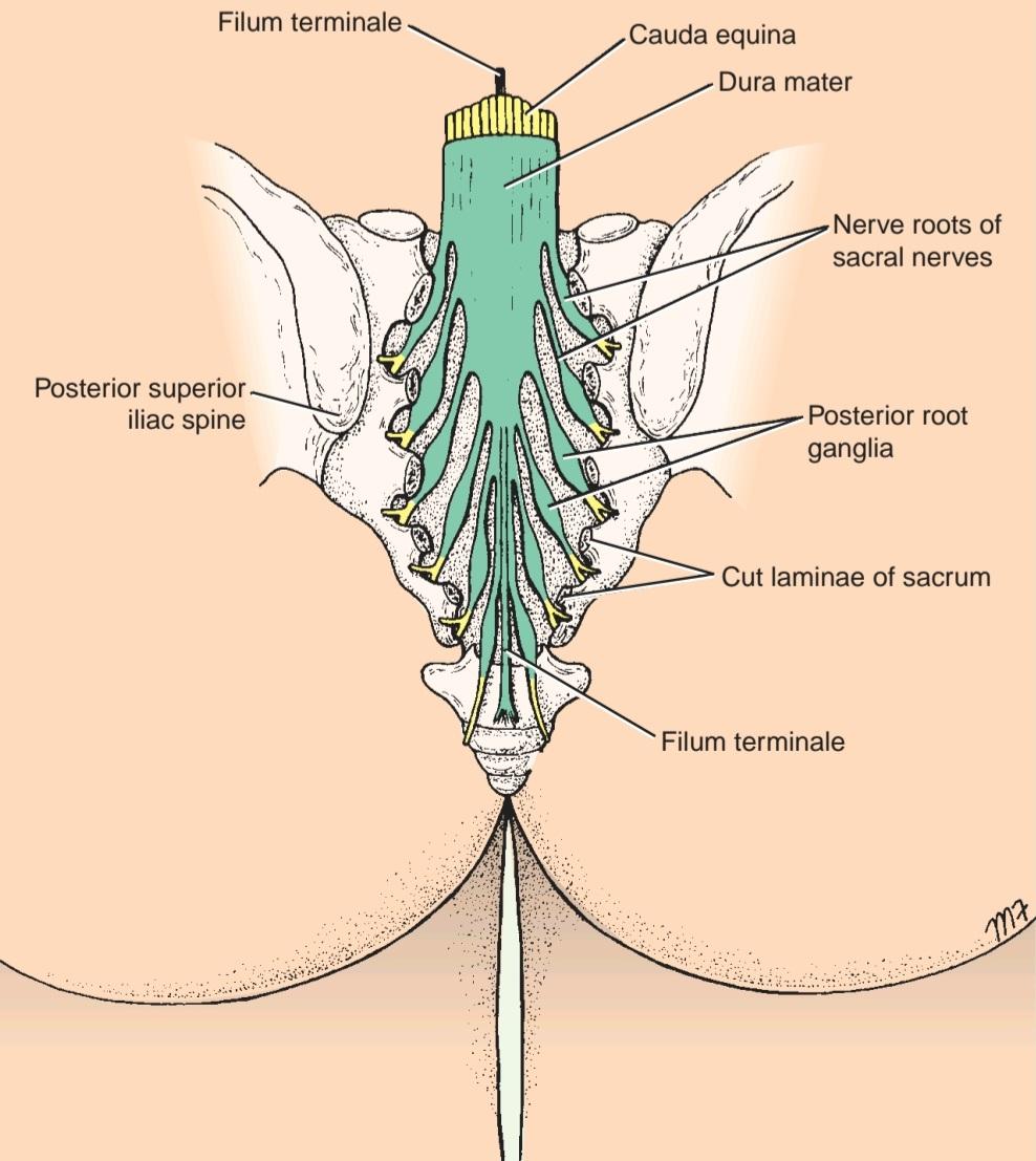
Figure 1-21 Posterior view of the sacrum. Laminae have been removed to show the sacral nerve roots lying within the sacral canal.
شکل ۱-۲۱ نمای خلفی استخوان خاجی. لامیناها برداشته شدهاند تا ریشههای عصبی خاجی که در داخل کانال خاجی قرار دارند، نشان داده شوند.
Head Injuries
A blow to the head may merely bruise the scalp; severe blows may tear or split the scalp. Even if the head is protected by a helmet, the brain can be severely damaged without clinical evidence of scalp injury.
آسیبهای سر
ضربه به سر ممکن است فقط پوست سر را کبود کند؛ ضربات شدید ممکن است پوست سر را پاره یا شکافته کند. حتی اگر سر توسط کلاه ایمنی محافظت شود، مغز میتواند بدون شواهد بالینی آسیب پوست سر، به شدت آسیب ببیند.
Skull Fractures
Severe blows to the head can change the shape of the skull at the point of impact. Small objects may penetrate the skull and lacerate the brain. Larger objects applied with great force may shatter the skull and drive fragments of bone into the brain at the site of impact.
شکستگی جمجمه
ضربات شدید به سر میتواند شکل جمجمه را در محل برخورد تغییر دهد. اشیاء کوچک ممکن است به جمجمه نفوذ کرده و مغز را پاره کنند. اشیاء بزرگتر که با نیروی زیاد وارد میشوند، ممکن است جمجمه را خرد کرده و قطعات استخوان را در محل برخورد به مغز فرو کنند.
In the adult, fractures of the skull are common, but they are less common in the young child. In the infant, the skull bones are more resilient, and they are separated by fibrous sutural ligaments. In the adult, the inner table of the skull is particularly brittle. Moreover, the sutural ligaments begin to ossify during middle age.
در بزرگسالان، شکستگی جمجمه شایع است، اما در کودکان خردسال کمتر دیده میشود. در نوزادان، استخوانهای جمجمه انعطافپذیرتر هستند و توسط رباطهای بخیهای فیبری از هم جدا میشوند. در بزرگسالان، سطح داخلی جمجمه به ویژه شکننده است. علاوه بر این، رباطهای بخیهای در میانسالی شروع به استخوانی شدن میکنند.
The type of fracture that occurs in the skull will depend on the age of the patient, the severity of the blow, and the area of the skull receiving the trauma. The adult skull resembles an eggshell, with limited resilience. A severe, localized blow will cause a local indentation, commonly with bone splintering. Blows to the vault can result in a series of linear fractures, which radiate out through the thin areas of the bone. The petrous parts of the temporal bones and the occipital crests (see Fig. 5-6) strongly reinforce the base of the skull and tend to deflect linear fractures.
نوع شکستگی که در جمجمه رخ میدهد به سن بیمار، شدت ضربه و ناحیهای از جمجمه که دچار ضربه شده است بستگی دارد. جمجمه بزرگسالان شبیه پوسته تخممرغ است و انعطافپذیری محدودی دارد. یک ضربه شدید و موضعی باعث ایجاد فرورفتگی موضعی میشود که معمولاً با خرد شدن استخوان همراه است. ضربات به طاق جمجمه میتواند منجر به مجموعهای از شکستگیهای خطی شود که از طریق نواحی نازک استخوان به بیرون منتشر میشوند. قسمتهای خارهای استخوانهای گیجگاهی و ستیغهای پسسری (شکل 5-6 را ببینید) به شدت پایه جمجمه را تقویت میکنند و تمایل دارند شکستگیهای خطی را منحرف کنند.
The young child’s skull resembles a ping-pong ball, because a localized blow produces a depression without splintering. This common type of circumscribed lesion is known as a “pond” fracture.
جمجمه کودک خردسال شبیه توپ پینگپنگ است، زیرا یک ضربه موضعی باعث ایجاد فرورفتگی بدون ترک خوردن میشود. این نوع رایج از ضایعه محدود به عنوان شکستگی “برکهای” شناخته میشود.

Figure 1-22 A: Mechanisms of acute cerebral injury when a blow is applied to the lateral side of the head. B: Varieties of intracranial hemorrhage. C: Mechanism of cerebral trauma following a blow on the chin. The movement of the brain within the skull can also tear the cerebral veins.
شکل ۱-۲۲ الف: مکانیسمهای آسیب حاد مغزی هنگام وارد شدن ضربه به قسمت جانبی سر. ب: انواع خونریزی داخل جمجمه. ج: مکانیسم ترومای مغزی پس از ضربه به چانه. حرکت مغز در داخل جمجمه همچنین میتواند باعث پارگی وریدهای مغزی شود.
Brain Injuries
Brain injuries are caused by displacement and distortion of the neuronal tissues at the moment of impact (Fig. 1-22). The brain, which is incompressible, is like a water-soaked log suspended in water. The brain is floating in the CSF in the subarachnoid space and is capable of a certain amount of anteroposterior and lateral gliding movement. The anteroposterior movement is limited by the attachment of the superior cerebral veins to the superior sagittal sinus. Lateral displacement of the brain is limited by the falx cerebri. The tentorium cerebelli and the falx cerebelli also restrict displacement of the brain.
آسیبهای مغزی
آسیبهای مغزی ناشی از جابجایی و تغییر شکل بافتهای عصبی در لحظه ضربه هستند (شکل ۱-۲۲). مغز که غیرقابل تراکم است، مانند یک کنده خیس خورده در آب است که در آب معلق است. مغز در مایع مغزی نخاعی (CSF) در فضای زیر عنکبوتیه شناور است و قادر به انجام مقداری حرکت لغزشی قدامی-خلفی و جانبی است. حرکت قدامی-خلفی توسط اتصال وریدهای مغزی فوقانی به سینوس ساژیتال فوقانی محدود میشود. جابجایی جانبی مغز توسط داس مغزی محدود میشود. چادرینه مخچه و داس مغزی نیز جابجایی مغز را محدود میکنند.
Thus, blows on the front or back of the head lead to displacement of the brain, which may cause severe cerebral damage, stretching and distortion of the brainstem, and stretching and even tearing of brain commissures. Blows to the side of the head cause less cerebral displacement, and the injuries to the brain consequently tend to be less severe. However, the falx cerebri is a tough structure and may cause considerable damage to the softer brain tissue in cases of a severe blow to the side of the head. Furthermore, remember that glancing blows to the head may cause considerable brain rotation, with shearing strains and brain distortion, particularly in areas where further rotation is prevented by bony prominences in the anterior and middle cranial fossae. Brain lacerations are very likely to occur when the brain is forcibly thrown against the sharp edges of bone within the skull (see pp. 191-192)-the lesser wings of the sphenoid, for example.
بنابراین، ضربات به جلو یا عقب سر منجر به جابجایی مغز میشود که ممکن است باعث آسیب شدید مغزی، کشش و تغییر شکل ساقه مغز و کشش و حتی پارگی اتصالات مغزی شود. ضربات به کنار سر باعث جابجایی کمتر مغز میشود و در نتیجه آسیبهای وارده به مغز معمولاً شدت کمتری دارند. با این حال، داس مغزی ساختاری سفت است و در صورت ضربه شدید به کنار سر، ممکن است آسیب قابل توجهی به بافت نرمتر مغز وارد کند. علاوه بر این، به یاد داشته باشید که ضربات ناگهانی به سر ممکن است باعث چرخش قابل توجه مغز، همراه با کشیدگیهای برشی و تغییر شکل مغز شود، به ویژه در مناطقی که برجستگیهای استخوانی در حفرههای جمجمهای قدامی و میانی مانع چرخش بیشتر میشوند. پارگی مغز زمانی که مغز به شدت به لبههای تیز استخوان درون جمجمه برخورد میکند (به صفحات ۱۹۱-۱۹۲ مراجعه کنید) – به عنوان مثال، بالهای کوچک اسفنوئید – بسیار محتمل است.
When the brain moves suddenly within the skull, the part of the brain that moves away from the skull wall is subjected to diminished pressure because the CSF has not had time to accommodate to the brain movement. This results in a suction effect on the brain surface, with rupture of surface blood vessels.
وقتی مغز به طور ناگهانی در داخل جمجمه حرکت میکند، بخشی از مغز که از دیواره جمجمه دور میشود، تحت فشار کمتری قرار میگیرد زیرا مایع مغزی نخاعی (CSF) زمان کافی برای تطبیق با حرکت مغز را نداشته است. این امر منجر به ایجاد یک اثر مکشی روی سطح مغز و پارگی رگهای خونی سطحی میشود.
A sudden severe blow to the head, as in an automobile accident, may result in damage to the brain at two sites: at the point of impact and at the pole of the brain opposite the point of impact, where the brain is thrown against the skull wall. This is referred to as contrecoup injury.
ضربه شدید ناگهانی به سر، مانند آنچه در یک تصادف رانندگی رخ میدهد، ممکن است منجر به آسیب به مغز در دو محل شود: در نقطه برخورد و در قطب مغز در مقابل نقطه برخورد، جایی که مغز به دیواره جمجمه پرتاب میشود. این آسیب، آسیب ضربهای نامیده میشود.
The movement of the brain within the skull at the time of head injuries not only is likely to cause avulsion of cranial nerves but commonly leads to rupture of tethering blood vessels. Fortunately, the large arteries found at the base of the brain are tortuous, and this, coupled with their strength, explains why they are rarely torn. The thin-walled cortical veins, which drain into the large dural venous sinuses, are very vulnerable and can cause severe subdural or subarachnoid hemorrhage.
حرکت مغز در داخل جمجمه در زمان آسیبهای سر، نه تنها احتمالاً باعث کندگی اعصاب جمجمهای میشود، بلکه معمولاً منجر به پارگی رگهای خونی متصل کننده میشود. خوشبختانه، شریانهای بزرگی که در پایه مغز یافت میشوند، پیچ و خم دارند و این، همراه با قدرت آنها، توضیح میدهد که چرا به ندرت پاره میشوند. وریدهای قشری با دیواره نازک که به سینوسهای وریدی بزرگ سختشامه تخلیه میشوند، بسیار آسیبپذیر هستند و میتوانند باعث خونریزی شدید سابدورال یا سابآراکنویید شوند.
Traumatic Brain Injury Following an Explosion or Blast
Deployed soldiers are frequently exposed to explosive devices, which may result in extensive injuries to the limbs, eyes, and ears. Open injuries to the skull, where shrapnel has entered the brain, are clearly visible and are dealt with accordingly.
آسیب تروماتیک مغزی پس از انفجار یا انفجار
سربازان مستقر اغلب در معرض مواد منفجره قرار میگیرند که ممکن است منجر به جراحات گسترده در اندامها، چشمها و گوشها شود. جراحات باز جمجمه، جایی که ترکشها وارد مغز شدهاند، به وضوح قابل مشاهده هستند و بر اساس آن با آنها برخورد میشود.
However, in closed injuries, in which the skull remains intact, the underlying brain may be damaged but left untreated. In these cases, the explosion produces a blast of air that strikes the skull and shakes up the brain, resulting in multiple injuries to the soft brain tissue as it is driven against the hard bony projections within the skull. The symptoms and signs will depend on the extent of the neurologic damage and will be mild, moderate, or severe. Although the moderate and severe cases are quickly recognized by medical personnel, the mild cases may be missed, and patients may later develop headaches, nausea, mood changes, and memory loss. Early diagnosis is imperative as studies of these patients have shown that mild neurologic damage can be successfully treated. Individuals who have been exposed to explosive devices should undergo computed tomography (CT) scan or magnetic resonance imaging (MRI) before returning to civilian life.
با این حال، در آسیبهای بسته، که در آن جمجمه سالم میماند، مغز زیرین ممکن است آسیب ببیند اما درمان نشود. در این موارد، انفجار باعث ایجاد انفجاری از هوا میشود که به جمجمه برخورد میکند و مغز را تکان میدهد و در نتیجه آسیبهای متعددی به بافت نرم مغز وارد میشود، زیرا به برآمدگیهای استخوانی سخت درون جمجمه برخورد میکند. علائم و نشانهها به میزان آسیب عصبی بستگی دارد و خفیف، متوسط یا شدید خواهد بود. اگرچه موارد متوسط و شدید به سرعت توسط پرسنل پزشکی تشخیص داده میشوند، اما موارد خفیف ممکن است نادیده گرفته شوند و بیماران بعداً دچار سردرد، حالت تهوع، تغییرات خلقی و از دست دادن حافظه شوند. تشخیص زودهنگام ضروری است زیرا مطالعات روی این بیماران نشان داده است که آسیب عصبی خفیف را میتوان با موفقیت درمان کرد. افرادی که در معرض مواد منفجره قرار گرفتهاند باید قبل از بازگشت به زندگی عادی تحت سیتیاسکن یا تصویربرداری رزونانس مغناطیسی (MRI) قرار گیرند.
Intracranial Hemorrhage
Although the brain is cushioned by the surrounding CSF in the subarachnoid space, any severe hemorrhage within the relatively rigid skull will ultimately exert pressure on the brain.
خونریزی داخل جمجمهای
اگرچه مغز توسط مایع مغزی نخاعی اطراف آن در فضای زیر عنکبوتیه محافظت میشود، اما هرگونه خونریزی شدید در داخل جمجمه نسبتاً سفت در نهایت به مغز فشار وارد میکند.
Four types of intracranial hemorrhage may result from trauma or cerebral vascular lesions (see Fig. 1-22).
چهار نوع خونریزی داخل جمجمهای ممکن است ناشی از تروما یا ضایعات عروقی مغزی باشد (شکل 1-22 را ببینید).
Epidural (extradural) hemorrhage results from injuries to the meningeal arteries (commonly the anterior division of the middle meningeal artery) or veins (see Fig. 15-6). A comparatively minor blow to the side of the head, resulting in fracture of the skull in the region of the anterior-inferior portion of the parietal bone, may sever the artery (see Fig. 1-22). Arterial or venous injury is especially likely to occur if the vessels enter a bony canal in this region. Bleeding occurs and strips the meningeal layer of dura from the internal surface of the skull. The intracranial pressure (ICP) rises, and the enlarging blood clot exerts local pressure on the underlying precentral gyrus (motor area). Blood may also pass laterally through the fracture line to form a soft swelling on the side of the head. To stop the hemorrhage, the torn artery must be ligated or plugged. The burr hole through the skull wall should be placed about 1.5 in (4 cm) above the midpoint of the zygomatic arch.
خونریزی اپیدورال (خارج سختشامه) ناشی از آسیب به شریانهای مننژیال (معمولاً بخش قدامی شریان مننژیال میانی) یا وریدها است (شکل 15-6 را ببینید). یک ضربه نسبتاً جزئی به کنار سر، که منجر به شکستگی جمجمه در ناحیه قسمت قدامی-تحتانی استخوان جداری میشود، ممکن است شریان را قطع کند (شکل 1-22 را ببینید). آسیب شریانی یا وریدی به ویژه در صورتی که رگها وارد کانال استخوانی در این ناحیه شوند، محتمل است. خونریزی رخ میدهد و لایه مننژیال سختشامه را از سطح داخلی جمجمه جدا میکند. فشار داخل جمجمه (ICP) افزایش مییابد و لخته خون بزرگ شده، فشار موضعی بر شکنج پیشمرکزی (ناحیه حرکتی) زیرین وارد میکند. خون همچنین ممکن است به صورت جانبی از خط شکستگی عبور کند و یک تورم نرم در کنار سر ایجاد کند. برای متوقف کردن خونریزی، شریان پاره شده باید مسدود یا مسدود شود. سوراخ مته از طریق دیواره جمجمه باید حدود ۴ سانتیمتر (۱.۵ اینچ) بالاتر از نقطه میانی قوس گونه قرار گیرد.
Subdural hemorrhage results from tearing of the superior cerebral veins where they enter the superior sagittal sinus (see Figs. 15-1 and 17-5). The cause is usually a blow to the front or back of the head resulting in excessive anteroposterior displacement of the brain within the skull. This condition, which is much more common than middle meningeal hemorrhage, can be caused by a sudden minor blow. Once the vein is torn, blood under low pressure begins to accumulate in the potential space between the dura and the arachnoid. The condition is rarely bilateral.
خونریزی سابدورال ناشی از پارگی وریدهای مغزی فوقانی در محل ورود آنها به سینوس ساژیتال فوقانی است (شکلهای 15-1 و 17-5 را ببینید). علت آن معمولاً ضربه به جلو یا عقب سر است که منجر به جابجایی بیش از حد قدامی-خلفی مغز در داخل جمجمه میشود. این وضعیت که بسیار شایعتر از خونریزی مننژی میانی است، میتواند در اثر یک ضربه ناگهانی جزئی ایجاد شود. پس از پاره شدن ورید، خون تحت فشار کم شروع به تجمع در فضای بالقوه بین سختشامه و عنکبوتیه میکند. این وضعیت به ندرت دو طرفه است.
Acute and chronic forms occur depending on the speed of accumulation of fluid in the subdural space. For example, if the patient starts to vomit, the venous pressure will rise as the result of a rise in the intrathoracic pressure. Under these circumstances, the subdural blood clot will rapidly increase in size, causing acute symptoms. In the chronic form, over a course of several months, the small blood clot will attract fluid by osmosis, in which case a hemorrhagic cyst forms and gradually expands, causing pressure symptoms. In both forms, the blood clot must be removed through burr holes in the skull.
اشکال حاد و مزمن بسته به سرعت تجمع مایع در فضای زیر سختشامه رخ میدهند. به عنوان مثال، اگر بیمار شروع به استفراغ کند، فشار وریدی در نتیجه افزایش فشار داخل قفسه سینه افزایش مییابد. در این شرایط، لخته خون زیر سختشامه به سرعت از نظر اندازه بزرگ میشود و باعث علائم حاد میشود. در شکل مزمن، طی یک دوره چند ماهه، لخته خون کوچک از طریق اسمز، مایع را جذب میکند، در این صورت یک کیست خونریزی دهنده تشکیل میشود و به تدریج گسترش مییابد و باعث علائم فشار میشود. در هر دو شکل، لخته خون باید از طریق سوراخهای ایجاد شده در جمجمه برداشته شود.
Subarachnoid hemorrhage results from nontraumatic leakage or rupture of a congenital aneurysm on the cerebral arterial circle (circle of Willis) or, less commonly, from an arteriovenous malformation. Sudden onset of severe headache, neck stiffness, and loss of consciousness occurs. The diagnosis is established by CT or MRI or by withdrawing heavily blood-stained CSF through a lumbar puncture.
خونریزی زیر عنکبوتیه ناشی از نشت یا پارگی غیرتروماتیک آنوریسم مادرزادی روی حلقه شریانی مغزی (حلقه ویلیس) یا، با شیوع کمتر، ناشی از یک ناهنجاری شریانی-وریدی است. شروع ناگهانی سردرد شدید، سفتی گردن و از دست دادن هوشیاری رخ میدهد. تشخیص با سیتیاسکن یا امآرآی یا با برداشتن مایع مغزی نخاعی (CSF) آغشته به خون از طریق پونکسیون کمری قطعی میشود.
With cerebral hemorrhage, spontaneous intracerebral hemorrhage (see Fig. 1-22) is most common in patients with hypertension. It is generally due to rupture of the thin- walled lenticulostriate artery (see Fig. 17-11), a branch of the middle cerebral artery (see Fig. 17-4). The hemorrhage involves important descending nerve fibers in the internal capsule and causes hemiplegia on the contralateral side. The patient immediately loses consciousness, and paralysis is evident when consciousness is regained. The diagnosis is established by brain CT or MRI.
در خونریزی مغزی، خونریزی داخل مغزی خود به خودی (شکل 1-22 را ببینید) در بیماران مبتلا به فشار خون بالا شایعتر است. این خونریزی عموماً به دلیل پارگی شریان عدسی-استریاتیک با دیواره نازک (شکل 17-11 را ببینید)، شاخهای از شریان مغزی میانی (شکل 17-4 را ببینید) رخ میدهد. خونریزی، رشتههای عصبی نزولی مهم در کپسول داخلی را درگیر میکند و باعث فلج ناقص در سمت مقابل میشود. بیمار بلافاصله هوشیاری خود را از دست میدهد و فلج هنگام بازیابی هوشیاری مشهود است. تشخیص با سیتیاسکن یا امآرآی مغز تأیید میشود.
Shaken Baby Syndrome
Inflicted head injury is the most common cause of traumatic death in infancy. Sudden deceleration, which occurs when an infant is held by the arms or trunk and shaken or the head is forcefully struck against a hard surface, is believed to be responsible for the brain injuries. Biomechanical studies have shown that the rotation of the floating brain around its center of gravity causes diffuse brain injuries, including diffuse axonal injury and subdural hematoma. In shaken baby syndrome, major rotational forces have to occur that clearly exceed those encountered in normal child play activities.
سندرم تکان دادن نوزاد
آسیب به سر شایعترین علت مرگ ناشی از ضربه در دوران نوزادی است. اعتقاد بر این است که کاهش ناگهانی سرعت، که زمانی رخ میدهد که نوزاد با بازوها یا تنه نگه داشته شده و تکان داده میشود یا سر به شدت به سطح سختی برخورد میکند، مسئول آسیبهای مغزی است. مطالعات بیومکانیکی نشان دادهاند که چرخش مغز شناور در اطراف مرکز ثقل خود باعث آسیبهای منتشر مغزی، از جمله آسیب آکسونی منتشر و هماتوم ساب دورال میشود. در سندرم تکان دادن نوزاد، نیروهای چرخشی عمدهای باید رخ دهند که به وضوح از نیروهایی که در فعالیتهای بازی معمولی کودک با آنها مواجه میشوند، فراتر میروند.
Most cases of shaken baby syndrome take place during the first year of life, and they are usually restricted to infants under 3 years of age. Common symptoms include lethargy, irritability, seizures, altered muscle tone, and symptoms indicating raised ICP, such as impaired consciousness, vomiting, breathing abnormalities, and apnea. In severe cases, the baby may be unresponsive, the fontanelles are bulging, and the child may have retinal hemor- rhages. Spinal tap may reveal blood in the CSF. Subdural or subarachnoid hemorrhages can be readily detected on CT or MRI scans. Autopsy findings commonly include localized subdural hemorrhage in the parietal-occipital region and subarachnoid blood, associated with massive cerebral swelling and widespread neuronal loss.
بیشتر موارد سندرم تکان دادن کودک در طول سال اول زندگی رخ میدهد و معمولاً به نوزادان زیر ۳ سال محدود میشود. علائم رایج شامل بیحالی، تحریکپذیری، تشنج، تغییر تون عضلانی و علائمی است که نشان دهنده افزایش فشار داخل جمجمه (ICP) مانند اختلال هوشیاری، استفراغ، ناهنجاریهای تنفسی و آپنه است. در موارد شدید، ممکن است نوزاد واکنشی نشان ندهد، فونتانلها برجسته باشند و کودک ممکن است خونریزی شبکیه داشته باشد. ضربه به نخاع ممکن است خون را در مایع مغزی نخاعی نشان دهد. خونریزیهای ساب دورال یا ساب آراکنوئید را میتوان به راحتی در سیتیاسکن یا امآرآی تشخیص داد. یافتههای کالبدشکافی معمولاً شامل خونریزی ساب دورال موضعی در ناحیه جداری-پسسری و خون ساب آراکنوئید است که با تورم شدید مغزی و از دست دادن گسترده نورونها همراه است.
Space-Occupying Lesions Within the Skull
Space-occupying or expanding lesions within the skull include tumor, hematoma, and abscess. Because the skull is a rigid container of fixed volume, these lesions will add to the normal bulk of the intracranial contents.
ضایعات فضاگیر درون جمجمه
ضایعات فضاگیر یا در حال گسترش درون جمجمه شامل تومور، هماتوم و آبسه میشوند. از آنجا که جمجمه یک محفظه سفت و سخت با حجم ثابت است، این ضایعات به حجم طبیعی محتویات داخل جمجمه اضافه میشوند.
An expanding lesion is first accommodated by the expulsion of CSF from the cranial cavity. Later, the veins become compressed, interference with the circulation of blood and CSF begins, and the ICP starts to rise. The venous congestion results in increased production and diminished absorption of CSF, the CSF volume begins to rise, and thus, a vicious circle is established.
یک ضایعه در حال گسترش ابتدا با خروج مایع مغزی نخاعی (CSF) از حفره جمجمه جبران میشود. بعداً، رگها فشرده میشوند، تداخل با گردش خون و مایع مغزی نخاعی آغاز میشود و فشار داخل جمجمه (ICP) شروع به افزایش میکند. احتقان وریدی منجر به افزایش تولید و کاهش جذب مایع مغزی نخاعی (CSF) میشود، حجم مایع مغزی نخاعی شروع به افزایش میکند و بنابراین، یک چرخه معیوب ایجاد میشود.
The position of the tumor within the brain may have a dramatic effect on the signs and symptoms. For example, a tumor that obstructs the outflow of CSF or directly presses on the great veins will cause a rapid increase in ICP. The signs and symptoms that enable the clinician to localize the lesion will depend on the degree of interference with brain function and nervous tissue destruction. Severe headache, possibly due to the stretching of the dura mater, and vomiting, due to pressure on the brainstem, are common complaints.
موقعیت تومور در مغز میتواند تأثیر چشمگیری بر علائم و نشانهها داشته باشد. به عنوان مثال، توموری که مانع خروج CSF میشود یا مستقیماً به رگهای بزرگ فشار میآورد، باعث افزایش سریع ICP میشود. علائم و نشانههایی که پزشک را قادر میسازد ضایعه را مشخص کند، به میزان تداخل با عملکرد مغز و تخریب بافت عصبی بستگی دارد. سردرد شدید، احتمالاً به دلیل کشیدگی سختشامه، و استفراغ، به دلیل فشار بر ساقه مغز، از شکایات شایع هستند.
A spinal tap should not be performed in patients with suspected intracranial tumor. The withdrawal of CSF may lead to a sudden displacement of the cerebral hemisphere through the notch in the tentorium cerebelli into the posterior cranial fossa (Fig. 1-23) or herniation of the medulla oblongata and cerebellum through the foramen magnum. CT scans or MRIS are used in diagnosis.
در بیماران مشکوک به تومور داخل جمجمه، نباید پونکسیون نخاعی انجام شود. خارج کردن CSF ممکن است منجر به جابجایی ناگهانی نیمکره مغزی از طریق بریدگی در چادرینه مخچه به داخل حفره جمجمه خلفی (شکل 1-23) یا فتق بصل النخاع و مخچه از طریق سوراخ بزرگ جمجمه شود. سی تی اسکن یا MRIS برای تشخیص استفاده میشوند.

Figure 1-23 Sudden displacement of the cerebral hemispheres through the tentorial notch into the posterior cranial fossa following a lumbar puncture; the cerebral tumor is situated in the right cerebral hemisphere. CT or MRI should be used rather than lumbar puncture when investigating a cerebral tumor.
شکل ۱-۲۳ جابجایی ناگهانی نیمکرههای مغزی از طریق بریدگی چادرینه به داخل حفره جمجمهای خلفی پس از پونکسیون کمری؛ تومور مغزی در نیمکره مغزی راست قرار دارد. هنگام بررسی تومور مغزی باید به جای پونکسیون کمری از سیتیاسکن یا امآرآی استفاده شود.
Computed Tomography
CT is used for the detection of intracranial lesions. The procedure is quick, safe, and accurate. The total dose of irradiation is no greater than for a conventional skull radiograph.
توموگرافی کامپیوتری
سیتیاسکن برای تشخیص ضایعات داخل جمجمهای استفاده میشود. این روش سریع، ایمن و دقیق است. دوز کل تابش بیشتر از رادیوگرافی جمجمه معمولی نیست.
CT relies on the same physics as conventional radiographs, in that structures are distinguished from one another by their ability to absorb energy from x-rays. The x-ray tube emits a narrow beam of radiation as it passes in a series of scanning movements through an arc of 180 degrees around the patient’s head. The x-rays having passed through the head are collected by a special x-ray detector. The information is fed to a computer that processes the information, which is then displayed as a reconstructed picture on a screen. Essentially, the observer sees an image of a thin slice through the head, which may then be photographed for later examination (Fig. 1-24).
سیتیاسکن بر اساس همان فیزیک رادیوگرافیهای معمولی عمل میکند، به این صورت که ساختارها با توانایی جذب انرژی از اشعه ایکس از یکدیگر متمایز میشوند. لوله اشعه ایکس، پرتو باریکی از تابش را در حین عبور از یک قوس ۱۸۰ درجهای در اطراف سر بیمار، ساطع میکند. اشعه ایکس عبور کرده از سر توسط یک آشکارساز مخصوص اشعه ایکس جمعآوری میشود. اطلاعات به رایانهای که اطلاعات را پردازش میکند، داده میشود و سپس به صورت یک تصویر بازسازی شده روی صفحه نمایش داده میشود. اساساً، ناظر تصویری از یک برش نازک از سر را میبیند که ممکن است برای بررسیهای بعدی از آن عکس گرفته شود (شکل ۱-۲۴).

Figure 1-24 CT scan showing the structure of the brain. A, B: Horizontal cuts (axial sections).
شکل ۱-۲۴ سیتیاسکن که ساختار مغز را نشان میدهد. الف، ب: برشهای افقی (برشهای محوری).
The sensitivity is such that small differences in x-ray absorption can be easily displayed. The gray matter of the cerebral cortex, white matter, internal capsule, corpus callosum, ventricles, and subarachnoid spaces can all be recognized. An iodine-containing medium can be injected intravascularly, which enhances greatly the contrast between tissues having a different blood flow.
حساسیت به حدی است که تفاوتهای کوچک در جذب اشعه ایکس به راحتی قابل نمایش است. ماده خاکستری قشر مغز، ماده سفید، کپسول داخلی، جسم پینهای، بطنها و فضاهای زیر عنکبوتیه، همگی قابل تشخیص هستند. میتوان یک ماده حاوی ید را به صورت داخل عروقی تزریق کرد که کنتراست بین بافتهایی که جریان خون متفاوتی دارند را به میزان زیادی افزایش میدهد.
Because a CT scan can be performed in 5 to 10 minutes, it is the method of choice in an emergency situation with patients with head trauma or suspected intracranial hemorrhage.
از آنجا که سیتیاسکن را میتوان در عرض ۵ تا ۱۰ دقیقه انجام داد، روش انتخابی در شرایط اضطراری برای بیمارانی است که دچار ضربه به سر یا خونریزی داخل جمجمهای مشکوک شدهاند.
Magnetic Resonance Imaging
MRI uses the magnetic properties of the hydrogen nucleus excited by radiofrequency radiation transmitted by a coil surrounding the head. The excited hydrogen nuclei emit a signal that is detected as induced electric currents in a receiver coil. MRI is absolutely safe to the patient, and because it provides better differentiation between gray and white matter, MRI can be more revealing than CT. The reason for this is that gray matter contains more hydrogen in the form of water than white matter, and the hydrogen atoms are less bound in fat (Fig. 1-25). MRI is the best imaging method for detecting low-contrast lesions such as brain tumors or small multiple sclerosis plaques. It is also capable of showing clear images of the brainstem, cerebellum, and the pituitary fossa, which, in the case of a CT scan, are overshadowed by the dense bones of the base of the skull.
تصویربرداری رزونانس مغناطیسی
MRI از خواص مغناطیسی هسته هیدروژن تحریک شده توسط تابش فرکانس رادیویی که توسط یک سیمپیچ اطراف سر منتقل میشود، استفاده میکند. هستههای هیدروژن تحریک شده سیگنالی را منتشر میکنند که به صورت جریانهای الکتریکی القایی در یک سیمپیچ گیرنده شناسایی میشود. MRI برای بیمار کاملاً بیخطر است و از آنجا که تمایز بهتری بین ماده خاکستری و سفید ایجاد میکند، MRI میتواند نسبت به سیتیاسکن اطلاعات بیشتری را آشکار کند. دلیل این امر این است که ماده خاکستری حاوی هیدروژن بیشتری به شکل آب نسبت به ماده سفید است و اتمهای هیدروژن کمتر در چربی محصور شدهاند (شکل 1-25). MRI بهترین روش تصویربرداری برای تشخیص ضایعات با کنتراست کم مانند تومورهای مغزی یا پلاکهای کوچک مولتیپل اسکلروزیس است. همچنین قادر به نمایش تصاویر واضح از ساقه مغز، مخچه و حفره هیپوفیز است که در مورد سیتیاسکن، تحت الشعاع استخوانهای متراکم قاعده جمجمه قرار میگیرند.

Figure 1-25 MRI showing the structure of the brain. A: Sagittal. B: Coronal. Compare with Figure 1-24. Note the better differentiation between gray and white matter.
شکل ۱-۲۵ MRI ساختار مغز را نشان میدهد. الف: ساژیتال. ب: کرونال. با شکل ۱-۲۴ مقایسه کنید. به تمایز بهتر بین ماده خاکستری و سفید توجه کنید.
The spinal cord structure is much more clearly visualized with MRI.
ساختار نخاع با MRI بسیار واضحتر قابل مشاهده است.
Unfortunately, an MRI takes longer and costs two-thirds more than a CT scan.
متاسفانه، MRI زمان بیشتری میبرد و دو سوم بیشتر از سیتیاسکن هزینه دارد.
Positron Emission Tomography
Positron emission tomography (PET) uses radioactive isotopes that decay with the emission of positively charged electrons (positrons) to map the biochemical, physiologic, and pharmacologic processes taking place in the brain.
توموگرافی گسیل پوزیترون
توموگرافی گسیل پوزیترون (PET) از ایزوتوپهای رادیواکتیو که با گسیل الکترونهای با بار مثبت (پوزیترون) واپاشی میشوند، برای نقشهبرداری از فرآیندهای بیوشیمیایی، فیزیولوژیکی و دارویی که در مغز رخ میدهند، استفاده میکند.
The appropriate isotope is incorporated into molecules of known biochemical behavior in the brain and then is injected into the patient. The metabolic activity of the compound can then be studied by making cross-sectional tomographic images of the brain using the same principles as in CT (Fig. 1-26). Making a series of time-lapse images at different anatomical sites allows variations in brain metabolism to be studied at these sites. This technique has been used to study the distribution and activity of neurotransmitters, variations in oxygen utilization, and cerebral blood flow.
ایزوتوپ مناسب در مولکولهایی با رفتار بیوشیمیایی شناخته شده در مغز گنجانده شده و سپس به بیمار تزریق میشود. سپس میتوان با تهیه تصاویر توموگرافی مقطعی از مغز با استفاده از همان اصول سیتیاسکن (شکل 1-26)، فعالیت متابولیکی ترکیب را مطالعه کرد. تهیه مجموعهای از تصاویر مرور زمان در مکانهای مختلف آناتومیک، امکان مطالعه تغییرات متابولیسم مغز را در این مکانها فراهم میکند. این تکنیک برای مطالعه توزیع و فعالیت انتقالدهندههای عصبی، تغییرات در استفاده از اکسیژن و جریان خون مغزی استفاده شده است.
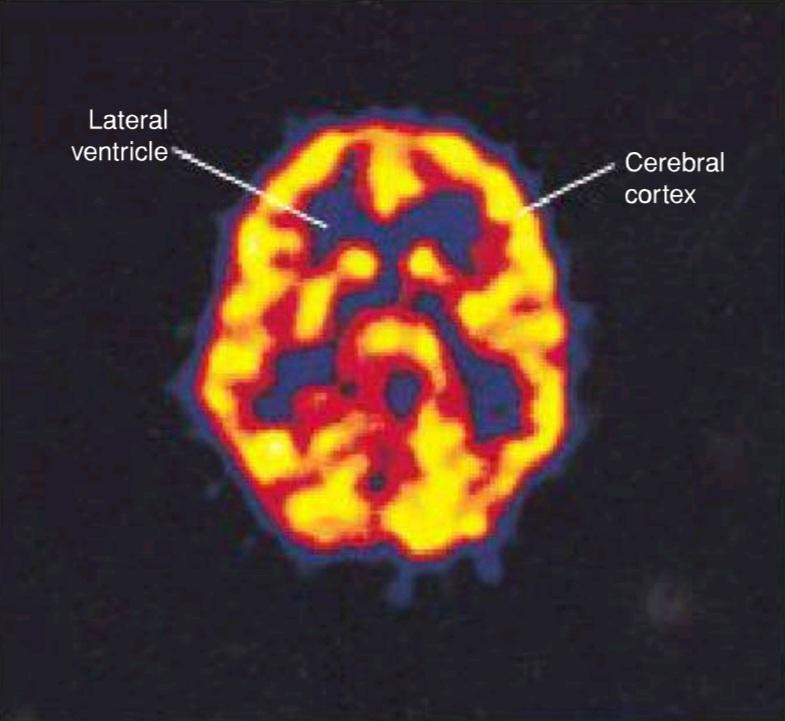
Figure 1-26 Axial (horizontal) PET scan of a normal brain following the injection of 18-fluorodeoxyglucose. Regions of active metabolism (yellow areas) are seen in the cerebral cortex. The lateral ventricles are also demonstrated. (Courtesy Dr. Holley Dey.)
شکل ۱-۲۶ اسکن PET محوری (افقی) از یک مغز طبیعی پس از تزریق ۱۸-فلوئورودئوکسی گلوکز. نواحی متابولیسم فعال (نواحی زرد) در قشر مغز دیده میشوند. بطنهای جانبی نیز نشان داده شدهاند. (با تشکر از دکتر هالی دی.)
PET has been successfully used in the evaluation of patients with brain tumors (Figs. 1-27 and 1-28), movement disorders, seizures, and schizophrenia.
PET با موفقیت در ارزیابی بیماران مبتلا به تومورهای مغزی (شکلهای ۱-۲۷ و ۱-۲۸)، اختلالات حرکتی، تشنج و اسکیزوفرنی مورد استفاده قرار گرفته است.
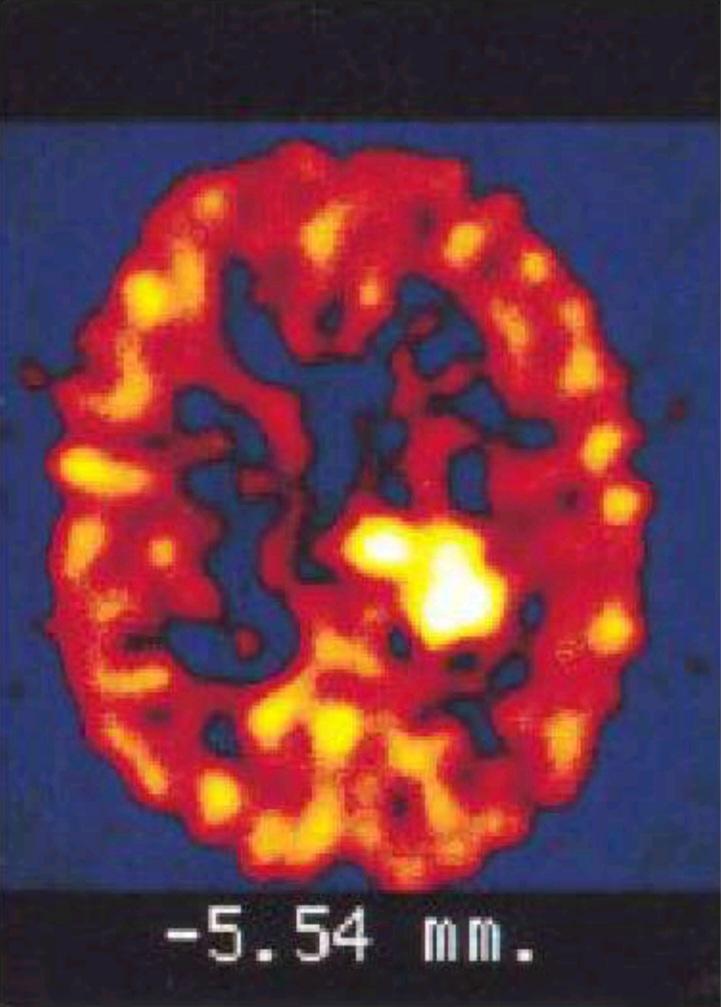
Figure 1-27 Axial (horizontal) PET scan of a 62-year- old male patient with a malignant glioma in the left parietal lobe following the injection of 18-fluorode- oxyglucose. A high concentration of the compound (circular yellow area) is seen in the region of the tumor. (Courtesy Dr. Holley Dey.)
شکل ۱-۲۷ اسکن PET محوری (افقی) از یک بیمار مرد 62 ساله مبتلا به گلیوما بدخیم در لوب آهیانه چپ پس از تزریق 18-فلوئورود-اکسی گلوکز. غلظت بالایی از این ترکیب (ناحیه زرد دایرهای) در ناحیه تومور مشاهده میشود. (با تشکر از دکتر هالی دی.)

Figure 1-28 Coronal PET scan of a 62-year-old male patient with a malignant glioma in the left parietal lobe following the injection of 18-fluorodeoxyglucose (same patient as in Fig. 1-25). A high concentration of the compound (circular yellow area) is seen in the region of the tumor. (Courtesy Dr. Holley Dey.)
شکل ۱-۲۸ اسکن PET کرونال از یک بیمار مرد 62 ساله مبتلا به گلیوما بدخیم در لوب آهیانه چپ پس از تزریق 18-فلوئورودئوکسی گلوکز (همان بیمار در شکل 1-25). غلظت بالایی از این ترکیب (ناحیه زرد دایرهای) در ناحیه تومور مشاهده میشود. (با تشکر از دکتر هالی دی.)
Key Concepts
Central and Peripheral Nervous Systems
مفاهیم کلیدی
سیستمهای عصبی مرکزی و محیطی
• The nervous system comprises the central nervous system (CNS) and peripheral nervous system (PNS).
• سیستم عصبی شامل سیستم عصبی مرکزی (CNS) و سیستم عصبی محیطی (PNS) است.
• The CNS consists of the brain and the spinal cord, both of which are surrounded by the meninges and the cerebrospinal fluid (CSF).
• سیستم عصبی مرکزی شامل مغز و نخاع است که هر دو توسط مننژ و مایع مغزی نخاعی (CSF) احاطه شدهاند.
• The PNS consists of all other nerves in the body.
• سیستم عصبی محیطی شامل تمام اعصاب دیگر بدن است.
• The autonomic nervous system (ANS) is concerned with involuntary structures and distributed throughout both CNS and PNS.
• سیستم عصبی خودکار (ANS) مربوط به ساختارهای غیرارادی است و در سراسر CNS و PNS توزیع شده است.
Major Divisions of the Central Nervous System
بخشهای اصلی سیستم عصبی مرکزی
• The brain has three major divisions: hindbrain, midbrain, and forebrain.
• مغز سه بخش اصلی دارد: مغز عقبی، مغز میانی و مغز جلویی.
• The hindbrain can be subdivided into the medulla oblongata, the pons, and the cerebellum; the forebrain into the diencephalon and the cerebrum.
• مغز پسین را میتوان به بصل النخاع، پل مغزی و مخچه تقسیم کرد؛ مغز پیشین به دیانسفالون و مخچه.
• The cerebrum is the largest component of the brain and consists of two hemispheres, covered by cerebral cortex, which is made up of a series of folds and fissures called gyri and sulci.
• مخ بزرگترین جزء مغز است و از دو نیمکره تشکیل شده است که توسط قشر مغز پوشیده شدهاند و قشر مغز از مجموعهای از چینها و شکافها به نام شکنجها و شیارها تشکیل شده است.
• The spinal cord is a cylindrical structure continuous with the medulla oblongata of the brainstem.
• نخاع یک ساختار استوانهای است که با بصل النخاع ساقه مغز در امتداد است.
• Along the length of the spinal cord, 31 pairs of spinal nerves are attached.
• در طول نخاع، ۳۱ جفت عصب نخاعی متصل هستند.
Major Divisions of the Peripheral Nervous System
بخشهای اصلی سیستم عصبی محیطی
• Motor and sensory roots connect the spinal nerve to the spinal cord.
• ریشههای حرکتی و حسی، عصب نخاعی را به نخاع متصل میکنند.
• The spinal nerves divide into anterior and posterior rami, both containing motor and sensory fibers.
• اعصاب نخاعی به شاخههای قدامی و خلفی تقسیم میشوند که هر دو حاوی فیبرهای حرکتی و حسی هستند.
• The posterior rami are distributed to the muscles and skin of the back.
• شاخههای خلفی در عضلات و پوست کمر توزیع شدهاند.
• Anterior rami supply the muscles and skin of the limbs and the anterolateral body wall.
• شاخههای قدامی، عضلات و پوست اندامها و دیواره قدامی-جانبی بدن را عصبدهی میکنند.
• Ganglia are collections of neuronal cell bodies that result in fusiform swellings within the dorsal roots, or as irregular swellings within the ANS.
• گانگلیونها مجموعهای از اجسام سلولی نورونی هستند که منجر به تورمهای دوکیشکل در ریشههای پشتی یا به صورت تورمهای نامنظم در ANS میشوند.
Early Development of the Nervous System
رشد اولیه سیستم عصبی
• During development, the embryo differentiates into three layers, entoderm, mesoderm, and ectoderm.
• در طول رشد، جنین به سه لایه آنتودرم، مزودرم و اکتودرم تمایز مییابد.
• The ectoderm gives rise to the entire nervous system, initially forming the neural plate, then neural folds, and subsequently fusing into the neural tube.
• اکتودرم باعث ایجاد کل سیستم عصبی میشود، ابتدا صفحه عصبی، سپس چینهای عصبی و متعاقباً ادغام آنها در لوله عصبی را تشکیل میدهد.
• The leading edge of the neural folds contains neural crest cells which differentiate into ganglion cells, Schwann cells, melanocytes, and cells of the suprarenal medulla.
• لبه جلویی چینهای عصبی حاوی سلولهای تاج عصبی است که به سلولهای گانگلیونی، سلولهای شوان، ملانوسیتها و سلولهای مدولای فوق کلیوی تمایز مییابند.
«فصل اول کتاب نورواناتومی بالینی اسنل»
🚀 با ما همراه شوید!
تازهترین مطالب و آموزشهای مغز و اعصاب را از دست ندهید. با فالو کردن کانال تلگرام آیندهنگاران مغز، از ما حمایت کنید!



