
گانگلیونهای سیستم خودمختار: ترنسمیترها، عوامل محرک و عوامل مسدود کننده


دعای مطالعه [ نمایش ]
بِسْمِ الله الرَّحْمنِ الرَّحیمِ
اَللّهُمَّ اَخْرِجْنى مِنْ ظُلُماتِ الْوَهْمِ
خدايا مرا بيرون آور از تاريكىهاى وهم،
وَ اَكْرِمْنى بِنُورِ الْفَهْمِ
و به نور فهم گرامى ام بدار،
اَللّهُمَّ افْتَحْ عَلَيْنا اَبْوابَ رَحْمَتِكَ
خدايا درهاى رحمتت را به روى ما بگشا،
وَانْشُرْ عَلَيْنا خَزائِنَ عُلُومِكَ بِرَحْمَتِكَ يا اَرْحَمَ الرّاحِمينَ
و خزانههاى علومت را بر ما باز كن به امید رحمتت اى مهربانترين مهربانان.
آیندهنگاران مغز: «ما مغز را میشناسیم، تا آینده را بسازیم.»
کتاب «نوروآناتومی بالینی اسنل» اثر دکتر ریچارد اس. اسنل، با ویرایش جدید دکتر رایان اسپلیتگربر، همچنان بهعنوان مرجع طلایی این حوزه شناخته میشود. این اثر با تلفیق تازهترین پژوهشها و کاربردهای بالینی، پلی استوار میان دانش پایه و عمل پزشکی بنا میکند.
تلاشهای علمی و سالها تدریس ارزشمند استاد گرانقدر آقای دکتر محمدتقی جغتایی الهامبخش ترجمه و گسترش این دانش در ایران بوده است.
ترجمه دقیق این کتاب توسط برند علمی آیندهنگاران مغز به مدیریت داریوش طاهری، امکان دسترسی فارسیزبانان به مرزهای نوین علوم اعصاب را فراهم کرده و رسالتی علمی در جهت شناخت عمیقتر مغز و ترسیم آیندهای روشنتر بر عهده گرفته است.
»» فصل ۱۴: سیستم عصبی خودکار؛ بخش مبانی و اصول؛ قسمت سوم
»» Chapter 14: Autonomic Nervous System
GANGLION-STIMULATING AGENTS
Stimulating drugs, such as nicotine, lobeline, and dimethylphenylpiperazinium, stimulate sympathetic and parasympathetic ganglia by activating the nicotinic receptors on the postsynaptic membrane and producing a fast EPSP.
عوامل محرک گانگلیون
داروهای محرک، مانند نیکوتین، لوبلین و دی متیل فنیل پیپرازینیوم، با فعال کردن گیرندههای نیکوتینی روی غشای پس سیناپسی و تولید EPSP سریع، گانگلیونهای سمپاتیک و پاراسمپاتیک را تحریک میکنند.
GANGLION-BLOCKING AGENTS
The two types of ganglion-blocking agents are depolarizing and nonpolarizing. Nicotine in high concentrations acts as a blocking agent by first stimulating the postganglionic neuron and causing depolarization and then by maintaining depolarization of the excitable membrane. During this latter phase, the postganglionic neuron will fail to respond to the administration of any stimulant, regardless of the type of receptor it activates.
عوامل مسدودکننده گانگلیون
دو نوع عامل مسدودکننده گانگلیون، دپلاریزهکننده و غیرپلاریزهکننده هستند. نیکوتین در غلظتهای بالا به عنوان یک عامل مسدودکننده عمل میکند، ابتدا با تحریک نورون پسگانگلیونی و ایجاد دپلاریزاسیون و سپس با حفظ دپلاریزاسیون غشای تحریکپذیر. در طول این مرحله اخیر، نورون پسگانگلیونی صرف نظر از نوع گیرندهای که فعال میکند، به تجویز هیچ محرکی پاسخ نخواهد داد.
Hexamethonium and tetraethylammonium block ganglia by competing with ACh at the nicotinic receptor sites.
هگزامتونیوم و تترااتیلآمونیوم با رقابت با استیل کولین در محل گیرندههای نیکوتینی، گانگلیونها را مسدود میکنند.
POSTGANGLIONIC NERVE ENDINGS
The postganglionic fibers terminate on the effector cells without special discrete endings. The axons run between the gland cells and the smooth and cardiac muscle fibers and lose their covering of Schwann cells. At sites where transmission occurs, clusters of vesicles are present within the axoplasm (see Fig. 3-35). The site on the axon may lie at some distance from the effector cell; thus, the transmission time may be slow at these endings. The diffusion of the transmitter through the large extracellular distance also permits a given nerve to have an action on a large number of effector cells.
انتهای عصبی پسگانگلیونی
فیبرهای پسگانگلیونی بدون انتهای مجزای خاص به سلولهای مؤثر ختم میشوند. آکسونها بین سلولهای غدهای و فیبرهای عضله صاف و قلبی قرار دارند و پوشش سلولهای شوان خود را از دست میدهند. در محلهایی که انتقال رخ میدهد، خوشههایی از وزیکولها در داخل آکسون وجود دارند (شکل 3-35 را ببینید). محل روی آکسون ممکن است در فاصلهای از سلول مؤثر قرار داشته باشد؛ بنابراین، زمان انتقال ممکن است در این انتهاها کند باشد. انتشار ترنسمیتر از طریق فاصله زیاد خارج سلولی همچنین به یک عصب معین اجازه میدهد تا روی تعداد زیادی از سلولهای مؤثر تأثیر بگذارد.
POSTGANGLIONIC TRANSMITTERS
Parasympathetic postganglionic nerve endings liberate ACh as their transmitter substance (Fig. 14-5). All neurons that release ACh at their endings are called cholinergic (work like ACh). The ACh traverses the synaptic cleft and binds reversibly with the cholinergic (muscarinic) receptor on the postsynaptic membrane. Within 2 to 3 msec, ACh is hydrolyzed into acetic acid and choline by the enzyme AChE, which is located on the surface of the nerve and receptor membranes. The choline is reabsorbed into the nerve ending and used again for synthesis of ACh.
انتقالدهندههای پسگانگلیونی
انتهای عصبی پسگانگلیونی پاراسمپاتیک، استیلکولین (ACh) را به عنوان ماده انتقالدهنده خود آزاد میکنند (شکل 14-5). تمام نورونهایی که استیلکولین را در انتهای خود آزاد میکنند، کولینرژیک نامیده میشوند (مانند استیلکولین عمل میکنند). استیلکولین از شکاف سیناپسی عبور میکند و به طور برگشتپذیر به گیرنده کولینرژیک (موسکارینی) روی غشای پسسیناپسی متصل میشود. در عرض 2 تا 3 میلیثانیه، استیلکولین توسط آنزیم AChE که در سطح غشای عصب و گیرنده قرار دارد، به اسید استیک و کولین هیدرولیز میشود. کولین دوباره به انتهای عصب جذب میشود و دوباره برای سنتز استیلکولین استفاده میشود.
Most sympathetic postganglionic nerve endings liberate norepinephrine as their transmitter substance. In addition, some sympathetic postganglionic nerve endings, particularly those that end on cells of sweat glands and the blood vessels in skeletal muscle, release ACh, which binds with muscarinic receptors on the postsynaptic membrane.
بیشتر پایانههای عصبی پسگانگلیونی سمپاتیک، نوراپی نفرین را به عنوان مادهی انتقالدهندهی خود آزاد میکنند. علاوه بر این، برخی از پایانههای عصبی پسگانگلیونی سمپاتیک، به ویژه آنهایی که به سلولهای غدد عرق و رگهای خونی در عضله اسکلتی ختم میشوند، استیلکولین آزاد میکنند که به گیرندههای موسکارینی روی غشای پسسیناپسی متصل میشود.
Sympathetic endings that use norepinephrine are called adrenergic endings. The two major kinds of receptors in the effector organs are called a and ẞ receptors. Two subgroups of a receptors (α, and a2 receptors) and two subgroups of ẞ receptors (B, and B2 receptors) have been described. Norepinephrine has a greater effect on a receptors than on ẞ receptors. Phenylephrine is a pure a stimulator. The bronchodilating drugs, such as metaproterenol and albuterol, mainly act on B2 receptors. As a general rule, a receptor sites are associated with most of the excitatory functions of the sympathetic system (e.g., smooth muscle contraction, vasoconstriction, diaphoresis), whereas the ẞ receptor sites are associated with most of the inhibitory functions (e.g., smooth muscle relaxation). B2 Receptors are mainly in the lung, and stimulation results in bronchodilation. ẞ, Receptors are in the myocardium, where they are associated with excitation.
پایانههای سمپاتیکی که از نوراپی نفرین استفاده میکنند، پایانههای آدرنرژیک نامیده میشوند. دو نوع اصلی گیرنده در اندامهای مؤثر، گیرندههای a و ẞ نامیده میشوند. دو زیرگروه از گیرندههای a (گیرندههای α و a2) و دو زیرگروه از گیرندههای ẞ (گیرندههای B و B2) شرح داده شدهاند. نوراپی نفرین تأثیر بیشتری بر گیرندههای a نسبت به گیرندههای ẞ دارد. فنیل افرین یک محرک خالص ẞ است. داروهای گشادکننده برونش، مانند متاپروترنول و آلبوترول، عمدتاً بر گیرندههای B2 عمل میکنند. به عنوان یک قاعده کلی، محلهای گیرنده a با بیشتر عملکردهای تحریکی سیستم سمپاتیک (مثلاً انقباض عضلات صاف، انقباض عروق، تعریق) مرتبط هستند، در حالی که محلهای گیرنده ẞ با بیشتر عملکردهای مهاری (مثلاً شل شدن عضلات صاف) مرتبط هستند. گیرندههای B2 عمدتاً در ریه هستند و تحریک آنها منجر به گشاد شدن برونشها میشود. گیرندههای ẞ در میوکارد هستند، جایی که با تحریک مرتبط هستند.
The action of norepinephrine on the receptor site of the effector cell is terminated by reuptake into the nerve terminal, where it is stored in presynaptic vesicles for reuse. Some of the norepinephrine escapes from the synaptic cleft into the general circulation and is subsequently metabolized in the liver.
عمل نوراپی نفرین در محل گیرنده سلول مؤثر با جذب مجدد به ترمینال عصبی خاتمه مییابد، جایی که در وزیکولهای پیش سیناپسی برای استفاده مجدد ذخیره میشود. مقداری از نوراپی نفرین از شکاف سیناپسی به گردش خون عمومی فرار میکند و متعاقباً در کبد متابولیزه میشود.

Figure 14-5 Efferent parts of the autonomic nervous system and the chemical transmitter substances released at the nerve endings. (ACh, acetylcholine; N. ep., norepinephrine.)
شکل ۱۴-۵ بخشهای وابران سیستم عصبی خودکار و مواد انتقالدهنده شیمیایی که در انتهای عصب آزاد میشوند. (ACh، استیل کولین؛ N. ep.، نوراپی نفرین.)
OTHER POSTGANGLIONIC TRANSMITTERS
Sympathetic and parasympathetic postganglionic neurons have been shown to liberate substances other than ACh or norepinephrine at their endings; these include adenosine triphosphate (ATP), neuropeptide Y, and substance P. These substances may be released alone or from neurons that release ACh or norepinephrine; they have their own specific receptors. The function of these transmitters is probably to modulate the effects of the primary transmitter.
سایر ترانسمیترهای پسگانگلیونی
نشان داده شده است که نورونهای پسگانگلیونی سمپاتیک و پاراسمپاتیک موادی غیر از استیل کولین یا نوراپی نفرین را در انتهای خود آزاد میکنند؛ این مواد شامل آدنوزین تری فسفات (ATP)، نوروپپتید Y و ماده P هستند. این مواد ممکن است به تنهایی یا از نورونهایی که استیل کولین یا نوراپی نفرین آزاد میکنند، آزاد شوند؛ آنها گیرندههای خاص خود را دارند. عملکرد این ترنسمیترها احتمالاً تعدیل اثرات ترنسمیتر اولیه است.
CHOLINERGIC RECEPTOR BLOCKADE
In the case of the parasympathetic and the sympathetic postganglionic nerve endings that liberate ACh as the transmitter substance, the receptors on the effector cells are muscarinic. This means that the action can be blocked by atropine. Atropine competitively antagonizes the muscarinic action by occupying the cholinergic receptor sites on the effector cells.
مسدود کردن گیرندههای کولینرژیک
در مورد انتهای اعصاب پسگانگلیونی پاراسمپاتیک و سمپاتیک که استیلکولین (ACh) را به عنوان مادهی انتقالدهنده آزاد میکنند، گیرندههای روی سلولهای مؤثر، موسکارینی هستند. این بدان معناست که این عمل میتواند توسط آتروپین مسدود شود. آتروپین با اشغال جایگاههای گیرندههای کولینرژیک روی سلولهای مؤثر، به طور رقابتی اثر موسکارینی را خنثی میکند.
ADRENERGIC RECEPTOR BLOCKADE
The a-adrenergic receptors can be blocked by agents such as phenoxybenzamine, and the ẞ-adrenergic receptors can be blocked by agents such as propranolol. The synthesis and storage of norepinephrine at sympathetic endings can be inhibited by reserpine.
مسدود کردن گیرندههای آدرنرژیک
گیرندههای آلفا-آدرنرژیک را میتوان توسط عواملی مانند فنوکسی بنزامین مسدود کرد و گیرندههای آلفا-آدرنرژیک را میتوان توسط عواملی مانند پروپرانولول مسدود کرد. سنتز و ذخیره نوراپی نفرین در انتهای سمپاتیک میتواند توسط رزرپین مهار شود.
HIGHER CONTROL
The hypothalamus has a controlling influence on the ANS and appears to integrate the autonomic and neuroendocrine systems, thus preserving body homeostasis (Fig. 14-6). Essentially, the hypothalamus should be regarded as a higher nervous center for the control of lower autonomic centers in the brainstem and spinal cord.
کنترل بالاتر
هیپوتالاموس تأثیر کنترلی بر ANS دارد و به نظر میرسد سیستمهای خودکار و نورواندوکرین را ادغام میکند و در نتیجه هموستاز بدن را حفظ میکند (شکل 14-6). اساساً، هیپوتالاموس باید به عنوان یک مرکز عصبی بالاتر برای کنترل مراکز خودکار پایینتر در ساقه مغز و نخاع در نظر گرفته شود.
Stimulation of the anterior region of the hypothalamus can influence parasympathetic responses, whereas stimulation of the posterior part of the hypothalamus gives rise to sympathetic responses. In addition, lower brainstem centers such as vasopressor, vasodilator, cardioaccelerator, cardiodecelerator, and respiratory centers have been found in the reticular formation as the result of experimental stimulation in more primitive animals. The various levels of control are believed to be exerted as the result of interconnections of the different regions by ascending and descending pathways. The neurons of the thoracolumbar outflow of the sympathetic part of the system and the neurons of the craniosacral outflow of the parasympathetic part of the system receive their control through the descending tracts of the reticular formation.
تحریک ناحیه قدامی هیپوتالاموس میتواند بر پاسخهای پاراسمپاتیک تأثیر بگذارد، در حالی که تحریک قسمت خلفی هیپوتالاموس باعث پاسخهای سمپاتیک میشود. علاوه بر این، مراکز پایینتر ساقه مغز مانند وازوپرسور، وازودیلاتور، کاردیواکسیلاتور، کاردیودکسیلاتور و مراکز تنفسی در نتیجه تحریک تجربی در حیوانات ابتداییتر در تشکیلات مشبک یافت شدهاند. اعتقاد بر این است که سطوح مختلف کنترل در نتیجه ارتباط متقابل نواحی مختلف از طریق مسیرهای صعودی و نزولی اعمال میشوند. نورونهای خروجی توراکولومبار بخش سمپاتیک سیستم و نورونهای خروجی کرانیوساکرال بخش پاراسمپاتیک سیستم، کنترل خود را از طریق مسیرهای نزولی تشکیلات مشبک دریافت میکنند.
Stimulation of different parts of the cerebral cortex and the limbic system produces autonomic effects, and this is presumably brought about through the hypothalamus. The ANS can be brought under voluntary control to some extent. This is seen, for example, in young individuals who may blush easily when embarrassed. As they mature, they are usually able to consciously train themselves to control this response. Also note that the higher centers of the brain can abnormally influence the activities of the ANS and induce diseases such as cardiac palpitations (arrhythmias) and even myocardial infarction.
تحریک بخشهای مختلف قشر مغز و سیستم لیمبیک اثرات خود مختار ایجاد میکند و این احتمالاً از طریق هیپوتالاموس انجام میشود. ANS را میتوان تا حدی تحت کنترل ارادی قرار داد. این مورد، برای مثال، در افراد جوانی دیده میشود که ممکن است هنگام خجالت کشیدن به راحتی سرخ شوند. با بالغ شدن، معمولاً میتوانند آگاهانه خود را برای کنترل این پاسخ آموزش دهند. همچنین توجه داشته باشید که مراکز بالاتر مغز میتوانند به طور غیرطبیعی بر فعالیتهای ANS تأثیر بگذارند و باعث بیماریهایی مانند تپش قلب (آریتمی) و حتی انفارکتوس میوکارد شوند.
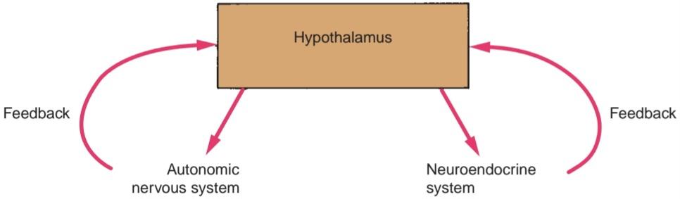
Figure 14-6 Hypothalamus as the control center for the autonomic nervous system and the neuroen- docrine system.
شکل ۱۴-۶ هیپوتالاموس به عنوان مرکز کنترل سیستم عصبی خودکار و سیستم نورواندوکرین.
» ادامه در قسمت چهارم مبانی فصل ۱۴
«فصل چهاردهم کتاب نورواناتومی بالینی اسنل»
🚀 با ما همراه شوید!
تازهترین مطالب و آموزشهای مغز و اعصاب را از دست ندهید. با فالو کردن کانال تلگرام آیندهنگاران مغز، از ما حمایت کنید!



