
عصبدهیهای خودکار: نعوظ آلت تناسلی و کلیتوریس، انزال و رحم


دعای مطالعه [ نمایش ]
بِسْمِ الله الرَّحْمنِ الرَّحیمِ
اَللّهُمَّ اَخْرِجْنى مِنْ ظُلُماتِ الْوَهْمِ
خدايا مرا بيرون آور از تاريكىهاى وهم،
وَ اَكْرِمْنى بِنُورِ الْفَهْمِ
و به نور فهم گرامى ام بدار،
اَللّهُمَّ افْتَحْ عَلَيْنا اَبْوابَ رَحْمَتِكَ
خدايا درهاى رحمتت را به روى ما بگشا،
وَانْشُرْ عَلَيْنا خَزائِنَ عُلُومِكَ بِرَحْمَتِكَ يا اَرْحَمَ الرّاحِمينَ
و خزانههاى علومت را بر ما باز كن به امید رحمتت اى مهربانترين مهربانان.
آیندهنگاران مغز: «ما مغز را میشناسیم، تا آینده را بسازیم.»
کتاب «نوروآناتومی بالینی اسنل» اثر دکتر ریچارد اس. اسنل، با ویرایش جدید دکتر رایان اسپلیتگربر، همچنان بهعنوان مرجع طلایی این حوزه شناخته میشود. این اثر با تلفیق تازهترین پژوهشها و کاربردهای بالینی، پلی استوار میان دانش پایه و عمل پزشکی بنا میکند.
تلاشهای علمی و سالها تدریس ارزشمند استاد گرانقدر آقای دکتر محمدتقی جغتایی الهامبخش ترجمه و گسترش این دانش در ایران بوده است.
ترجمه دقیق این کتاب توسط برند علمی آیندهنگاران مغز به مدیریت داریوش طاهری، امکان دسترسی فارسیزبانان به مرزهای نوین علوم اعصاب را فراهم کرده و رسالتی علمی در جهت شناخت عمیقتر مغز و ترسیم آیندهای روشنتر بر عهده گرفته است.
»» فصل ۱۴: سیستم عصبی خودکار؛ بخش مبانی و اصول
»» Chapter 14: Autonomic Nervous System
Anal Canal Involuntary Internal Sphincter
The circular smooth muscle coat is thickened at the upper end of the anal canal to form the involuntary internal sphincter. The sphincter is innervated by postganglionic sympathetic fibers from the hypogastric plexuses (Fig. 14-12). Each hypogastric plexus receives sympathetic fibers from the aortic plexus and from the lumbar and pelvic parts of the sympathetic trunks. The sympathetic nerves cause the internal anal sphincter to contract.
اسفنکتر داخلی غیرارادی کانال مقعد
پوشش عضله صاف دایرهای در انتهای فوقانی کانال مقعد ضخیم میشود تا اسفنکتر داخلی غیرارادی را تشکیل دهد. اسفنکتر توسط فیبرهای سمپاتیک پسگانگلیونی از شبکههای هیپوگاستریک عصبدهی میشود (شکل 14-12). هر شبکه هیپوگاستریک فیبرهای سمپاتیک را از شبکه آئورت و از قسمتهای کمری و لگنی تنههای سمپاتیک دریافت میکند. اعصاب سمپاتیک باعث انقباض اسفنکتر داخلی مقعد میشوند.
Urinary Bladder
The muscular coat of the bladder is composed of smooth muscle, which at the bladder neck is thickened to form the sphincter vesicae. The nerve supply of the smooth muscle is from the hypogastric plexuses (see Fig. 14-12). The sympathetic postganglionic fibers originate in the first and second lumbar ganglia of the sympathetic trunk and travel to the hypogastric plexuses.
مثانه ادراری
پوشش عضلانی مثانه از عضله صاف تشکیل شده است که در گردن مثانه ضخیم شده و وزیکای اسفنکتر را تشکیل میدهد. عصبرسانی عضله صاف از شبکههای هیپوگاستریک است (شکل 14-12 را ببینید). فیبرهای پسگانگلیونی سمپاتیک از گانگلیونهای کمری اول و دوم تنه سمپاتیک سرچشمه میگیرند و به شبکههای هیپوگاستریک میروند.
The parasympathetic preganglionic fibers arise as the pelvic splanchnic nerves from the second, third, and fourth sacral nerves; they pass through the hypogastric plexuses to reach the bladder wall, where they synapse with postganglionic neurons.
فیبرهای پیشگانگلیونی پاراسمپاتیک به عنوان اعصاب اسپلانکنیک لگنی از اعصاب خاجی دوم، سوم و چهارم منشأ میگیرند. آنها از شبکههای هیپوگاستریک عبور میکنند تا به دیواره مثانه برسند، جایی که با نورونهای پسگانگلیونی سیناپس میکنند.
The sympathetic nerves to the detrusor muscle have little or no action on the smooth muscle of the bladder wall and are distributed mainly to the blood vessels. The sympathetic nerves to the sphincter vesicae play only a minor role in causing contraction of the sphincter in maintaining urinary continence. However, in the male, the sympathetic innervation of the sphincter causes active contraction of the bladder neck during ejaculation (brought about by sympathetic action), thus preventing seminal fluid from entering the bladder. The parasympathetic nerves stimulate the contraction of the smooth muscle of the bladder wall and, in some way, inhibit the contraction of the sphincter vesicae.
اعصاب سمپاتیک عضله دترسور تأثیر کمی بر عضله صاف دیواره مثانه دارند یا اصلاً تأثیری ندارند و عمدتاً در رگهای خونی توزیع میشوند. اعصاب سمپاتیک وزیکای اسفنکتر تنها نقش جزئی در ایجاد انقباض اسفنکتر در حفظ اختیار ادرار دارند. با این حال، در مردان، عصبدهی سمپاتیک اسفنکتر باعث انقباض فعال گردن مثانه در هنگام انزال (ناشی از عمل سمپاتیک) میشود و بنابراین از ورود مایع منی به مثانه جلوگیری میکند. اعصاب پاراسمپاتیک انقباض عضله صاف دیواره مثانه را تحریک میکنند و به نوعی انقباض وزیکای اسفنکتر را مهار میکنند.

Figure 14-12 Autonomic innervation of the sphincters of the anal canal and urinary bladder.
شکل ۱۴-۱۲ عصبدهی خودکار اسفنکترهای کانال مقعد و مثانه.
Penile and Clitoral Erection
In erection, the genital erectile tissue becomes engorged with blood. The initial vascular engorgement is controlled by the parasympathetic part of the ANS. The parasympathetic preganglionic fibers originate in the gray matter of the second, third, and fourth sacral segments of the spinal cord (Fig. 14-13). The fibers enter the hypogastric plexuses and synapse on the postganglionic neurons. The postganglionic fibers join the internal pudendal arteries and are distributed along their branches, which enter the erectile tissue. The parasympathetic nerves cause vasodilatation of the arteries and greatly increase the blood flow to the erectile tissue.
نعوظ آلت تناسلی و کلیتوریس
در نعوظ، بافت نعوظی تناسلی پر از خون میشود. پرخونی اولیه عروقی توسط بخش پاراسمپاتیک ANS کنترل میشود. فیبرهای پیش عقدهای پاراسمپاتیک از ماده خاکستری بخشهای دوم، سوم و چهارم خاجی نخاع سرچشمه میگیرند (شکل ۱۴-۱۳). این فیبرها وارد شبکههای هیپوگاستریک شده و روی نورونهای پس عقدهای سیناپس برقرار میکنند. فیبرهای پس عقدهای به شریانهای پودندال داخلی میپیوندند و در امتداد شاخههای خود که وارد بافت نعوظی میشوند، توزیع میشوند. اعصاب پاراسمپاتیک باعث گشاد شدن عروق شریانها شده و جریان خون به بافت نعوظی را به میزان زیادی افزایش میدهند.
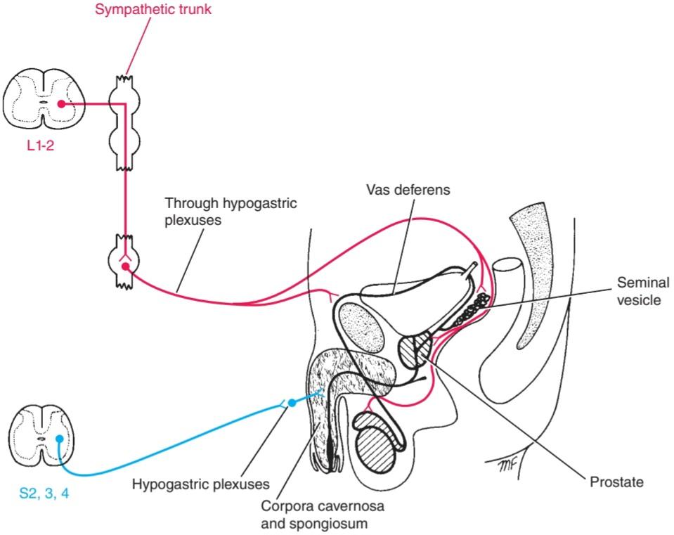
Figure 14-13 Autonomic innervation of the male reproductive tract.
شکل ۱۴-۱۳ عصبدهی خودکار دستگاه تناسلی مردان.
Ejaculation
During the increasing sexual excitement that occurs during sex play, the external urinary meatus of the glans penis becomes moist as a result of the secretions of the bulbourethral glands. Friction on the glans penis, reinforced by other afferent nervous impulses, results in a discharge along the sympathetic nerve fibers to the smooth muscle of the duct of the epididymis and the vas deferens on each side, the seminal vesicles, and the pros- tate. The smooth muscle contracts and the spermatozoa, together with the secretions of the seminal vesicles and prostate, are discharged into the prostatic urethra. The fluid now joins the secretions of the bulbourethral glands and penile urethral glands and is then ejected from the penile urethra as a result of the rhythmic contractions of the bulbospongiosus muscles, which compress the urethra. Meanwhile, the sphincter of the bladder contracts and prevents a reflux of the spermatozoa into the bladder. The spermatozoa and the secretions of the several accessory glands constitute the seminal fluid, or semen. At the climax of male sexual excitement, a mass discharge of nervous impulses takes place in the CNS. Impulses pass down the spinal cord to the sympathetic outflow (T1-L2). The nervous impulses that pass to the genital organs are thought to leave the cord at the first and second lumbar segments in the preganglionic sympathetic fibers (see Fig. 14-13). Many of these fibers synapse with postganglionic neurons in the first and second lumbar ganglia. Other fibers may synapse in ganglia in the lower lumbar or pelvic parts of the sympathetic trunks. The postganglionic fibers are then distributed to the vas deferens, the seminal vesicles, and the prostate through the hypogastric plexuses. The sympathetic nerves stimulate the contractions of the smooth muscle in the walls of these structures and cause the spermatozoa, together with the secretions of the seminal vesicles and prostate, to be discharged into the urethra.
انزال
در طول هیجان جنسی فزایندهای که در طول بازی جنسی رخ میدهد، مجرای ادرار خارجی سر آلت تناسلی در نتیجه ترشحات غدد پیازی-میزی مرطوب میشود. اصطکاک روی سر آلت تناسلی، که توسط سایر تکانههای عصبی آوران تقویت میشود، منجر به ترشح در امتداد الیاف عصبی سمپاتیک به عضله صاف مجرای اپیدیدیم و مجرای دفران در هر طرف، کیسههای منی و پروستات میشود. عضله صاف منقبض میشود و اسپرمها، همراه با ترشحات کیسههای منی و پروستات، به مجرای ادرار پروستات تخلیه میشوند. اکنون مایع به ترشحات غدد پیازی-میزی و غدد مجرای ادرار آلت تناسلی میپیوندد و سپس در نتیجه انقباضات ریتمیک عضلات پیازی-اسفنجی که مجرای ادرار را فشرده میکنند، از مجرای ادرار آلت تناسلی خارج میشود. در همین حال، اسفنکتر مثانه منقبض میشود و از رفلاکس اسپرمها به مثانه جلوگیری میکند. اسپرم و ترشحات چندین غده فرعی، مایع منی یا منی را تشکیل میدهند. در اوج هیجان جنسی مردان، تخلیه انبوهی از تکانههای عصبی در سیستم عصبی مرکزی (CNS) رخ میدهد. تکانهها از طریق نخاع به خروجی سمپاتیک (T1-L2) منتقل میشوند. تصور میشود تکانههای عصبی که به اندامهای تناسلی منتقل میشوند، در بخشهای کمری اول و دوم در فیبرهای سمپاتیک پیش عقدهای از نخاع خارج میشوند (شکل 14-13 را ببینید). بسیاری از این فیبرها با نورونهای پس عقدهای در گانگلیونهای کمری اول و دوم سیناپس میکنند. سایر فیبرها ممکن است در گانگلیونهای قسمتهای کمری تحتانی یا لگنی تنههای سمپاتیک سیناپس ایجاد کنند. سپس فیبرهای پس عقدهای از طریق شبکههای هیپوگاستریک به مجرای دفران، کیسههای منی و پروستات توزیع میشوند. اعصاب سمپاتیک انقباضات عضلات صاف دیوارههای این ساختارها را تحریک میکنند و باعث میشوند اسپرمها به همراه ترشحات کیسههای منی و پروستات به داخل مجرای ادرار تخلیه شوند.
Uterus
Preganglionic sympathetic nerve fibers leave the spinal cord at segmental levels T12 and L1 and are believed to synapse with ganglion cells in the sympathetic trunk or possibly in the inferior hypogastric plexuses (Fig. 14-14). The postganglionic fibers supply the smooth muscle of the uterus. Parasympathetic preganglionic fibers leave the spinal cord at levels $2-$4 and synapse with ganglion cells in the inferior hypogastric plexuses. Although the uterine muscle is largely under hormonal control, sym- pathetic innervation may cause uterine contraction and vasoconstriction, whereas parasympathetic fibers have the opposite effect.
رحم
فیبرهای عصبی سمپاتیک پیش عقدهای نخاع را در سطوح قطعهای T12 و L1 ترک میکنند و اعتقاد بر این است که با سلولهای گانگلیونی در تنه سمپاتیک یا احتمالاً در شبکههای هیپوگاستریک تحتانی سیناپس میدهند (شکل 14-14). فیبرهای پس عقدهای عضله صاف رحم را تغذیه میکنند. فیبرهای پیش عقدهای پاراسمپاتیک نخاع را در سطوح 2 تا 4 ترک میکنند و با سلولهای گانگلیونی در شبکههای هیپوگاستریک تحتانی سیناپس میدهند. اگرچه عضله رحم تا حد زیادی تحت کنترل هورمونی است، عصبدهی سمپاتیک ممکن است باعث انقباض رحم و انقباض عروق شود، در حالی که فیبرهای پاراسمپاتیک اثر معکوس دارند.
Afferent pain fibers from the fundus and the body of the uterus ascend to the spinal cord through the hypogastric plexuses, entering it through the posterior roots of the 10th, 11th, and 12th thoracic spinal nerves. Fibers from the cervix run in the pelvic splanchnic nerves and enter the spinal cord through the posterior roots of the second, third, and fourth sacral nerves.
فیبرهای آوران درد از فوندوس و بدنه رحم از طریق شبکههای هیپوگاستریک به نخاع صعود میکنند و از طریق ریشههای خلفی اعصاب نخاعی سینهای دهم، یازدهم و دوازدهم وارد آن میشوند. رشتههایی از دهانه رحم در اعصاب اسپلانکنیک لگنی عبور میکنند و از طریق ریشههای خلفی اعصاب خاجی دوم، سوم و چهارم وارد نخاع میشوند.
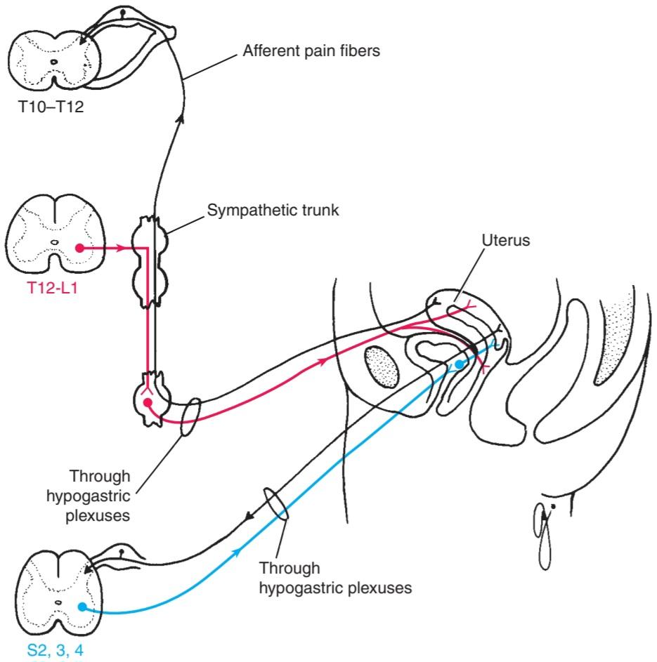
Figure 14-14 Autonomic innervation of the uterus; the pathway taken by the afferent sensory fibers is shown as well.
شکل ۱۴-۱۴ عصبدهی خودکار رحم؛ مسیر طی شده توسط فیبرهای حسی آوران نیز نشان داده شده است.
Upper Limb Arteries
The arteries of the upper limb are innervated by sympathetic nerves. The preganglionic fibers originate from cell bodies in the second to the eighth thoracic segments of the spinal cord (Fig. 14-15). They pass to the sympathetic trunk through white rami and ascend in the trunk to synapse in the middle cervical, inferior cervical, first thoracic, or stellate ganglia. The postganglionic fibers join the nerves that form the brachial plexus and are distributed to the arteries within the branches of the plexus. The sympathetic nerves cause vasoconstriction of cutaneous arteries and vasodilatation of arteries that supply skeletal muscle.
شریانهای اندام فوقانی
شریانهای اندام فوقانی توسط اعصاب سمپاتیک عصبدهی میشوند. فیبرهای پیش عقدهای از اجسام سلولی در قطعات دوم تا هشتم سینهای نخاع منشأ میگیرند (شکل ۱۴-۱۵). آنها از طریق شاخههای سفید به تنه سمپاتیک میروند و در تنه بالا میروند تا در گانگلیونهای گردنی میانی، گردنی تحتانی، سینهای اول یا ستارهای سیناپس برقرار کنند. فیبرهای پس عقدهای به اعصابی که شبکه بازویی را تشکیل میدهند، میپیوندند و در شریانهای درون شاخههای شبکه توزیع میشوند. اعصاب سمپاتیک باعث انقباض عروق شریانهای پوستی و گشاد شدن عروق شریانهایی میشوند که به عضلات اسکلتی خونرسانی میکنند.

Figure 14-15 Sympathetic innervation of the arteries of the upper limb (A) and lower limb (B).
شکل ۱۴-۱۵ عصبدهی سمپاتیک شریانهای اندام فوقانی (A) و اندام تحتانی (B).
Lower Limb Arteries
The arteries of the lower limb are also innervated by sympathetic nerves (see Fig. 14-15). The preganglionic fibers originate from cell bodies in the lower three thoracic and upper two or three lumbar segments of the spinal cord. The preganglionic fibers pass to the lower thoracic and upper lumbar ganglia of the sympathetic trunk through white rami. The fibers synapse in the lumbar and sacral ganglia and the postganglionic fibers reach the arteries through branches of the lumbar and sacral plexuses.
شریانهای اندام تحتانی
شریانهای اندام تحتانی نیز توسط اعصاب سمپاتیک عصبدهی میشوند (شکل 14-15 را ببینید). فیبرهای پیشگانگلیونی از اجسام سلولی در سه بخش پایینی قفسه سینه و دو یا سه بخش بالایی کمری نخاع منشأ میگیرند. فیبرهای پیشگانگلیونی از طریق شاخههای سفید به گانگلیونهای پایینی قفسه سینه و بالایی کمری تنه سمپاتیک عبور میکنند. فیبرها در گانگلیونهای کمری و خاجی سیناپس میدهند و فیبرهای پسگانگلیونی از طریق شاخههای شبکه کمری و خاجی به شریانها میرسند.
ANS PHYSIOLOGIC REFLEXES
The ANS is involved in important visual and cardiovascular reflexes.
رفلکسهای فیزیولوژیکی ANS
ANS در رفلکسهای مهم بینایی و قلبی عروقی نقش دارد.
Direct and Consensual Light Reflexes
Afferent nervous impulses travel from the retina through the optic nerve, optic chiasma, and optic tract (see Fig. 11-3). A small number of fibers leave the optic tract and synapse on nerve cells in the pretectal nucleus, which lies close to the superior colliculus. The impulses are passed by axons of the pretectal nerve cells to the parasympathetic nuclei (Edinger-Westphal nuclei) of the oculomotor nerve on both sides. Here, the fibers synapse and the parasympathetic nerves travel through the oculomotor nerve to the ciliary ganglion in the orbit. Finally, postganglionic parasympathetic fibers pass through the short ciliary nerves to the eyeball and to the constrictor pupillae muscle of the iris. Both pupils constrict in the consensual light reflex because the pretectal nucleus sends fibers to the parasympathetic nuclei on both sides of the midbrain.
رفلکسهای نوری مستقیم و توافقی
پیامهای عصبی آوران از شبکیه از طریق عصب بینایی، کیاسمای بینایی و دستگاه بینایی حرکت میکنند (شکل 11-3 را ببینید). تعداد کمی از فیبرها دستگاه بینایی را ترک میکنند و روی سلولهای عصبی در هسته پرهتکتال، که نزدیک به کولیکولوس فوقانی قرار دارد، سیناپس میکنند. این تکانهها توسط آکسونهای سلولهای عصبی پرهتکتال به هستههای پاراسمپاتیک (هستههای ادینگر-وستفال) عصب حرکتی چشم در هر دو طرف منتقل میشوند. در اینجا، فیبرها سیناپس میکنند و اعصاب پاراسمپاتیک از طریق عصب حرکتی چشم به گانگلیون مژگانی در حدقه میروند. در نهایت، فیبرهای پاراسمپاتیک پسگانگلیونی از طریق اعصاب مژگانی کوتاه به کره چشم و به عضله تنگکننده مردمک عنبیه میروند. هر دو مردمک در رفلکس نوری توافقی منقبض میشوند زیرا هسته پرهتکتال فیبرهایی را به هستههای پاراسمپاتیک در دو طرف مغز میانی میفرستد.
Accommodation Reflex
When the eyes are directed from a distant to a near object, contraction of the medial recti brings about convergence of the ocular axes, the lenses thicken to increase their refractive power by contraction of the ciliary muscle, and the pupils constrict to restrict the light waves to the thickest central part of the lenses. The afferent impulses travel through the optic nerve, the optic chiasma, the optic tract, the lateral geniculate body, and the optic radiation to the visual cortex (see Fig. 11-3). The visual cortex is connected to the eye field of the frontal cortex. From here, cortical fibers descend through the internal capsule to the oculomotor nuclei in the midbrain. The oculomotor nerve travels to the medial recti muscles. Some of the descending cortical fibers synapse with the parasympathetic nuclei (Edinger-Westphal nuclei) of the oculomotor nerve on both sides. The parasympathetic preganglionic fibers then travel through the oculomotor nerve to the ciliary ganglion in the orbit where they synapse. Finally, postganglionic parasympathetic fibers pass through the short ciliary nerves to the ciliary muscle and the constrictor pupillae muscle of the iris.
رفلکس تطابق
هنگامی که چشمها از یک شیء دور به یک شیء نزدیک هدایت میشوند، انقباض عضله راست داخلی باعث همگرایی محورهای چشمی میشود، عدسیها ضخیم میشوند تا قدرت انکساری خود را با انقباض عضله مژگانی افزایش دهند و مردمکها منقبض میشوند تا امواج نور را به ضخیمترین قسمت مرکزی عدسیها محدود کنند. تکانههای آوران از طریق عصب بینایی، کیاسمای بینایی، راه بینایی، جسم زانویی جانبی و تابش نوری به قشر بینایی منتقل میشوند (شکل 11-3 را ببینید). قشر بینایی به میدان بینایی قشر پیشانی متصل است. از اینجا، الیاف قشری از طریق کپسول داخلی به هستههای حرکتی چشم در مغز میانی پایین میروند. عصب حرکتی چشم به عضلات مستقیم داخلی میرود. برخی از الیاف قشری نزولی با هستههای پاراسمپاتیک (هستههای ادینگر-وستفال) عصب حرکتی چشم در هر دو طرف سیناپس میکنند. سپس فیبرهای پیش عقدهای پاراسمپاتیک از طریق عصب حرکتی چشم به گانگلیون مژگانی در حدقه چشم میروند و در آنجا سیناپس تشکیل میدهند. در نهایت، فیبرهای پس عقدهای پاراسمپاتیک از طریق اعصاب مژگانی کوتاه به عضله مژگانی و عضله تنگکننده مردمک عنبیه میرسند.
Carotid Sinus and Aortic Arch Reflexes
The carotid sinus, located in the bifurcation of the common carotid artery, and the aortic arch serve as baro- receptors. As the blood pressure rises, nerve endings situated in the walls of these vessels are stimulated. The afferent fibers from the carotid sinus ascend in the glossopharyngeal nerve and terminate in the nucleus solitarius (see Figs. 11-16 and 11-17). The afferent fibers from the aortic arch ascend in the vagus nerve. Connector neurons in the medulla oblongata activate the parasympathetic nucleus (dorsal nucleus) of the vagus, which slows the heart rate. At the same time, reticulospinal fibers descend to the spinal cord and inhibit the preganglionic sympathetic outflow to the heart and cutaneous arterioles. The combined effect of stimulation of the parasympathetic action on the heart and inhibition of the sympathetic action on the heart and peripheral blood vessels reduces the rate and force of contraction of the heart and reduces the peripheral resistance of the blood vessels. Consequently, the blood pressure falls. The blood pressure of the individual is thus modified by the afferent information received from the baroreceptors. The modulator of the ANS, namely, the hypothalamus, in turn, can be influenced by other, higher centers in the CNS.
رفلکسهای سینوس کاروتید و قوس آئورت
سینوس کاروتید، که در محل دو شاخه شدن شریان کاروتید مشترک قرار دارد، و قوس آئورت به عنوان گیرندههای فشار عمل میکنند. با افزایش فشار خون، انتهای عصبی واقع در دیوارههای این رگها تحریک میشوند. فیبرهای آوران از سینوس کاروتید در عصب زبانی-حلقی بالا میروند و در هسته انفرادی خاتمه مییابند (شکلهای 11-16 و 11-17 را ببینید). فیبرهای آوران از قوس آئورت در عصب واگ بالا میروند. نورونهای رابط در بصل النخاع، هسته پاراسمپاتیک (هسته پشتی) واگ را فعال میکنند که ضربان قلب را کاهش میدهد. در همان زمان، فیبرهای رتیکولواسپینال به نخاع پایین میآیند و جریان سمپاتیک پیش عقدهای را به قلب و شریانچههای پوستی مهار میکنند. اثر ترکیبی تحریک عمل پاراسمپاتیک بر قلب و مهار عمل سمپاتیک بر قلب و رگهای خونی محیطی، سرعت و نیروی انقباض قلب را کاهش داده و مقاومت محیطی رگهای خونی را کم میکند. در نتیجه، فشار خون پایین میآید. بنابراین فشار خون فرد توسط اطلاعات آوران دریافتی از گیرندههای فشاری تغییر میکند. تعدیلکننده ANS، یعنی هیپوتالاموس، به نوبه خود میتواند تحت تأثیر سایر مراکز بالاتر در سیستم عصبی مرکزی قرار گیرد.
Bainbridge Right Atrial Reflex
This reflex is initiated when the nerve endings in the wall of the right atrium and in the walls of the venae cavae are stimulated by a rise of venous pressure. The afferent fibers ascend in the vagus to the medulla oblongata and terminate on the nucleus of the tractus solitarius (see Fig. 11-18). Connector neurons inhibit the parasympathetic nucleus (dorsal) of the vagus, and reticulospinal fibers stimulate the thoracic sympathetic outflow to the heart, resulting in cardiac acceleration.
رفلکس دهلیز راست بینبریج
این رفلکس زمانی آغاز میشود که انتهای اعصاب در دیواره دهلیز راست و دیوارههای وریدهای اجوف تحتانی توسط افزایش فشار وریدی تحریک میشوند. الیاف آوران در واگ به بصل النخاع صعود میکنند و در هسته عضله منفرد (شکل 11-18 را ببینید) خاتمه مییابند. نورونهای رابط، هسته پاراسمپاتیک (پشتی) واگ را مهار میکنند و الیاف رتیکولواسپینال، جریان خروجی سمپاتیک قفسه سینه به قلب را تحریک میکنند و در نتیجه باعث تسریع ضربان قلب میشوند.
«فصل چهاردهم کتاب نورواناتومی بالینی اسنل»
🚀 با ما همراه شوید!
تازهترین مطالب و آموزشهای مغز و اعصاب را از دست ندهید. با فالو کردن کانال تلگرام آیندهنگاران مغز، از ما حمایت کنید!



 ورود/ ثبت نام با جیمیل
