اعصاب مغزی؛ عصب مغزی X: عصب واگ


دعای مطالعه [ نمایش ]
بِسْمِ الله الرَّحْمنِ الرَّحیمِ
اَللّهُمَّ اَخْرِجْنى مِنْ ظُلُماتِ الْوَهْمِ
خدايا مرا بيرون آور از تاريكىهاى وهم،
وَ اَكْرِمْنى بِنُورِ الْفَهْمِ
و به نور فهم گرامى ام بدار،
اَللّهُمَّ افْتَحْ عَلَيْنا اَبْوابَ رَحْمَتِكَ
خدايا درهاى رحمتت را به روى ما بگشا،
وَانْشُرْ عَلَيْنا خَزائِنَ عُلُومِكَ بِرَحْمَتِكَ يا اَرْحَمَ الرّاحِمينَ
و خزانههاى علومت را بر ما باز كن به امید رحمتت اى مهربانترين مهربانان.
آیندهنگاران مغز: «ما مغز را میشناسیم، تا آینده را بسازیم.»
کتاب «نوروآناتومی بالینی اسنل» اثر دکتر ریچارد اس. اسنل، با ویرایش جدید دکتر رایان اسپلیتگربر، همچنان بهعنوان مرجع طلایی این حوزه شناخته میشود. این اثر با تلفیق تازهترین پژوهشها و کاربردهای بالینی، پلی استوار میان دانش پایه و عمل پزشکی بنا میکند.
تلاشهای علمی و سالها تدریس ارزشمند استاد گرانقدر دکتر محمدتقی جغتایی الهامبخش ترجمه و گسترش این دانش در ایران بوده است.
ترجمه دقیق این کتاب توسط برند علمی آیندهنگاران مغز به مدیریت داریوش طاهری، امکان دسترسی فارسیزبانان به مرزهای نوین علوم اعصاب را فراهم کرده و رسالتی علمی در جهت شناخت عمیقتر مغز و ترسیم آیندهای روشنتر بر عهده گرفته است.
»» فصل ۱۱: هستههای اعصاب جمجمهای؛ بخش دهم مبانی و اصول
»» Chapter 11: Cranial Nerve Nuclei
VAGUS NERVE (CRANIAL NERVE X)
The vagus nerve is a motor and a sensory nerve.
عصب واگ (عصب جمجمهای X)
عصب واگ یک عصب حرکتی و حسی است.
Vagus Nerve Nuclei
The vagus nerve has three nuclei: (1) the main motor nucleus, (2) the parasympathetic nucleus, and (3) the sensory nucleus.
هستههای عصب واگ
عصب واگ دارای سه هسته است: (1) هسته اصلی حرکتی، (2) هسته پاراسمپاتیک و (3) هسته حسی.
Main Motor Nucleus
The main motor nucleus lies deep in the reticular formation of the medulla oblongata and is formed by the nucleus ambiguus (Fig. 11-18). It receives corticonuclear fibers from both cerebral hemispheres. The efferent fibers supply the constrictor muscles of the pharynx and the intrinsic muscles of the larynx (Fig. 11-19).
هسته حرکتی اصلی
هسته حرکتی اصلی در عمق تشکیلات مشبک بصل النخاع قرار دارد و توسط هسته مبهم (nucleus ambiguus) تشکیل میشود (شکل 11-18). این هسته فیبرهای قشری-هستهای را از هر دو نیمکره مغزی دریافت میکند. فیبرهای وابران، عضلات منقبض کننده حلق و عضلات داخلی حنجره را تغذیه میکنند (شکل 11-19).

Figure 11-18 Vagus nerve nuclei and their central connections.
شکل ۱۱-۱۸ هستههای عصب واگ و اتصالات مرکزی آنها.
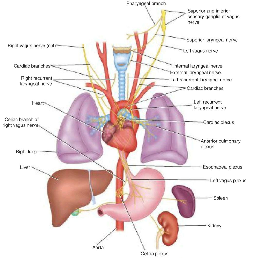
Figure 11-19 Distribution of the vagus nerve.
شکل ۱۱-۱۹ توزیع عصب واگ.
Parasympathetic Nucleus
The parasympathetic nucleus forms the dorsal nucleus of the vagus and lies beneath the floor of the lower part of the fourth ventricle posterolateral to the hypoglossal nucleus (see Fig. 11-18). It receives afferent fibers from the hypothalamus through the descending autonomic pathways. It also receives other afferents, including those from the glossopharyngeal nerve (carotid sinus reflex). The efferent fibers are distributed to the involuntary muscle of the bronchi, heart, esophagus, stomach, small intestine, and large intestine as far as the distal third of the transverse colon (see Fig. 11-19).
هسته پاراسمپاتیک
هسته پاراسمپاتیک، هسته پشتی عصب واگ را تشکیل میدهد و در زیر کف قسمت پایینی بطن چهارم، در قسمت خلفی-جانبی هسته هیپوگلوسال قرار دارد (شکل 11-18 را ببینید). این هسته، فیبرهای آوران را از هیپوتالاموس از طریق مسیرهای اتونوم نزولی دریافت میکند. همچنین سایر آورانها، از جمله آورانهای عصب زبانی-حلقی (رفلکس سینوس کاروتید) را نیز دریافت میکند. فیبرهای وابران در عضله غیرارادی برونشها، قلب، مری، معده، روده کوچک و روده بزرگ تا یک سوم انتهایی کولون عرضی توزیع میشوند (شکل 11-19 را ببینید).
Sensory Nucleus
The sensory nucleus is the lower part of the nucleus of the tractus solitarius. Sensations of taste travel through the peripheral axons of nerve cells situated in the inferior ganglion on the vagus nerve. The central processes of those cells synapse on nerve cells in the nucleus (see Fig. 11-18). Efferent fibers cross the median plane and ascend to the ventral group of nuclei of the opposite thalamus as well as to a number of hypothalamic nuclei. From the thalamus, the axons of the thalamic cells pass through the internal capsule and corona radiata to end in the postcentral gyrus.
هسته حسی
هسته حسی قسمت پایینی هسته دستگاه عصبی منزوی است. حس چشایی از طریق آکسونهای محیطی سلولهای عصبی واقع در گانگلیون تحتانی روی عصب واگ منتقل میشود. فرآیندهای مرکزی این سلولها روی سلولهای عصبی موجود در هسته سیناپس برقرار میکنند (شکل 11-18 را ببینید). فیبرهای وابران از صفحه میانی عبور کرده و به گروه شکمی هستههای تالاموس مقابل و همچنین به تعدادی از هستههای هیپوتالاموس صعود میکنند. از تالاموس، آکسونهای سلولهای تالاموس از کپسول داخلی و تاج شعاعی عبور میکنند تا در شکنج پس مرکزی به پایان برسند.
Afferent information concerning common sensation enters the brainstem through the superior ganglion of the vagus nerve but ends in the spinal nucleus of the trigeminal nerve.
اطلاعات آوران مربوط به حس مشترک از طریق گانگلیون فوقانی عصب واگ وارد ساقه مغز میشود اما در هسته نخاعی عصب سه قلو به پایان میرسد.
Vagus Nerve Course
The vagus nerve leaves the anterolateral surface of the upper part of the medulla oblongata as a series of rootlets in a groove between the olive and the inferior cerebellar peduncle (see Fig. 11-18). The nerve passes laterally through the posterior cranial fossa and leaves the skull through the jugular foramen. The vagus nerve possesses two sensory ganglia, a rounded superior ganglion, situated on the nerve within the jugular foramen, and a cylindrical inferior ganglion, which lies on the nerve just below the foramen. Below the inferior ganglion, the cranial root of the accessory nerve joins the vagus nerve and is distributed mainly in its pharyngeal and recurrent laryngeal branches.
مسیر عصب واگ
عصب واگ از سطح قدامی-جانبی قسمت فوقانی بصل النخاع به صورت مجموعهای از ریشههای کوچک در شیاری بین زیتون و پایه مخچه تحتانی خارج میشود (شکل 11-18 را ببینید). این عصب به صورت جانبی از حفره جمجمهای خلفی عبور میکند و از طریق سوراخ ژوگولار از جمجمه خارج میشود. عصب واگ دارای دو گانگلیون حسی است، یک گانگلیون فوقانی گرد که روی عصب درون سوراخ ژوگولار قرار دارد و یک گانگلیون تحتانی استوانهای که درست زیر سوراخ ژوگولار روی عصب قرار دارد. در زیر گانگلیون تحتانی، ریشه جمجمهای عصب فرعی به عصب واگ میپیوندد و عمدتاً در شاخههای حلقی و حنجرهای مکرر آن توزیع میشود.
The vagus nerve descends vertically in the neck within the carotid sheath with the internal jugular vein and the internal and common carotid arteries.
عصب واگ به صورت عمودی در گردن در داخل غلاف کاروتید به همراه ورید ژوگولار داخلی و شریانهای کاروتید داخلی و مشترک نزول میکند.
The right vagus nerve enters the thorax and passes posterior to the root of the right lung, contributing to the pulmonary plexus. It then passes on to the posterior surface of the esophagus and contributes to the esophageal plexus. It enters the abdomen through the esophageal opening of the diaphragm. The posterior vagal trunk (which is the name now given to the right vagus) is distributed to the posterior surface of the stomach and, by a large celiac branch, to the duodenum, liver, kidneys, and small and large intestines as far as the distal third of the transverse colon. This wide distribution is accomplished through the celiac, superior mesenteric, and renal plexuses.
عصب واگ راست وارد قفسه سینه میشود و از پشت ریشه ریه راست عبور میکند و به شبکه ریوی میپیوندد. سپس به سطح خلفی مری میرود و به شبکه مری میپیوندد. از طریق دهانه مری دیافراگم وارد شکم میشود. تنه خلفی واگ (که اکنون نامی است که به واگ راست داده شده است) به سطح خلفی معده و توسط یک شاخه بزرگ سلیاک به دوازدهه، کبد، کلیهها و رودههای کوچک و بزرگ تا یک سوم انتهایی روده بزرگ عرضی توزیع میشود. این توزیع گسترده از طریق شبکههای سلیاک، مزانتریک فوقانی و کلیوی انجام میشود.
The left vagus nerve enters the thorax and crosses the left side of the aortic arch and descends behind the root of the left lung, contributing to the pulmonary plexus. The left vagus then descends on the anterior surface of the esophagus, contributing to the esophageal plexus. It enters the abdomen through the esophageal opening of the diaphragm. The anterior vagal trunk (which is the name now given to the left vagus) divides into several branches, which are distributed to the stomach, liver, upper part of the duodenum, and head of the pancreas.
عصب واگ چپ وارد قفسه سینه میشود و از سمت چپ قوس آئورت عبور میکند و از پشت ریشه ریه چپ پایین میآید و به شبکه ریوی میپیوندد. سپس واگ چپ روی سطح قدامی مری پایین میآید و به شبکه مری میپیوندد. از طریق دهانه مری دیافراگم وارد شکم میشود. تنه واگ قدامی (که اکنون نامی است که به واگ چپ داده شده است) به چندین شاخه تقسیم میشود که در معده، کبد، قسمت فوقانی دوازدهه و سر پانکراس توزیع میشوند.
«فصل یازدهم کتاب نورواناتومی بالینی اسنل»
🚀 با ما همراه شوید!
تازهترین مطالب و آموزشهای مغز و اعصاب را از دست ندهید. با فالو کردن کانال تلگرام آیندهنگاران مغز، از ما حمایت کنید!