
سیستم لیمبیک: تشکیلات هیپوکامپ، هسته آمیگدالوئید، مسیرهای ارتباطی و عملکردها


دعای مطالعه [ نمایش ]
بِسْمِ الله الرَّحْمنِ الرَّحیمِ
اَللّهُمَّ اَخْرِجْنى مِنْ ظُلُماتِ الْوَهْمِ
خدايا مرا بيرون آور از تاريكىهاى وهم،
وَ اَكْرِمْنى بِنُورِ الْفَهْمِ
و به نور فهم گرامى ام بدار،
اَللّهُمَّ افْتَحْ عَلَيْنا اَبْوابَ رَحْمَتِكَ
خدايا درهاى رحمتت را به روى ما بگشا،
وَانْشُرْ عَلَيْنا خَزائِنَ عُلُومِكَ بِرَحْمَتِكَ يا اَرْحَمَ الرّاحِمينَ
و خزانههاى علومت را بر ما باز كن به امید رحمتت اى مهربانترين مهربانان.
آیندهنگاران مغز: «ما مغز را میشناسیم، تا آینده را بسازیم.»
کتاب «نوروآناتومی بالینی اسنل» اثر دکتر ریچارد اس. اسنل، با ویرایش جدید دکتر رایان اسپلیتگربر، همچنان بهعنوان مرجع طلایی این حوزه شناخته میشود. این اثر با تلفیق تازهترین پژوهشها و کاربردهای بالینی، پلی استوار میان دانش پایه و عمل پزشکی بنا میکند.
تلاشهای علمی و سالها تدریس ارزشمند استاد گرانقدر آقای دکتر محمدتقی جغتایی الهامبخش ترجمه و گسترش این دانش در ایران بوده است.
ترجمه دقیق این کتاب توسط برند علمی آیندهنگاران مغز به مدیریت داریوش طاهری، امکان دسترسی فارسیزبانان به مرزهای نوین علوم اعصاب را فراهم کرده و رسالتی علمی در جهت شناخت عمیقتر مغز و ترسیم آیندهای روشنتر بر عهده گرفته است.
»» فصل ۹: تشکیلات مشبک و سیستم لیمبیک؛ قسمت دوم
»» Chapter 9: Reticular Formation and Limbic System
LIMBIC SYSTEM
The word limbic means border or margin, and the term limbic system was loosely used to include a group of structures that lie in the border zone between the cerebral cortex and the hypothalamus. Research has now shown that the limbic system is involved with many other structures beyond the border zone in the control of emotion, behavior, and drive; it also appears to be important to memory.
سیستم لیمبیک
کلمه لیمبیک به معنای مرز یا حاشیه است و اصطلاح سیستم لیمبیک به طور کلی برای شامل گروهی از ساختارهایی که در ناحیه مرزی بین قشر مغز و هیپوتالاموس قرار دارند، استفاده میشد. اکنون تحقیقات نشان داده است که سیستم لیمبیک با بسیاری از ساختارهای دیگر فراتر از ناحیه مرزی در کنترل احساسات، رفتار و انگیزهها درگیر است. همچنین به نظر میرسد که برای حافظه مهم است.
Anatomically, the limbic structures include the subcallosal, the cingulate, and the parahippocampal gyri, the hippocampal formation, the amygdaloid nucleus, the mammillary bodies, and the anterior thalamic nucleus (Fig. 9-3). The alveus, the fimbria, the fornix, the mammillothalamic tract, and the stria terminalis constitute the connecting pathways of this system.
از نظر آناتومیکی، ساختارهای لیمبیک شامل زیرکالوزال، سینگولیت و شکنج پاراهیپوکامپ، تشکیلات هیپوکامپ، هسته آمیگدالوئید، اجسام پستانی و هسته تالاموس قدامی هستند (شکل 9-3). آلوئوس، فیمبریا، فورنیکس، مسیر پستانی تالاموس و استریا ترمینالیس مسیرهای اتصال این سیستم را تشکیل میدهند.
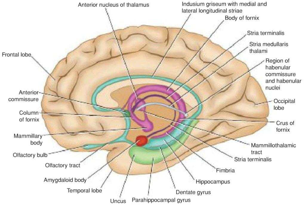
Figure 9-3 Medial aspect of the right cerebral hemisphere showing structures that form the limbic system.
شکل ۹-۳ نمای داخلی نیمکره راست مغز که ساختارهایی را نشان میدهد که سیستم لیمبیک را تشکیل میدهند.
Hippocampal Formation
The hippocampal formation consists of the hippocampus, the dentate gyrus, and the parahippocampal gyrus.
تشکیلات هیپوکامپ
تشکیلات هیپوکامپ شامل هیپوکامپ، شکنج دندانهدار و شکنج پاراهیپوکامپ است.
The hippocampus is a curved elevation of gray matter that extends throughout the entire length of the floor of the inferior horn of the lateral ventricle (Fig. 9-4). Its anterior end is expanded to form the pes hippocampus. It is named hippocampus because it resembles a seahorse in coronal section. The convex ventricular surface is covered with ependyma, beneath which lies a thin layer of white matter called the alveus (Fig. 9-5). The alveus consists of nerve fibers that have originated in the hippocampus, and these converge medially to form a bundle called the fimbria. The fimbria, in turn, becomes continuous with the crus of the fornix. The hippocampus terminates posteriorly beneath the splenium of the corpus callosum.
هیپوکامپ یک برآمدگی خمیده از ماده خاکستری است که در تمام طول کف شاخ تحتانی بطن جانبی امتداد دارد (شکل 9-4). انتهای قدامی آن گسترش یافته و pes hippocampus را تشکیل میدهد. به این دلیل هیپوکامپ نامیده میشود که در برش کرونال شبیه اسب دریایی است. سطح محدب بطن با اپندیم پوشیده شده است که در زیر آن یک لایه نازک از ماده سفید به نام آلوئوس قرار دارد (شکل 9-5). آلوئوس از الیاف عصبی تشکیل شده است که از هیپوکامپ منشأ گرفتهاند و این الیاف به صورت داخلی همگرا میشوند تا دستهای به نام فیمبریا را تشکیل دهند. فیمبریا به نوبه خود با ساق فورنیکس امتداد مییابد. هیپوکامپ در خلف زیر اسپلنیوم جسم پینهای خاتمه مییابد.

Figure 9-4 Dissection of the right cerebral hemisphere exposing the cavity of the lateral ventricle, showing the hippocampus, the dentate gyrus, and the fornix.
شکل ۹-۴ تشریح نیمکره راست مغز که حفره بطن جانبی را نمایان میکند و هیپوکامپ، شکنج دندانهای و فورنیکس را نشان میدهد.

Figure 9-5 Coronal section of the hippocampus and related structures.
شکل ۹-۵ برش کرونال از هیپوکامپ و ساختارهای مرتبط.
The dentate gyrus is a narrow, notched band of gray matter that lies between the fimbria of the hippo- campus and the parahippocampal gyrus (see Fig. 9-4). Posteriorly, the gyrus accompanies the fimbria almost to the splenium of the corpus callosum and becomes continuous with the indusium griseum. The indusium griseum is a thin, vestigial layer of gray matter that covers the superior surface of the corpus callosum (Fig. 9-6). Embedded in the superior surface of the indusium griseum are two slender bundles of white fibers on each side called the medial and lateral longitudinal striae. The striae are the remains of the white matter of the vestigial indusium griseum. Anteriorly, the dentate gyrus is continued into the uncus.
شکنج دندانهدار یک نوار باریک و بریده از ماده خاکستری است که بین فیمبریای هیپوکامپ و شکنج پاراهیپوکامپ قرار دارد (شکل 9-4 را ببینید). در قسمت خلفی، شکنج تقریباً تا اسپلنیوم جسم پینهای، فیمبریا را همراهی میکند و در امتداد ایندوسیوم گریزئوم قرار میگیرد. ایندوسیوم گریزئوم یک لایه نازک و باقیمانده از ماده خاکستری است که سطح فوقانی جسم پینهای را میپوشاند (شکل 9-6). در سطح فوقانی ایندوسیوم گریزئوم، دو دسته باریک از الیاف سفید در هر طرف به نام نوارهای طولی داخلی و جانبی قرار دارند. این نوارها بقایای ماده سفید ایندوسیوم گریزئوم باقیمانده هستند. در قسمت قدامی، شکنج دندانهدار به داخل آنکوس ادامه مییابد.
The parahippocampal gyrus lies between the hippocampal fissure and the collateral sulcus and is continuous with the hippocampus along the medial edge of the temporal lobe (see Figs. 9-4 and 9-5).
شکنج پاراهیپوکامپ بین شیار هیپوکامپ و شیار جانبی قرار دارد و در امتداد لبه داخلی لوب گیجگاهی با هیپوکامپ در ارتباط است (شکلهای 9-4 و 9-5 را ببینید).

Figure 9-6 Dissection of both cerebral hemispheres showing the superior surface of the corpus callosum.
شکل ۹-۶ تشریح هر دو نیمکره مغزی که سطح فوقانی جسم پینهای را نشان میدهد.
Amygdaloid Nucleus
The amygdaloid nucleus is so named because it resembles an almond. It is situated partly anterior and partly superior to the tip of the inferior horn of the lateral ventricle (see Fig. 7-14). It is fused with the tip of the tail of the caudate nucleus, which has passed anteriorly in the roof of the inferior horn of the lateral ventricle. The stria terminalis emerges from its posterior aspect. The amygdaloid nucleus consists of a complex of nuclei that can be grouped into a larger basolateral group and smaller corticomedial group.
هسته آمیگدالوئید
هسته آمیگدالوئید به این دلیل به این نام خوانده میشود که شبیه بادام است. این هسته تا حدی در جلو و تا حدی در بالای نوک شاخ تحتانی بطن جانبی قرار دارد (شکل 7-14 را ببینید). این هسته با نوک دم هسته دمدار که به صورت قدامی در سقف شاخ تحتانی بطن جانبی عبور کرده است، جوش میخورد. استریا ترمینالیس از وجه خلفی آن بیرون میآید. هسته آمیگدالوئید از مجموعهای از هستهها تشکیل شده است که میتوان آنها را در یک گروه بزرگتر قاعدهای-جانبی و گروه کوچکتر قشری-میانی گروهبندی کرد.
The mammillary bodies and the anterior nucleus of the thalamus are considered elsewhere in this text.
اجسام پستانی و هسته قدامی تالاموس در جای دیگری از این متن مورد بررسی قرار گرفتهاند.
Connecting Pathways of the Limbic System
The connecting pathways of the limbic system are the alveus, the fimbria, the fornix, the mammillothalamic tract, and the stria terminalis.
مسیرهای ارتباطی سیستم لیمبیک
مسیرهای ارتباطی سیستم لیمبیک عبارتند از: آلوئوس، فیمبریا، فورنیکس، مسیر مامیلوتالامیک و استریا ترمینالیس.
The alveus consists of a thin layer of white matter that lies on the superior or ventricular surface of the hippocampus (see Fig. 9-5). It is composed of nerve fibers that originate in the hippocampal cortex. The fibers converge on the medial border of the hippocampus to form a bundle called the fimbria.
آلوئوس از یک لایه نازک ماده سفید تشکیل شده است که روی سطح فوقانی یا بطنی هیپوکامپ قرار دارد (شکل 9-5 را ببینید). این لایه از فیبرهای عصبی تشکیل شده است که از قشر هیپوکامپ سرچشمه میگیرند. فیبرها در مرز داخلی هیپوکامپ به هم میرسند و دستهای به نام فیمبریا را تشکیل میدهند.
The fimbria now leaves the posterior end of the hippocampus as the crus of the fornix (see Fig. 9-4). The crus from each side curves posteriorly and superiorly beneath the splenium of the corpus callosum and around the posterior surface of the thalamus. The two crura now converge to form the body of the fornix, which is applied closely to the undersurface of the corpus callosum (see Fig. 9-3). As the two crura come together, they are connected by transverse fibers called the commissure of the fornix (see Fig. 7-16). These fibers decussate and join the hippocampi of the two sides.
فیمبریا اکنون انتهای خلفی هیپوکامپ را به عنوان ساق فورنیکس ترک میکند (شکل 9-4 را ببینید). ساق از هر طرف به سمت خلف و بالا در زیر اسپلنیوم جسم پینهای و در اطراف سطح خلفی تالاموس انحنا پیدا میکند. دو ساق اکنون به هم میرسند و بدنه فورنیکس را تشکیل میدهند که نزدیک به سطح زیرین جسم پینهای قرار دارد (شکل 9-3 را ببینید). هنگامی که دو ساق به هم میرسند، توسط فیبرهای عرضی به نام رابط فورنیکس به هم متصل میشوند (شکل 7-16 را ببینید). این فیبرها به صورت متقاطع در دو طرف مغز قرار گرفته و به هیپوکامپهای دو طرف مغز متصل میشوند.
Anteriorly, the body of the fornix is connected to the undersurface of the corpus callosum by the septum pellucidum. Inferiorly, the body of the fornix is related to the tela choroidea and the ependymal roof of the third ventricle.
در قسمت قدامی، بدنه فورنیکس توسط دیواره شفاف به سطح زیرین جسم پینهای متصل میشود. در قسمت تحتانی، بدنه فورنیکس به تلا مشیمیه و سقف اپاندیمی بطن سوم مرتبط است.
The body of the fornix splits anteriorly into two anterior columns of the fornix, each of which curves anteriorly and inferiorly over the interventricular foramen (foramen of Monro). Then, each column disappears into the lateral wall of the third ventricle to reach the mammillary body (see Fig. 9-3).
بدنه فورنیکس در قسمت قدامی به دو ستون قدامی فورنیکس تقسیم میشود که هر کدام به صورت قدامی و تحتانی روی سوراخ بین بطنی (سوراخ مونرو) انحنا دارند. سپس، هر ستون در دیواره جانبی بطن سوم ناپدید میشود تا به جسم پستانی برسد (شکل 9-3 را ببینید).
The mammillothalamic tract provides important connections between the mammillary body and the anterior nuclear group of the thalamus.
راه پستانی تالاموس ارتباطات مهمی بین جسم پستانی و گروه هستهای قدامی تالاموس برقرار میکند.
The stria terminalis emerges from the posterior aspect of the amygdaloid nucleus and runs as a bundle of nerve fibers posteriorly in the roof of the inferior horn of the lateral ventricle on the medial side of the tail of the caudate nucleus. It follows the curve of the caudate nucleus and comes to lie in the floor of the body of the lateral ventricle.
استریا ترمینالیس از قسمت خلفی هسته آمیگدالوئید خارج میشود و به صورت دستهای از الیاف عصبی در پشت سقف شاخ تحتانی بطن جانبی در سمت داخلی دم هسته دمی امتداد مییابد. این رشته عصبی منحنی هسته دمی را دنبال میکند و در کف جسم بطن جانبی قرار میگیرد.
Hippocampal Structure and the Dentate Gyrus
The cortical structure of the parahippocampal gyrus is six layered (see Fig. 9-5). As the cortex is traced into the hippocampus, there is a gradual transition from a six-to a three-layered arrangement. These three layers are the superficial molecular layer, consisting of nerve fibers and scattered small neurons; the pyramidal layer, consisting of many large pyramid-shaped neurons; and the inner polymorphic layer, which is similar in structure to the polymorphic layer of the cortex seen elsewhere.
ساختار هیپوکامپ و شکنج دندانهدار
ساختار قشری شکنج پاراهیپوکامپ شش لایه است (شکل 9-5 را ببینید). با ردیابی قشر مغز به هیپوکامپ، یک گذار تدریجی از آرایش شش لایه به آرایش سه لایه رخ میدهد. این سه لایه عبارتند از: لایه مولکولی سطحی، متشکل از فیبرهای عصبی و نورونهای کوچک پراکنده؛ لایه هرمی، متشکل از بسیاری از نورونهای بزرگ هرمی شکل؛ و لایه چندشکلی داخلی، که از نظر ساختار مشابه لایه چندشکلی قشر مغز است که در جاهای دیگر دیده میشود.
The dentate gyrus also has three layers, but the pyramidal layer is replaced by the granular layer. The granular layer is composed of densely arranged rounded or oval neurons that give rise to axons that terminate on the dendrites of the pyramidal cells in the hippocampus. A few of the axons join the fimbria and enter the fornix.
شکنج دندانهدار نیز سه لایه دارد، اما لایه هرمی با لایه دانهای جایگزین شده است. لایه دانهای از نورونهای گرد یا بیضی شکل متراکم تشکیل شده است که باعث ایجاد آکسونهایی میشوند که به دندریتهای سلولهای هرمی در هیپوکامپ ختم میشوند. تعداد کمی از آکسونها به فیمبریا میپیوندند و وارد فورنیکس میشوند.
Afferent Connections of the Hippocampus
Afferent connections of the hippocampus may be divided into six groups (Fig. 9-7):
اتصالات آوران هیپوکامپ
اتصالات آوران هیپوکامپ را میتوان به شش گروه تقسیم کرد (شکل 9-7):

Figure 9-7 Diagram showing some important afferent and efferent connections of the limbic system.
شکل ۹-۷ نموداری که برخی از ارتباطات مهم آوران و وابران سیستم لیمبیک را نشان میدهد.
1. Fibers arising in the cingulate gyrus pass to the hippocampus.
۱. فیبرهایی که از شکنج سینگولیت منشأ میگیرند به هیپوکامپ میروند.
2. Fibers arising from the septal nuclei (nuclei lying within the midline close to the anterior commissure) pass posterior in the fornix to the hippocampus.
۲. فیبرهایی که از هستههای سپتوم (هستههایی که در خط میانی نزدیک به رابط قدامی قرار دارند) منشأ میگیرند، از خلف فورنیکس به هیپوکامپ میروند.
3. Fibers arising from one hippocampus pass across the midline to the opposite hippocampus in the com- missure of the fornix.
۳. فیبرهایی که از یک هیپوکامپ منشأ میگیرند، از خط میانی به هیپوکامپ مقابل در رابط فورنیکس میروند.
4. Fibers from the indusium griseum pass posteriorly in the longitudinal striae to the hippocampus.
۴. فیبرهایی که از ایندوسیوم گریزئوم منشأ میگیرند، از خلف در استریای طولی به هیپوکامپ میرسند.
5. Fibers from the entorhinal area or olfactory-associated cortex pass to the hippocampus.
۵. فیبرهایی که از ناحیه انتورینال یا قشر مرتبط با بویایی منشأ میگیرند، به هیپوکامپ میروند.
6. Fibers arising from the dentate and parahippocampal gyri travel to the hippocampus.
۶. فیبرهایی که از شکنجهای دندانهای و پاراهیپوکامپ منشأ میگیرند، به هیپوکامپ میروند.
Efferent Connections of the Hippocampus
Axons of the large pyramidal cells of the hippocampus emerge to form the alveus and the fimbria. The fimbria continues as the crus of the fornix. The two crura converge to form the body of the fornix. The body of the fornix splits into the two columns of the fornix, which curve downward and forward in front of the interven- tricular foramina. The fibers within the fornix are distributed to the following regions (see Fig. 9-7):
اتصالات وابران هیپوکامپ
آکسونهای سلولهای هرمی بزرگ هیپوکامپ برای تشکیل آلوئوس و فیمبریا بیرون میآیند. فیمبریا به عنوان ساق فورنیکس ادامه مییابد. دو ساق به هم میرسند و بدنه فورنیکس را تشکیل میدهند. بدنه فورنیکس به دو ستون فورنیکس تقسیم میشود که در مقابل سوراخهای بین بطنی به سمت پایین و جلو خم میشوند. الیاف درون فورنیکس در مناطق زیر توزیع شدهاند (شکل 9-7 را ببینید):
1. Fibers pass posterior to the anterior commissure to enter the mammillary body, where they end in the medial nucleus.
۱. فیبرها از خلف رابط قدامی عبور میکنند تا وارد جسم پستانی شوند، جایی که در هسته میانی به پایان میرسند.
2. Fibers pass posterior to the anterior commissure to end in the anterior nuclei of the thalamus.
۲. فیبرها از خلف رابط قدامی عبور میکنند تا در هستههای قدامی تالاموس به پایان برسند.
3. Fibers pass posterior to the anterior commissure to enter the tegmentum of the midbrain.
۳. فیبرها از خلف رابط قدامی عبور میکنند تا وارد تگمنتوم مغز میانی شوند.
4. Fibers pass anterior to the anterior commissure to end in the septal nuclei, the lateral preoptic area, and the anterior part of the hypothalamus.
۴. فیبرها از قدام رابط قدامی عبور میکنند تا در هستههای سپتوم، ناحیه پیشاپتیک جانبی و قسمت قدامی هیپوتالاموس به پایان برسند.
5. Fibers join the stria medullaris thalami to reach the habenular nuclei.
۵. فیبرها به استریا مدولا تالاموس میپیوندند تا به هستههای هابنولار برسند.
Consideration of the above complex anatomical pathways indicates that the structures comprising the limbic system not only are interconnected but also send projection fibers to many different parts of the nervous system. Physiologists now recognize the importance of the hypothalamus as being the major output pathway of the limbic system.
بررسی مسیرهای آناتومیکی پیچیده فوق نشان میدهد که ساختارهای تشکیلدهنده سیستم لیمبیک نه تنها به هم پیوستهاند، بلکه فیبرهای عصبی را به بخشهای مختلف سیستم عصبی میفرستند. فیزیولوژیستها اکنون به اهمیت هیپوتالاموس به عنوان مسیر خروجی اصلی سیستم لیمبیک پی بردهاند.
Limbic System Functions
The limbic system, via the hypothalamus and its connections with outflow of the ANS and its control of the endocrine system, is able to influence many aspects of emotional behavior. These include particularly the reactions of fear and anger and the emotions associated with sexual behavior.
عملکردهای سیستم لیمبیک
سیستم لیمبیک، از طریق هیپوتالاموس و ارتباطات آن با خروجی ANS و کنترل آن بر سیستم غدد درون ریز، قادر به تأثیر بر بسیاری از جنبههای رفتار عاطفی است. این موارد به ویژه شامل واکنشهای ترس و خشم و احساسات مرتبط با رفتار جنسی است.
Evidence also suggests that the hippocampus is concerned with converting recent memory to longterm memory. A lesion of the hippocampus results in the individual being unable to store long-term memory. Memory of remote past events before the lesion developed is unaffected. This condition is called anterograde amnesia. Note that injury to the amygdaloid nucleus and the hippocampus produces greater memory loss than injury to either one of these structures alone.
شواهد همچنین نشان میدهد که هیپوکامپ با تبدیل حافظه اخیر به حافظه بلندمدت مرتبط است. ضایعه هیپوکامپ منجر به ناتوانی فرد در ذخیره حافظه بلندمدت میشود. حافظه رویدادهای گذشته دور قبل از ایجاد ضایعه تحت تأثیر قرار نمیگیرد. این وضعیت فراموشی پیشرونده نامیده میشود. توجه داشته باشید که آسیب به هسته آمیگدالوئید و هیپوکامپ باعث از دست دادن حافظه بیشتری نسبت به آسیب به هر یک از این ساختارها به تنهایی میشود.
Evidence does not show that the limbic system has an olfactory function. Its various afferent and efferent connections provide pathways for the integration and effective homeostatic responses to a wide variety of environmental stimuli.
شواهد نشان نمیدهد که سیستم لیمبیک عملکرد بویایی دارد. اتصالات مختلف آوران و وابران آن مسیرهایی را برای ادغام و پاسخهای هموستاتیک مؤثر به طیف گستردهای از محرکهای محیطی فراهم میکند.
«فصل نهم کتاب نورواناتومی بالینی اسنل»
🚀 با ما همراه شوید!
تازهترین مطالب و آموزشهای مغز و اعصاب را از دست ندهید. با فالو کردن کانال تلگرام آیندهنگاران مغز، از ما حمایت کنید!



