
فیزیولوژی پزشکی گایتون و هال؛ کنترل هومورال و موضعی جریان خون بافتها


دعای مطالعه [ نمایش ]
بِسْمِ الله الرَّحْمنِ الرَّحیمِ
اَللّهُمَّ اَخْرِجْنى مِنْ ظُلُماتِ الْوَهْمِ
خدايا مرا بيرون آور از تاريكىهاى وهم،
وَ اَكْرِمْنى بِنُورِ الْفَهْمِ
و به نور فهم گرامى ام بدار،
اَللّهُمَّ افْتَحْ عَلَيْنا اَبْوابَ رَحْمَتِكَ
خدايا درهاى رحمتت را به روى ما بگشا،
وَانْشُرْ عَلَيْنا خَزائِنَ عُلُومِكَ بِرَحْمَتِكَ يا اَرْحَمَ الرّاحِمينَ
و خزانههاى علومت را بر ما باز كن به امید رحمتت اى مهربانترين مهربانان.
کتاب «فیزیولوژی پزشکی گایتون و هال» بهعنوان یکی از جامعترین و معتبرترین منابع در حوزه علوم پزشکی، همچنان مرجع کلیدی برای درک عملکرد پیچیده بدن انسان است. این اثر با تکیه بر تازهترین پژوهشها و توضیحات دقیق از سازوکارهای فیزیولوژیک، پلی میان علوم پایه پزشکی و کاربردهای بالینی ایجاد میکند و نقشی بیبدیل در آموزش، پژوهش و ارتقای دانش سلامت ایفا مینماید.
ترجمه دقیق و علمی این شاهکار توسط برند علمی آیندهنگاران مغز به مدیریت داریوش طاهری، دسترسی فارسیزبانان به مرزهای نوین دانش فیزیولوژی را ممکن ساخته و رسالتی علمی برای ارتقای آموزش پزشکی، فهم عمیقتر سازوکارهای بدن و توسعه روشهای نوین در حوزه سلامت فراهم آورده است.
» کتاب فیزیولوژی پزشکی گایتون و هال
» » فصل ۱۷: کنترل هومورال و موضعی جریان خون بافتها
در حال ویرایش
» Guyton and Hall Textbook of Medical Physiology
»» CHAPTER 17: Local and Humoral Control of Tissue Blood Flow
LOCAL CONTROL OF BLOOD FLOW IN RESPONSE TO TISSUE NEEDS
A fundamental principle of circulatory function is that most tissues have the ability to control their own local blood flow in proportion to their specific metabolic needs. Some of the specific needs of the tissues for blood flow include the following:
کنترل موضعی جریان خون در پاسخ به نیازهای بافتی
یک اصل اساسی عملکرد گردش خون این است که اکثر بافت ها توانایی کنترل جریان خون محلی خود را متناسب با نیازهای متابولیکی خاص خود دارند. برخی از نیازهای خاص بافت ها برای جریان خون عبارتند از:
1. Delivery of oxygen to the tissues
2. Delivery of other nutrients such as glucose, amino acids, and fatty acids
3. Removal of carbon dioxide from the tissues
4. Removal of hydrogen ions from the tissues
5. Maintenance of proper concentrations of ions in the tissues
6. Transport of various hormones and other substanc- es to the different tissue.
1. رساندن اکسیژن به بافت ها
2. تحویل سایر مواد مغذی مانند گلوکز، اسیدهای آمینه و اسیدهای چرب
3. حذف دی اکسید کربن از بافت ها
4. حذف یون های هیدروژن از بافت ها
5. حفظ غلظت مناسب یون ها در بافت ها
6. انتقال هورمون های مختلف و سایر مواد به بافت های مختلف.
Certain organs have special requirements. For example, blood flow to the skin determines heat loss from the body and, in this way, helps control body temperature. Also, delivery of adequate quantities of blood plasma to the kidneys allows the kidneys to filter and excrete the waste products of the body and to regulate body fluid volumes and electrolytes.
اندام های خاصی نیازهای خاصی دارند. به عنوان مثال، جریان خون به پوست، از دست دادن گرما از بدن را تعیین می کند و به این ترتیب، به کنترل دمای بدن کمک می کند. همچنین، تحویل مقادیر کافی پلاسمای خون به کلیه ها به کلیه ها اجازه می دهد تا مواد زائد بدن را فیلتر و دفع کنند و حجم مایعات و الکترولیت های بدن را تنظیم کنند.
We shall see that these factors exert extreme degrees of local blood flow control and that different tissues place different levels of importance on these factors in control- ling blood flow.
خواهیم دید که این عوامل درجات شدیدی از کنترل جریان خون موضعی را اعمال میکنند و بافتهای مختلف سطوح مختلفی را برای کنترل جریان خون به این عوامل اهمیت میدهند.
Variations in Blood Flow in Different Tissues and Organs. Note the very large blood flows listed in Table 17-1 for some organs-for example, several hundred milliliters per minute per 100 grams of thyroid or adrenal gland tissue and a total blood flow of 1350 ml/min in the liver, which is 95 ml/min/100 g of liver tissue.
تغییرات در جریان خون در بافت ها و اندام های مختلف. به جریان خون بسیار زیاد فهرست شده در جدول 17-1 برای برخی از اندام ها توجه کنید – برای مثال، چند صد میلی لیتر در دقیقه به ازای هر 100 گرم بافت تیروئید یا غده فوق کلیوی و مجموع جریان خون 1350 میلی لیتر در دقیقه در کبد که 95 میلی لیتر در دقیقه / 100 گرم بافت کبد است.
Also note the extremely large blood flow through the kidneys-1100 ml/min. This extreme amount of flow is required for the kidneys to perform their function of cleansing the blood of waste products and regulating composition of the body fluids precisely.
همچنین به جریان بسیار زیاد خون در کلیه ها توجه کنید – 1100 میلی لیتر در دقیقه. این مقدار شدید جریان برای کلیه ها لازم است تا عملکرد خود را در پاکسازی خون از مواد زائد و تنظیم دقیق ترکیب مایعات بدن انجام دهند.
Conversely, most surprising is the low blood flow to all the inactive muscles of the body-only a total of 750 ml/ min-even though the muscles constitute between 30% and 40% of the total body mass. In the resting state, the metabolic activity of the muscles is low, as is the blood flow-only 4 ml/min/100 g. Yet, during heavy exercise, muscle metabolic activity can increase more than 60-fold and the blood flow as much as 20-fold, increasing to as high as 16,000 ml/min in the body’s total muscle vascular bed (or 80 ml/min/100 g of muscle).
برعکس، غافلگیرکننده ترین جریان خون پایین به تمام عضلات غیرفعال بدن است – در مجموع 750 میلی لیتر در دقیقه – حتی اگر ماهیچه ها بین 30 تا 40 درصد از کل توده بدن را تشکیل دهند. در حالت استراحت، فعالیت متابولیک عضلات کم است، همانطور که جریان خون تنها 4 میلی لیتر در دقیقه در 100 گرم است. با این حال، در طول تمرینات سنگین، فعالیت متابولیک ماهیچه می تواند بیش از 60 برابر و جریان خون تا 20 برابر افزایش یابد و به 16000 میلی لیتر در دقیقه در کل بستر عروقی عضلانی بدن (یا 80 میلی لیتر در دقیقه / 100 گرم عضله) افزایش یابد.
Importance of Blood Flow Control by the Local Tissues. The following question might be asked: Why not continuously provide a very large blood flow through every tissue of the body that would always be enough to sup- ply the tissue’s needs, regardless of whether the activity of the tissue is small or large? The answer is equally simple; such a mechanism would require many times more blood flow than the heart can pump.
اهمیت کنترل جریان خون توسط بافت های محلی. ممکن است سوال زیر مطرح شود: چرا به طور مداوم جریان خون بسیار زیادی را در تمام بافت های بدن فراهم نمی کنیم که همیشه برای تامین نیازهای بافت کافی باشد، صرف نظر از اینکه فعالیت بافت کوچک یا بزرگ باشد؟ پاسخ به همان اندازه ساده است. چنین مکانیسمی نیاز به جریان خون چند برابر بیشتر از آنچه قلب می تواند پمپاژ کند، دارد.
Experiments have shown that the blood flow to each tissue usually is regulated at the minimal level that will supply the tissue’s requirements-no more, no less. For example, in tissues for which the most important requirement is delivery of oxygen, the blood flow is always controlled at a level only slightly more than that required to maintain full tissue oxygenation but no more than this. By controlling local blood flow in such an exact way, the tissues almost never experience oxygen nutritional deficiency, and the workload on the heart is kept at a minimum.
آزمایشها نشان دادهاند که جریان خون به هر بافت معمولاً در حداقل سطحی تنظیم میشود که نیازهای بافت را تامین میکند – نه بیشتر، نه کمتر. به عنوان مثال، در بافت هایی که مهم ترین نیاز برای آنها تحویل اکسیژن است، جریان خون همیشه در سطحی فقط کمی بیشتر از سطح مورد نیاز برای حفظ اکسیژن کامل بافت کنترل می شود، اما نه بیشتر از این. با کنترل جریان خون موضعی به این روش دقیق، بافت ها تقریباً هیچ گاه کمبود تغذیه ای اکسیژن را تجربه نمی کنند و بار کاری بر روی قلب به حداقل می رسد.
MECHANISMS OF BLOOD FLOW CONTROL
Local blood flow control can be divided into two phases, acute control and long-term control. Acute control is achieved by rapid changes in local vasodilation or vasoconstriction of the arterioles, metarterioles, and precapillary sphincters that occur within seconds to minutes to provide rapid maintenance of appropriate local tissue blood flow. Long-term control means slow, controlled changes in flow over a period of days, weeks, or even months. In general, these long-term changes provide even better control of the flow in proportion to the needs of the tissues. These changes come about as a result of an increase or decrease in the physical sizes and numbers of blood vessels supplying the tissues.
مکانیسم های کنترل جریان خون
کنترل جریان خون موضعی را می توان به دو مرحله کنترل حاد و کنترل طولانی مدت تقسیم کرد. کنترل حاد با تغییرات سریع در گشاد شدن عروق موضعی یا انقباض عروق شریانها، متارتریولها و اسفنکترهای پیش مویرگی که در عرض چند ثانیه تا چند دقیقه رخ میدهد به دست میآید تا جریان خون بافتی مناسب را حفظ کند. کنترل طولانی مدت به معنای تغییرات آهسته و کنترل شده در جریان در طی یک دوره چند روزه، هفتهای یا حتی ماههاست. به طور کلی، این تغییرات طولانی مدت کنترل بهتر جریان را متناسب با نیاز بافت ها فراهم می کند. این تغییرات در نتیجه افزایش یا کاهش اندازه فیزیکی و تعداد رگ های خونی تامین کننده بافت ها ایجاد می شود.
Table 17-1 Blood Flow to Different Organs and Tissues Under Basal Conditions.
جدول 17-1 جریان خون به اندام ها و بافت های مختلف تحت شرایط پایه.

ACUTE CONTROL OF LOCAL BLOOD FLOW
کنترل حاد جریان خون محلی
Increases in Tissue Metabolism Increase Tissue Blood Flow
Figure 17-1 shows the approximate acute effect on blood flow of increasing the rate of metabolism in a local tissue, such as in a skeletal muscle. Note that an increase in metabolism up to eight times normal increases the blood flow acutely about fourfold.
افزایش متابولیسم بافت، جریان خون بافت را افزایش می دهد
شکل 17-1 اثر حاد تقریبی را بر جریان خون افزایش سرعت متابولیسم در یک بافت محلی، مانند عضله اسکلتی نشان می دهد. توجه داشته باشید که افزایش متابولیسم تا هشت برابر طبیعی، جریان خون را به طور حاد حدود چهار برابر افزایش می دهد.
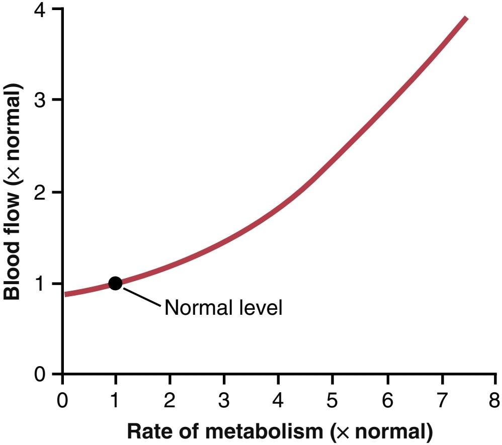
Figure 17-1. Effect of increasing rate of metabolism on tissue blood flow.
شکل 17-1. تأثیر افزایش سرعت متابولیسم بر جریان خون بافتی.
Reduced Oxygen Availability Increases Tissue Blood Flow. One of the most necessary of the metabolic nutrients is oxygen. Whenever the availability of oxygen to the tissues decreases, such as during the following: (1) at a high altitude at the top of a high mountain; (2) in pneumonia; (3) in carbon monoxide poisoning (which poisons the ability of hemoglobin to transport oxygen); or (4) in cyanide poisoning (which poisons the ability of the tissues to use oxygen), the blood flow through the tissues increases markedly. Figure 17-2 shows that as the arterial oxygen saturation decreases to about 25% of normal, the blood flow through an isolated leg increases about threefold; that is, the blood flow increases almost enough, but not quite enough, to make up for the decreased amount of oxygen in the blood, thus almost maintaining a relatively constant supply of oxygen to the tissues.
کاهش در دسترس بودن اکسیژن جریان خون بافت را افزایش می دهد. یکی از ضروری ترین مواد مغذی متابولیک، اکسیژن است. هر زمان که در دسترس بودن اکسیژن به بافت ها کاهش یابد، مانند موارد زیر: (1) در ارتفاع بالا در بالای یک کوه بلند. (2) در پنومونی. (3) در مسمومیت با مونوکسید کربن (که توانایی هموگلوبین برای انتقال اکسیژن را مسموم می کند). یا (4) در مسمومیت با سیانید (که توانایی بافت ها برای استفاده از اکسیژن را مسموم می کند)، جریان خون در بافت ها به طور قابل توجهی افزایش می یابد. شکل 17-2 نشان می دهد که با کاهش اشباع اکسیژن شریانی به حدود 25 درصد طبیعی، جریان خون از طریق یک پای جدا شده حدود سه برابر افزایش می یابد. یعنی جریان خون تقریباً به اندازه کافی افزایش مییابد، اما نه به اندازه کافی، تا مقدار کاهش اکسیژن خون را جبران کند، بنابراین تقریباً یک عرضه نسبتاً ثابت اکسیژن به بافتها حفظ میشود.
Total cyanide poisoning of oxygen usage by a local tissue area can cause local blood flow to increase as much as sevenfold, thus demonstrating the extreme effect of oxygen deficiency to increase blood flow. The mechanisms whereby changes in tissue metabolism or oxygen availability alter tissue blood flow are not fully understood, but two main theories have been proposed, the vasodilator theory and the oxygen demand theory.
مسمومیت کل سیانید ناشی از مصرف اکسیژن توسط یک ناحیه بافت محلی می تواند باعث افزایش جریان خون موضعی تا 7 برابر شود، بنابراین تأثیر شدید کمبود اکسیژن در افزایش جریان خون را نشان می دهد. مکانیسم هایی که به موجب آن تغییرات در متابولیسم بافت یا در دسترس بودن اکسیژن باعث تغییر جریان خون بافت می شود به طور کامل شناخته نشده است، اما دو نظریه اصلی ارائه شده است، نظریه گشادکننده عروق و نظریه تقاضای اکسیژن.

Figure 17-2. Effect of decreasing arterial oxygen saturation on blood flow through an isolated dog leg.
شکل 17-2. اثر کاهش اشباع اکسیژن شریانی بر جریان خون از طریق یک پای سگ جدا شده
Vasodilator Theory for Acute Local Blood Flow Regulation-Possible Special Role of Adenosine. According to the vasodilator theory, the greater the rate of metabolism or the less the availability of oxygen or some other nutrients to a tissue, the greater the rate of formation of vasodilator substances in the tissue cells. The vasodilator substances are then believed to diffuse through the tissues to the precapillary sphincters, metarterioles, and arterioles to cause dilation. Some of the different vasodilator substances that have been suggested are adenosine, carbon dioxide, adenosine phosphate compounds, histamine, potassium ions, and hydrogen ions.
تئوری وازودیلاتور برای تنظیم جریان خون موضعی حاد-نقش ویژه احتمالی آدنوزین. بر اساس تئوری گشادکننده عروق، هر چه میزان متابولیسم بیشتر باشد یا اکسیژن یا برخی مواد مغذی دیگر برای بافت کمتر باشد، سرعت تشکیل مواد گشادکننده عروق در سلولهای بافت بیشتر میشود. سپس اعتقاد بر این است که مواد گشادکننده عروق از طریق بافت ها به اسفنکترهای پیش مویرگی، متارتریول ها و شریان ها منتشر می شوند و باعث اتساع می شوند. برخی از مواد مختلف گشادکننده عروقی که پیشنهاد شدهاند عبارتند از آدنوزین، دی اکسید کربن، ترکیبات آدنوزین فسفات، هیستامین، یونهای پتاسیم و یونهای هیدروژن.
Vasodilator substances may be released from the tissue in response to oxygen deficiency. For example, experiments have shown that decreased oxygen availability can cause adenosine and lactic acid (containing hydrogen ions) to be released into the spaces between the tissue cells; these substances then cause intense acute vasodilation and therefore are responsible, or partially responsible, for the local blood flow regulation. Other vasodilator substances, such as carbon dioxide, lactic acid, and potassium ions, also tend to increase in the tissues when blood flow is reduced and cell metabolism continues at the same rate, or when cell metabolism is suddenly increased. An increase in the concentration of vasodilator metabolites causes vasodilation of the arterioles, thus increasing the tissue blood flow and returning the tissue concentration of the metabolites toward normal.
مواد گشادکننده عروق ممکن است در پاسخ به کمبود اکسیژن از بافت آزاد شوند. برای مثال، آزمایشها نشان دادهاند که کاهش دسترسی به اکسیژن میتواند باعث آزاد شدن آدنوزین و اسید لاکتیک (حاوی یونهای هیدروژن) در فضاهای بین سلولهای بافت شود. این مواد سپس باعث اتساع حاد عروق می شوند و بنابراین مسئول تنظیم جریان خون موضعی هستند یا تا حدی مسئول هستند. سایر مواد گشادکننده عروق، مانند دی اکسید کربن، اسید لاکتیک و یونهای پتاسیم نیز در بافتها افزایش مییابند که جریان خون کاهش مییابد و متابولیسم سلولی با همان سرعت ادامه مییابد، یا زمانی که متابولیسم سلولی به طور ناگهانی افزایش مییابد. افزایش غلظت متابولیت های گشادکننده عروق باعث اتساع عروق شریان ها می شود، در نتیجه جریان خون بافت افزایش می یابد و غلظت متابولیت های بافتی به حالت عادی برمی گردد.
Many physiologists believe that adenosine is an important local vasodilator for controlling local blood flow. For example, minute quantities of adenosine are released from heart muscle cells when coronary blood flow becomes too little, and this release of adenosine causes enough local vasodilation in the heart to return coronary blood flow to normal. Also, whenever the heart becomes more active than normal, the heart’s metabolism increases, causing increased utilization of oxygen, followed by (1) decreased oxygen concentration in the heart muscle cells with (2) consequent degradation of adenosine triphosphate (ATP), which (3) increases the release of adenosine. It is believed that much of this adenosine leaks out of the heart muscle cells to cause coronary vasodilation, providing increased coronary blood flow to supply the increased nutrient demands of the active heart.
بسیاری از فیزیولوژیست ها معتقدند که آدنوزین یک گشادکننده عروق موضعی مهم برای کنترل جریان خون موضعی است. به عنوان مثال، زمانی که جریان خون کرونر خیلی کم می شود، مقادیر اندکی آدنوزین از سلول های ماهیچه قلب آزاد می شود و این آزاد شدن آدنوزین باعث اتساع عروق موضعی کافی در قلب می شود تا جریان خون کرونر به حالت عادی برگردد. همچنین، هر زمان که قلب بیش از حد طبیعی فعال شود، متابولیسم قلب افزایش مییابد و باعث افزایش استفاده از اکسیژن و به دنبال آن (1) کاهش غلظت اکسیژن در سلولهای عضله قلب و (2) تخریب متعاقب آدنوزین تری فسفات (ATP) میشود که (3) آزادسازی آدنوزین را افزایش میدهد. اعتقاد بر این است که بسیاری از این آدنوزین از سلولهای عضله قلب نشت میکند و باعث اتساع عروق کرونر میشود و جریان خون کرونری را افزایش میدهد تا نیازهای غذایی بیشتر قلب فعال را تامین کند.
Although research evidence is less clear, many physiologists also have suggested that the same adenosine mechanism is an important controller of blood flow in skeletal muscle and many other tissues, as well as in the heart. However, it has been difficult to prove that sufficient quantities of any single vasodilator substance, including adenosine, are formed in the tissues to cause all the measured increase in blood flow. It is likely that a combination of several different vasodilators released by the tissues contributes to blood flow regulation.
اگرچه شواهد تحقیقاتی کمتر واضح است، بسیاری از فیزیولوژیستها نیز پیشنهاد کردهاند که همان مکانیسم آدنوزین یک کنترلکننده مهم جریان خون در ماهیچههای اسکلتی و بسیاری از بافتهای دیگر و همچنین در قلب است. با این حال، اثبات اینکه مقادیر کافی از هر ماده گشادکننده عروق، از جمله آدنوزین، در بافت ها تشکیل می شود تا باعث افزایش اندازه گیری شده در جریان خون شود، مشکل بوده است. این احتمال وجود دارد که ترکیبی از چندین گشادکننده عروقی مختلف که توسط بافت ها آزاد می شوند به تنظیم جریان خون کمک کنند.
Oxygen Demand Theory for Local Blood Flow Control. Although the vasodilator theory is widely accepted, several critical facts have made other physiologists favor another theory, which can be called the oxygen demand theory or, more accurately, the nutrient demand theory (because other nutrients besides oxygen are involved). Oxygen is one of the metabolic nutrients required to cause vascular muscle contraction, with other nutrients required as well. Therefore, in the absence of adequate oxygen, it is reasonable to believe that the blood vessels would relax and therefore dilate. Also, increased utilization of oxygen in the tissues as a result of increased metabolism theoretically could decrease the availability of oxygen to the smooth muscle fibers in the local blood vessels, causing local vasodilation.
تئوری تقاضای اکسیژن برای کنترل جریان خون محلی. اگرچه تئوری گشادکننده عروق به طور گسترده پذیرفته شده است، چندین واقعیت حیاتی باعث شده است که فیزیولوژیست های دیگر از نظریه دیگری حمایت کنند، که می توان آن را نظریه تقاضای اکسیژن یا به طور دقیق تر، نظریه تقاضای مواد مغذی نامید (زیرا مواد مغذی دیگری غیر از اکسیژن در آن نقش دارند). اکسیژن یکی از مواد مغذی متابولیکی است که برای ایجاد انقباض عضله عروقی مورد نیاز است و همچنین سایر مواد مغذی مورد نیاز است. بنابراین، در غیاب اکسیژن کافی، منطقی است که باور کنیم رگ های خونی شل شده و در نتیجه منبسط می شوند. همچنین، افزایش استفاده از اکسیژن در بافتها در نتیجه افزایش متابولیسم از لحاظ نظری میتواند دسترسی اکسیژن به فیبرهای عضلانی صاف در رگهای خونی موضعی را کاهش دهد و باعث اتساع موضعی عروق شود.
A mechanism whereby oxygen availability could operate is shown in Figure 17-3. This figure shows a tissue vascular unit, consisting of a metarteriole with a single sidearm capillary and its surrounding tissue. At the origin of the capillary is a precapillary sphincter and around the metarteriole are several other smooth muscle fibers. When observing such a tissue under a microscope, the precapillary sphincters are normally completely open or completely closed. The number of precapillary sphincters that are open at any given time is roughly proportional to the requirements of the tissue for nutrition. The precapillary sphincters and metarterioles open and close cyclically several times per minute, with the duration of the open phases being proportional to the metabolic needs of the tissues for oxygen. The cyclical opening and closing is called vasomotion.
مکانیزمی که به موجب آن در دسترس بودن اکسیژن می تواند عمل کند در شکل 17-3 نشان داده شده است. این شکل یک واحد عروقی بافتی را نشان میدهد که از یک متارتریول با یک مویرگ بازوی کناری و بافت اطراف آن تشکیل شده است. در منشا مویرگ یک اسفنکتر پیش مویرگی و در اطراف متارتریول چندین رشته ماهیچه صاف دیگر قرار دارد. هنگام مشاهده چنین بافتی در زیر میکروسکوپ، اسفنکترهای پیش مویرگی معمولاً کاملاً باز یا کاملاً بسته هستند. تعداد اسفنکترهای پیش مویرگی که در هر زمان معین باز هستند تقریباً متناسب با نیاز بافت برای تغذیه است. اسفنکترهای پیش مویرگی و متارتریول ها به صورت چرخه ای چندین بار در دقیقه باز و بسته می شوند و مدت زمان فازهای باز متناسب با نیاز متابولیکی بافت ها به اکسیژن است. باز و بسته شدن چرخه ای وازوموشن نامیده می شود.
Because smooth muscle requires oxygen to remain contracted, one might assume that the strength of contraction of the sphincters would increase with an increase in oxygen concentration. Consequently, when the oxygen concentration in the tissue rises above a certain level, the precapillary and metarteriole sphincters presumably would close until the tissue cells consume the excess oxygen. However, when the excess oxygen is gone and the oxygen concentration falls low enough, the sphincters open once more to begin the cycle again.
از آنجایی که ماهیچه های صاف برای منقبض ماندن به اکسیژن نیاز دارند، می توان تصور کرد که قدرت انقباض اسفنکترها با افزایش غلظت اکسیژن افزایش می یابد. در نتیجه، هنگامی که غلظت اکسیژن در بافت از یک سطح معین بالاتر می رود، احتمالاً اسفنکترهای پیش مویرگی و متارتریول بسته می شوند تا زمانی که سلول های بافتی اکسیژن اضافی را مصرف کنند. با این حال، هنگامی که اکسیژن اضافی از بین رفت و غلظت اکسیژن به اندازه کافی پایین آمد، اسفنکترها یک بار دیگر باز می شوند تا چرخه دوباره شروع شود.
Thus, on the basis of available data, either the vasodilator substance theory or oxygen demand theory could explain acute local blood flow regulation in response to the metabolic needs of the tissues. It is probably a combination of the two mechanisms.
بنابراین، بر اساس دادههای موجود، نظریه ماده گشادکننده عروق یا نظریه تقاضای اکسیژن میتواند تنظیم حاد جریان خون موضعی را در پاسخ به نیازهای متابولیکی بافتها توضیح دهد. احتمالاً ترکیبی از این دو مکانیسم است.
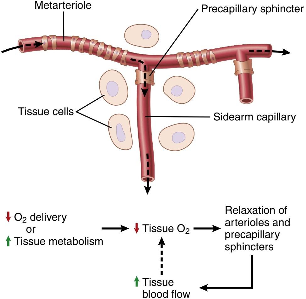
Figure 17-3. Diagram of a tissue unit area for an explanation of acute local feedback control of blood flow, showing a metarteriole passing through the tissue and a sidearm capillary with its precapillary sphincter for controlling capillary blood flow.
شکل 17-3. نمودار یک ناحیه واحد بافت برای توضیح کنترل بازخورد موضعی حاد جریان خون، نشان دهنده عبور یک متارتریول از بافت و یک مویرگ بازو با اسفنکتر پیش مویرگی آن برای کنترل جریان خون مویرگی.
Possible Role of Other Nutrients Besides Oxygen in Control of Local Blood Flow. Under special conditions, it has been shown that the lack of glucose in the perfusing blood can cause local tissue vasodilation. It also is possible that this same effect occurs when other nutrients, such as amino acids or fatty acids, are deficient, although this is still uncertain. In addition, vasodilation occurs in the vitamin deficiency disease beriberi, in which the patient has deficiencies of the vitamin B substances thiamine, niacin, and riboflavin. In this disease, the peripheral vascular blood flow almost everywhere in the body often increases twofold to threefold. Because all these vitamins are necessary for oxygen-induced phosphorylation, which is required to produce ATP in the tissue cells, one can understand how deficiency of these vitamins might lead to diminished smooth muscle contractile ability and therefore local vasodilation as well.
نقش احتمالی سایر مواد مغذی علاوه بر اکسیژن در کنترل جریان خون موضعی. در شرایط خاص، نشان داده شده است که کمبود گلوکز در خون پرفیوژن می تواند باعث اتساع موضعی عروق بافتی شود. همچنین ممکن است این اثر مشابه زمانی رخ دهد که سایر مواد مغذی، مانند اسیدهای آمینه یا اسیدهای چرب، کمبود داشته باشند، اگرچه هنوز مشخص نیست. علاوه بر این، اتساع عروق در بیماری بری بری ناشی از کمبود ویتامین رخ می دهد که در آن بیمار کمبود مواد ویتامین B تیامین، نیاسین و ریبوفلاوین را دارد. در این بیماری، جریان خون عروق محیطی تقریباً در همه جای بدن اغلب دو تا سه برابر افزایش می یابد. از آنجا که همه این ویتامین ها برای فسفوریلاسیون ناشی از اکسیژن، که برای تولید ATP در سلول های بافت مورد نیاز است، ضروری هستند، می توان فهمید که چگونه کمبود این ویتامین ها ممکن است منجر به کاهش توانایی انقباض ماهیچه صاف و در نتیجه اتساع موضعی عروق شود.
Special Examples of Acute Metabolic Control of Local Blood Flow
The mechanisms we have described thus far for local blood flow control are called metabolic mechanisms because they all function in response to the metabolic needs of the tissues. Two additional special examples of metabolic control of local blood flow are reactive hyperemia and active hyperemia (Figure 17-4).
نمونه های ویژه کنترل متابولیک حاد جریان خون موضعی
مکانیسمهایی که تاکنون برای کنترل جریان خون موضعی توضیح دادهایم، مکانیسمهای متابولیک نامیده میشوند زیرا همگی در پاسخ به نیازهای متابولیکی بافتها عمل میکنند. دو نمونه خاص دیگر از کنترل متابولیک جریان خون موضعی عبارتند از هایپرمی واکنشی و پرخونی فعال (شکل 17-4).

Figure 17-4. Reactive hyperemia in a tissue after temporary occlusion of the artery supplying blood flow and active hyperemia following increased tissue metabolic activity.
شکل 17-4. پرخونی واکنشی در بافت پس از انسداد موقت شریان تامین کننده جریان خون و پرخونی فعال بدنبال افزایش فعالیت متابولیکی بافت.
Reactive Hyperemia Occurs After Tissue Blood Sup- ply Is Blocked for a Short Time. When the blood sup- ply to a tissue is blocked for a few seconds to as long as 1 hour or more and then is unblocked, blood flow through the tissue usually increases immediately to four to seven times normal. This increased flow will continue for a few seconds if the block has lasted only a few seconds but sometimes continues for as long as many hours if the blood flow has been stopped for an hour or more. This phenomenon is called reactive hyperemia.
هیپرمی واکنشی پس از مسدود شدن خون رسانی بافتی برای مدت کوتاهی رخ می دهد. هنگامی که خون رسانی به بافت برای چند ثانیه تا یک ساعت یا بیشتر مسدود می شود و سپس مسدود می شود، جریان خون در بافت معمولاً بلافاصله به چهار تا هفت برابر طبیعی افزایش می یابد. اگر بلوک فقط چند ثانیه طول بکشد، این افزایش جریان برای چند ثانیه ادامه خواهد داشت، اما اگر جریان خون برای یک ساعت یا بیشتر متوقف شده باشد، گاهی اوقات تا چند ساعت ادامه مییابد. این پدیده پرخونی واکنشی نامیده می شود.
Reactive hyperemia is another manifestation of the local metabolic blood flow regulation mechanism-that is, lack of flow sets into motion all the factors that cause vasodilation. After short periods of vascular occlusion, the extra blood flow during the reactive hyperemia phase lasts long enough to repay almost exactly the tissue oxygen deficit that has accrued during the period of occlusion. This mechanism emphasizes the close connection between local blood flow regulation and delivery of oxygen and other nutrients to the tissues.
پرخونی واکنشی یکی دیگر از تظاهرات مکانیسم تنظیم جریان خون متابولیک موضعی است – یعنی کمبود جریان تمام عواملی را که باعث اتساع عروق می شوند به حرکت در می آورد. پس از دورههای کوتاه انسداد عروق، جریان خون اضافی در مرحله پرخونی واکنشی به اندازهای طول میکشد که تقریباً دقیقاً کمبود اکسیژن بافتی را که در طول دوره انسداد ایجاد شده است، جبران کند. این مکانیسم بر ارتباط نزدیک بین تنظیم جریان خون موضعی و تحویل اکسیژن و سایر مواد مغذی به بافت ها تاکید می کند.
Active Hyperemia Occurs When Tissue Metabolic Rate Increases. When a tissue becomes highly active, such as an exercising muscle, a gastrointestinal gland during a hypersecretory period, or even the brain during increased mental activity, the rate of blood flow through the tissue increases (see Figure 17-4). The increase in lo- cal metabolism causes the cells to devour tissue fluid nutrients rapidly and release large quantities of vasodilator substances. The result is dilation of local blood vessels and increased local blood flow. In this way, the active tissue receives the additional nutrients required to sustain its new level of function. As noted earlier, active hyperemia in skeletal muscle can increase local muscle blood flow as much as 20-fold during intense exercise.
هیپرمی فعال زمانی رخ می دهد که میزان متابولیک بافت افزایش می یابد. هنگامی که یک بافت به شدت فعال می شود، مانند یک ماهیچه در حال ورزش، یک غده گوارشی در طول یک دوره ترشح بیش از حد، یا حتی مغز در طول افزایش فعالیت ذهنی، سرعت جریان خون در بافت افزایش می یابد (شکل 17-4 را ببینید). افزایش متابولیسم موضعی باعث می شود سلول ها مواد مغذی مایع بافتی را به سرعت می بلعند و مقادیر زیادی از مواد گشادکننده عروق آزاد می کنند. نتیجه اتساع عروق خونی موضعی و افزایش جریان خون موضعی است. به این ترتیب، بافت فعال مواد مغذی اضافی مورد نیاز برای حفظ سطح جدید عملکرد خود را دریافت می کند. همانطور که قبلا ذکر شد، پرخونی فعال در ماهیچه های اسکلتی می تواند جریان خون موضعی ماهیچه ها را در طول ورزش شدید تا 20 برابر افزایش دهد.
Autoregulation of Blood Flow During Changes in Arterial Pressure-Metabolic and Myogenic Mechanisms
In any tissue of the body, a rapid increase in arterial pressure causes an immediate rise in blood flow. However, within less than 1 minute, the blood flow in most tissues returns almost to the normal level, even though the arterial pressure is kept elevated. This return of flow toward normal is called autoregulation. After autoregulation has occurred, the local blood flow in most tissues will be related to arterial pressure approximately in accord with the solid acute curve in Figure 17-5. Note that between arterial pressures of about 70 and 175 mm Hg, the blood flow increases only 20% to 30%, even though the arterial pressure increases 150%. In some tissues, such as the brain and heart, this autoregulation is even more precise.
خودتنظیمی جریان خون در طول تغییرات فشار شریانی – مکانیسمهای متابولیک و میوژنیک
در هر بافتی از بدن، افزایش سریع فشار شریانی باعث افزایش فوری جریان خون می شود. با این حال، در کمتر از 1 دقیقه، جریان خون در اکثر بافت ها تقریباً به سطح طبیعی باز می گردد، حتی اگر فشار شریانی بالا نگه داشته شود. این بازگشت جریان به سمت نرمال خود تنظیم نامیده می شود. پس از وقوع خودتنظیمی، جریان خون موضعی در بیشتر بافتها تقریباً مطابق با منحنی حاد جامد در شکل 17-5 با فشار شریانی مرتبط خواهد بود. توجه داشته باشید که بین فشارهای شریانی حدود 70 تا 175 میلی متر جیوه، جریان خون تنها 20 تا 30 درصد افزایش می یابد، حتی اگر فشار شریانی 150 درصد افزایش یابد. در برخی از بافت ها، مانند مغز و قلب، این خودتنظیمی حتی دقیق تر است.

Figure 17-5. Effect of different levels of arterial pressure on blood flow through a muscle. The solid red curve shows the effect if the arterial pressure is raised over a period of a few minutes. The dashed green curve shows the effect if the arterial pressure is raised slowly over many weeks.
شکل 17-5. تأثیر سطوح مختلف فشار شریانی بر جریان خون از طریق عضله. اگر فشار شریانی در مدت چند دقیقه افزایش یابد، منحنی قرمز جامد تأثیر را نشان می دهد. اگر فشار شریانی به آهستگی در طی چند هفته افزایش یابد، منحنی سبز بریده بریده اثر را نشان می دهد.
For almost a century, two views have been proposed to explain this acute autoregulation mechanism. They have been called the metabolic theory and the myogenic theory.
برای تقریبا یک قرن، دو دیدگاه برای توضیح این مکانیسم خودتنظیمی حاد ارائه شده است. آنها را نظریه متابولیک و نظریه میوژنیک نامیده اند.
The metabolic theory can be understood easily by applying the basic principles of local blood flow regulation discussed in previous sections. Thus, when the arterial pressure becomes too great, the excess flow provides too much oxygen and too many other nutrients to the tissues and washes out the vasodilators released by the tissues. These nutrients (especially oxygen) and decreased tissue levels of vasodilators then cause the blood vessels to constrict and return flow to nearly normal, despite the increased pressure.
تئوری متابولیک را می توان با به کارگیری اصول اولیه تنظیم جریان خون موضعی که در بخش های قبلی مورد بحث قرار گرفت، به راحتی درک کرد. بنابراین، هنگامی که فشار شریانی بیش از حد زیاد میشود، جریان اضافی اکسیژن و مواد مغذی زیادی را به بافتها میرساند و گشادکنندههای عروق آزاد شده توسط بافتها را میشوید. این مواد مغذی (مخصوصاً اکسیژن) و کاهش سطوح بافتی گشادکنندههای عروقی باعث انقباض رگهای خونی و بازگشت جریان تقریباً به حالت عادی، علیرغم افزایش فشار میشوند.
The myogenic theory, however, suggests that another mechanism not related to tissue metabolism explains the phenomenon of autoregulation. This theory is based on the observation that a sudden stretch of small blood vessels causes the smooth muscle of the vessel wall to contract. Therefore, it has been proposed that when high arterial pressure stretches the vessel, reactive vascular constriction results, which reduces blood flow nearly back to normal. Conversely, at low pressures, the degree of stretch of the vessel is less, so the smooth muscle relaxes, reducing vascular resistance and helping to return flow toward normal.
با این حال، نظریه میوژنیک نشان می دهد که مکانیسم دیگری که به متابولیسم بافت مربوط نیست، پدیده خودتنظیمی را توضیح می دهد. این نظریه مبتنی بر این مشاهدات است که کشش ناگهانی رگهای خونی کوچک باعث انقباض عضله صاف دیواره عروق میشود. بنابراین، پیشنهاد شده است که وقتی فشار شریانی بالا رگ را کشیده، انقباض عروقی واکنشی ایجاد میکند که جریان خون را تقریباً به حالت عادی کاهش میدهد. برعکس، در فشارهای کم، میزان کشش رگ کمتر است، بنابراین عضله صاف شل می شود، مقاومت عروقی کاهش می یابد و به بازگشت جریان به حالت طبیعی کمک می کند.
The myogenic response is inherent to vascular smooth muscle and can occur in the absence of neural or hormonal influences. It is most pronounced in arterioles but can also be observed in arteries, venules, veins, and even lymphatic vessels. Myogenic contraction is initiated by stretch-induced vascular depolarization, which then rapidly increases calcium ion entry from the extracellular fluid into the cells, causing them to contract. Changes in vascular pressure may also open or close other ion channels that influence vascular contraction. The precise mechanisms whereby changes in pressure cause opening or closing of vascular ion channels are still uncertain but likely involve mechanical effects of pressure on extracellular proteins that are tethered to cytoskeleton elements of the vascular wall or to the ion channels themselves.
پاسخ میوژنیک ذاتی ماهیچه صاف عروق است و می تواند در غیاب تأثیرات عصبی یا هورمونی رخ دهد. بیشتر در شریانها آشکار میشود، اما در شریانها، وریدها، سیاهرگها و حتی عروق لنفاوی نیز قابل مشاهده است. انقباض میوژنیک با دپلاریزاسیون عروقی ناشی از کشش آغاز می شود، که سپس به سرعت ورود یون کلسیم از مایع خارج سلولی به سلول ها را افزایش می دهد و باعث انقباض آنها می شود. تغییرات در فشار عروقی همچنین ممکن است کانالهای یونی دیگری را که بر انقباض عروقی تأثیر میگذارند باز یا ببندد. مکانیسمهای دقیقی که به موجب آن تغییرات فشار باعث باز یا بسته شدن کانالهای یونی عروقی میشود هنوز نامشخص است، اما احتمالاً شامل اثرات مکانیکی فشار روی پروتئینهای خارج سلولی است که به عناصر اسکلت سلولی دیواره عروقی یا خود کانالهای یونی متصل هستند.
The myogenic mechanism appears to be important in preventing excessive stretching of blood vessels when blood pressure is increased. However, the role of the myogenic mechanism in blood flow regulation is unclear because this pressure-sensing mechanism cannot detect changes in blood flow in the tissue directly. Metabolic factors appear to override the myogenic mechanism in circumstances in which the metabolic demands of the tissues are significantly increased, such as during vigorous muscle exercise, which causes dramatic increases in skeletal muscle blood flow.
به نظر می رسد مکانیسم میوژنیک در جلوگیری از کشش بیش از حد رگ های خونی هنگام افزایش فشار خون مهم باشد. با این حال، نقش مکانیسم myogenic در تنظیم جریان خون نامشخص است، زیرا این مکانیسم حسگر فشار نمی تواند تغییرات در جریان خون در بافت را مستقیماً تشخیص دهد. به نظر می رسد عوامل متابولیک در شرایطی که نیازهای متابولیکی بافت ها به طور قابل توجهی افزایش می یابد، مانند هنگام تمرین شدید عضلانی، که باعث افزایش چشمگیر جریان خون ماهیچه های اسکلتی می شود، بر مکانیسم میوژنیک غلبه می کنند.
Special Mechanisms for Acute Blood Flow Control in Specific Tissues
Although the general mechanisms for local blood flow control discussed thus far are present in almost all tissues of the body, distinctly different mechanisms operate in a few special areas. All mechanisms are discussed through- out this text in relation to specific organs, but two notable mechanisms are as follows:
مکانیسم های ویژه برای کنترل جریان خون حاد در بافت های خاص
اگرچه مکانیسمهای کلی برای کنترل جریان خون موضعی که تاکنون مورد بحث قرار گرفت تقریباً در تمام بافتهای بدن وجود دارد، مکانیسمهای کاملاً متفاوتی در چند ناحیه خاص عمل میکنند. همه مکانیسم ها در این متن در رابطه با اندام های خاص مورد بحث قرار می گیرند، اما دو مکانیسم قابل توجه به شرح زیر است:
1.In the kidneys, blood flow control is significantly vested in a mechanism called tubuloglomerular feedback, in which the composition of the fluid in the early distal tubule is detected by an epithelial structure of the distal tubule, called the macula densa. This structure is located where the distal tubule lies adjacent to the afferent and efferent arterioles at the nephron juxtaglomerular apparatus. When too much fluid filters from the blood through the glomerulus into the tubular system, feedback signals from the macula densa cause constriction of the afferent arterioles, thereby reducing renal blood flow and glomerular filtration rate back to nearly normal. The details of this mechanism are discussed in Chapter 27.
1. در کلیه ها، کنترل جریان خون به طور قابل توجهی تحت مکانیزمی به نام فیدبک توبولوگلومرولی است که در آن ترکیب مایع در لوله دیستال اولیه توسط ساختار اپیتلیالی لوله دیستال به نام ماکولا دنسا تشخیص داده می شود. این ساختار در جایی قرار دارد که توبول دیستال در مجاورت شریان های آوران و وابران در دستگاه juxtaglomerular نفرون قرار دارد. هنگامی که مایع بیش از حد از خون از طریق گلومرول به سیستم لولهای فیلتر میشود، سیگنالهای بازخوردی از ماکولا دنسا باعث انقباض شریانهای آور میشود و در نتیجه جریان خون کلیوی و نرخ فیلتراسیون گلومرولی را تقریباً به حالت طبیعی کاهش میدهد. جزئیات این مکانیسم در فصل 27 مورد بحث قرار گرفته است.
2.In the brain, in addition to control of blood flow by tissue oxygen concentration, the concentrations of carbon dioxide and hydrogen ions play prominent roles. An increase of either or both of these substances dilates the cerebral vessels and allows rapid washout of the excess carbon dioxide or hydrogen ions from the brain tissues. This mechanism is important because the level of ex- citability of the brain is highly dependent on exact control of both carbon dioxide concentration and hydrogen ion concentration. This special mechanism for cerebral blood flow control is presented in Chapter 62.
2. در مغز، علاوه بر کنترل جریان خون توسط غلظت اکسیژن بافت، غلظت دی اکسید کربن و یون های هیدروژن نقش برجسته ای دارند. افزایش هر دو یا هر یک از این مواد باعث گشاد شدن عروق مغزی می شود و باعث می شود که یون های دی اکسید کربن یا هیدروژن اضافی از بافت های مغز به سرعت پاک شوند. این مکانیسم مهم است زیرا سطح تحریک پذیری مغز به شدت به کنترل دقیق غلظت دی اکسید کربن و غلظت یون هیدروژن بستگی دارد. این مکانیسم ویژه برای کنترل جریان خون مغزی در فصل 62 ارائه شده است.
3.In the skin, blood flow control is closely linked to body temperature regulation. Cutaneous and subcutaneous flow regulates heat loss from the body by metering the flow of heat from the core to the surface of the body, where heat is lost to the environment. Skin blood flow is controlled largely by the central nervous system through the sympathetic nerves, as discussed in Chapter 74. Although skin blood flow is only about 3 ml/min/100 g of tissue in cool weather, large changes from that value can occur as needed. When humans are exposed to body heating, skin blood flow may increase greatly, to as high as 7 to 8 L/min for the entire body. When body temperature is reduced, skin blood flow decreases, falling to barely above zero at very low temperatures. Even with severe vasoconstriction, skin blood flow is usually great enough to meet the basic metabolic demands of the skin.
3. در پوست، کنترل جریان خون ارتباط نزدیکی با تنظیم دمای بدن دارد. جریان پوستی و زیر جلدی اتلاف گرما از بدن را با اندازه گیری جریان گرما از هسته به سطح بدن تنظیم می کند، جایی که گرما به محیط تلف می شود. جریان خون پوست عمدتاً توسط سیستم عصبی مرکزی از طریق اعصاب سمپاتیک کنترل میشود، همانطور که در فصل 74 بحث شد. اگرچه جریان خون پوست در هوای خنک تنها حدود 3 میلیلیتر/دقیقه/100 گرم بافت است، تغییرات زیادی از این مقدار میتواند در صورت نیاز رخ دهد. هنگامی که انسان در معرض گرمای بدن قرار می گیرد، جریان خون پوست ممکن است به شدت افزایش یابد و به 7 تا 8 لیتر در دقیقه برای کل بدن برسد. هنگامی که دمای بدن کاهش می یابد، جریان خون پوست کاهش می یابد و در دمای بسیار پایین به سختی به بالای صفر می رسد. حتی با انقباض عروق شدید، جریان خون پوست معمولا به اندازه کافی عالی است که نیازهای متابولیکی اولیه پوست را برآورده کند.
Control of Tissue Blood Flow: Endothelium-Derived Relaxing or Constricting Factors
The endothelial cells lining the blood vessels synthesize several substances that when released, can affect the degree of relaxation or contraction of the vascular wall. For many of these endothelium-derived relaxing or constrictor factors, the physiological roles are just beginning to be understood.
کنترل جریان خون بافت: عوامل آرامشدهنده یا منقبض کننده مشتق از اندوتلیوم
سلول های اندوتلیال پوشاننده رگ های خونی مواد مختلفی را سنتز می کنند که وقتی آزاد می شوند، می توانند بر میزان شل شدن یا انقباض دیواره عروق تأثیر بگذارند. برای بسیاری از این عوامل آرامشبخش یا منقبضکننده مشتق از اندوتلیوم، نقشهای فیزیولوژیکی تازه در حال درک شدن هستند.
Nitric Oxide Is a Vasodilator Released from Healthy Endothelial Cells. The most important of the endotheliumderived relaxing factors is nitric oxide (NO), a lipophilic gas that is released from endothelial cells in response to a variety of chemical and physical stimuli. Endothelial- derived nitric oxide synthase (eNOS) enzymes synthesize NO from arginine and oxygen and by reduction of inorganic nitrate. After diffusing out of the endothelial cell, NO has a half-life in the blood of only about 6 seconds and acts mainly in the local tissues, where it is released. NO activates soluble guanylate cyclases in vascular smooth muscle cells (Figure 17-6), resulting in the conversion of cyclic guanosine triphosphate (CGTP) to cyclic guanosine monophosphate (cGMP) and activation of cGMP- dependent protein kinase (PKG), which has several actions that cause the blood vessels to relax.
نیتریک اکسید یک گشادکننده عروق است که از سلول های اندوتلیال سالم آزاد می شود. مهمترین عامل آرامش بخش مشتق از اندوتلیوم، اکسید نیتریک (NO)، یک گاز چربی دوست است که در پاسخ به انواع محرک های شیمیایی و فیزیکی از سلول های اندوتلیال آزاد می شود. آنزیم های نیتریک اکسید سنتاز مشتق از اندوتلیال (eNOS) NO را از آرژنین و اکسیژن و با کاهش نیترات معدنی سنتز می کنند. پس از انتشار به خارج از سلول اندوتلیال، NO در خون تنها حدود 6 ثانیه نیمه عمر دارد و عمدتاً در بافت های موضعی، جایی که آزاد می شود، عمل می کند. NO سیکلازهای گوانیلات محلول را در سلولهای ماهیچه صاف عروق فعال میکند (شکل 17-6)، و در نتیجه تبدیل گوانوزین تری فسفات حلقوی (CGTP) به گوانوزین مونوفسفات حلقوی (cGMP) و فعال شدن cGMP وابسته به cGMP باعث شل شدن چندین فعالیت پروتئین کیناز خون (PKG) میشود.

Figure 17-6. Nitric oxide synthase (eNOS) enzyme in endothelial cells synthesizes nitric oxide (NO) from arginine and oxygen. NO activates soluble guanylate cyclases in vascular smooth muscle cells, resulting in conversion of cyclic guanosine triphosphate (CGTP) to cyclic guano- sine monophosphate (cGMP), which ultimately causes the blood vessels to relax.
شکل 17-6. آنزیم نیتریک اکسید سنتاز (eNOS) در سلول های اندوتلیال، اکسید نیتریک (NO) را از آرژنین و اکسیژن سنتز می کند. NO سیکلازهای گوانیلات محلول را در سلولهای ماهیچه صاف عروق فعال میکند و در نتیجه گوانوزین تری فسفات حلقوی (CGTP) به گوانوزین مونوفسفات حلقوی (cGMP) تبدیل میشود که در نهایت باعث شل شدن رگهای خونی میشود.
The flow of blood through the arteries and arterioles causes shear stress on the endothelial cells because of viscous drag of the blood against the vascular walls. This stress contorts the endothelial cells in the direction of flow and causes significant increase in NO release. The NO then relaxes the blood vessels, fortunately, because the local metabolic mechanisms for controlling tissue blood flow mainly dilate the very small arteries and arterioles in each tissue. Yet, when blood flow through a microvascular portion of the circulation increases, this action secondarily stimulates the release of NO from larger vessels as a result of increased flow and shear stress in these vessels. The released NO increases the diameters of the larger upstream blood vessels whenever microvascular blood flow increases downstream. Without such a response, the effectiveness of local blood flow control would be decreased because a significant part of the resistance to blood flow is in the upstream small arteries.
جریان خون از طریق شریان ها و شریان ها به دلیل کشش چسبناک خون در برابر دیواره های عروقی باعث ایجاد فشار برشی بر روی سلول های اندوتلیال می شود. این تنش سلول های اندوتلیال را در جهت جریان منقبض می کند و باعث افزایش قابل توجهی در انتشار NO می شود. خوشبختانه NO رگهای خونی را شل میکند، زیرا مکانیسمهای متابولیک موضعی برای کنترل جریان خون بافتی عمدتاً شریانها و شریانهای بسیار کوچک در هر بافت را گشاد میکنند. با این حال، هنگامی که جریان خون از طریق یک بخش میکروواسکولار گردش خون افزایش مییابد، این عمل به طور ثانویه باعث آزاد شدن NO از عروق بزرگتر در نتیجه افزایش جریان و تنش برشی در این عروق میشود. NO آزاد شده قطر رگ های خونی بزرگتر بالادست را هر زمان که جریان خون میکروواسکولار در پایین دست افزایش می یابد افزایش می دهد. بدون چنین پاسخی، اثربخشی کنترل جریان خون موضعی کاهش مییابد زیرا بخش قابل توجهی از مقاومت در برابر جریان خون در شریانهای کوچک بالادست است.
NO synthesis and release from endothelial cells are also stimulated by some vasoconstrictors, such as angiotensin II, which bind to specific receptors on endothelial cells. The increased NO release protects against excessive vasoconstriction.
سنتز و آزادسازی NO از سلول های اندوتلیال نیز توسط برخی از منقبض کننده های عروقی مانند آنژیوتانسین II تحریک می شود که به گیرنده های خاصی روی سلول های اندوتلیال متصل می شود. افزایش آزادسازی NO از انقباض عروق بیش از حد محافظت می کند.
When endothelial cells are damaged by chronic hyper- tension or atherosclerosis, impaired NO synthesis may contribute to excessive vasoconstriction and worsening of the hypertension and endothelial damage. If untreated, this may eventually cause vascular injury and damage to vulnerable tissues such as the heart, kidneys, and brain.
هنگامی که سلول های اندوتلیال توسط فشار خون مزمن یا آترواسکلروز آسیب می بینند، اختلال در سنتز NO ممکن است به انقباض بیش از حد عروق و بدتر شدن فشار خون بالا و آسیب اندوتلیال کمک کند. در صورت عدم درمان، ممکن است در نهایت باعث آسیب عروقی و آسیب به بافت های آسیب پذیر مانند قلب، کلیه ها و مغز شود.
Even before NO was discovered, clinicians used nitro- glycerin, amyl nitrate, and other nitrate derivatives to treat patients who had angina pectoris—that is, severe chest pain caused by ischemia of the heart muscle. These drugs, when broken down chemically, release NO and cause dilation of blood vessels throughout the body, including the coronary blood vessels.
حتی قبل از کشف NO، پزشکان از نیتروگلیسیرین، آمیل نیترات و سایر مشتقات نیترات برای درمان بیمارانی که آنژین صدری داشتند – یعنی درد شدید قفسه سینه ناشی از ایسکمی عضله قلب – استفاده می کردند. این داروها وقتی به صورت شیمیایی تجزیه می شوند، NO آزاد می کنند و باعث گشاد شدن رگ های خونی در سراسر بدن از جمله عروق کرونر می شوند.
Other important applications of NO physiology and pharmacology are the development and clinical use of drugs (e.g., sildenafil) that inhibit cGMP-specific phospho- diesterase-5 (PDE-5), an enzyme that degrades cGMP. By preventing the degradation of cGMP, the PDE-5 inhibitors effectively prolong the actions of NO to cause vasodilation. The primary clinical use of the PDE-5 inhibitors is to treat erectile dysfunction. Penile erection is caused by parasym- pathetic nerve impulses through the pelvic nerves to the penis, where the neurotransmitters acetylcholine and NO are released. By preventing the degradation of NO, the PDE-5 inhibitors enhance the dilation of the blood vessels in the penis and aid in erection, as discussed in Chapter 81.
دیگر کاربردهای مهم فیزیولوژی و فارماکولوژی NO، توسعه و استفاده بالینی از داروهایی (به عنوان مثال، سیلدنافیل) است که فسفودی استراز-5 (PDE-5) اختصاصی cGMP را مهار می کند، آنزیمی که cGMP را تجزیه می کند. با جلوگیری از تخریب cGMP، مهارکنندههای PDE-5 به طور موثری فعالیتهای NO را طولانی میکنند تا باعث اتساع عروق شوند. استفاده بالینی اولیه از مهارکننده های PDE-5 برای درمان اختلال نعوظ است. نعوظ آلت تناسلی توسط تکانه های عصبی پاراسمپاتیک از طریق اعصاب لگن به آلت تناسلی ایجاد می شود، جایی که انتقال دهنده های عصبی استیل کولین و NO آزاد می شوند. همانطور که در فصل 81 بحث شد، با جلوگیری از تخریب NO، مهارکنندههای PDE-5 گشاد شدن رگهای خونی آلت تناسلی را افزایش میدهند و به نعوظ کمک میکنند.
Endothelin Is a Powerful Vasoconstrictor Released From Damaged Endothelium. Endothelial cells also re- lease vasoconstrictor substances. The most important of these is endothelin, a large, 27-amino acid peptide that requires only minute amounts (nanograms) to cause powerful vasoconstriction. This substance is present in the endothelial cells of all or most blood vessels but greatly increases when the vessels are injured. The usual stimulus for release is damage to the endothelium, such as that caused by crushing the tissues or injecting a traumatizing chemical into the blood vessel. After severe blood vessel damage, local release of endothelin and subsequent vasoconstriction helps prevent extensive bleeding from arteries as large as 5 millimeters in diameter that might have been torn open by crushing injury.
اندوتلین یک تنگ کننده عروق قوی است که از اندوتلیوم آسیب دیده رها می شود. سلول های اندوتلیال نیز مواد منقبض کننده عروق آزاد می کنند. مهمترین آنها اندوتلین است، یک پپتید بزرگ با 27 اسید آمینه که برای ایجاد انقباض قوی عروق تنها به مقادیر بسیار کمی (نانوگرم) نیاز دارد. این ماده در سلولهای اندوتلیال همه یا اکثر رگهای خونی وجود دارد اما زمانی که رگها آسیب میبینند، به شدت افزایش مییابد. محرک معمول برای آزادسازی آسیب به اندوتلیوم است، مانند آسیبی که در اثر له کردن بافت ها یا تزریق یک ماده شیمیایی آسیب زا به رگ خونی ایجاد می شود. پس از آسیب شدید رگ های خونی، آزادسازی موضعی اندوتلین و متعاقب آن انقباض عروقی به جلوگیری از خونریزی گسترده از شریان هایی به قطر 5 میلی متر کمک می کند که ممکن است در اثر آسیب له شده باز شده باشند.
Increased endothelin release is also believed to contribute to vasoconstriction when the endothelium is damaged by hypertension. Drugs that block endothelin receptors have been used to treat pulmonary hypertension but generally have not been used for lowering blood pressure in patients with systemic arterial hypertension.
همچنین اعتقاد بر این است که افزایش ترشح اندوتلین در انقباض عروقی که اندوتلیوم توسط فشار خون بالا آسیب می بیند، نقش دارد. داروهایی که گیرندههای اندوتلین را مسدود میکنند برای درمان پرفشاری خون ریوی استفاده شدهاند، اما عموماً برای کاهش فشار خون در بیماران مبتلا به فشار خون شریانی سیستمیک استفاده نشدهاند.
LONG-TERM BLOOD FLOW REGULATION
Thus far, most of the mechanisms for local blood flow regulation that we have discussed act within a few seconds to a few minutes after the local tissue conditions have changed. Yet, even after full activation of these acute mechanisms, the blood flow usually is adjusted only about three quarters of the way to the exact additional requirements of the tissues. For example, when the arterial pressure suddenly increases from 100 to 150 mm Hg, the blood flow increases almost instantaneously, by about 100%. Then, within 30 seconds to 2 minutes, the flow decreases back to about 10% to 15% above the original control value. This example illustrates the rapidity of the acute mechanisms for local blood flow regulation, but also demonstrates that the regulation is still incomplete because a 10% to 15% excess blood flow remains in some tissues.
تنظیم طولانی مدت جریان خون
تا کنون، بیشتر مکانیسمهای تنظیم جریان خون موضعی که در مورد آنها بحث کردیم، در عرض چند ثانیه تا چند دقیقه پس از تغییر شرایط بافت محلی عمل میکنند. با این حال، حتی پس از فعالسازی کامل این مکانیسمهای حاد، جریان خون معمولاً تنها در حدود سه چهارم مسیر مطابق با نیازهای اضافی بافتها تنظیم میشود. به عنوان مثال، هنگامی که فشار شریانی به طور ناگهانی از 100 به 150 میلیمتر جیوه افزایش مییابد، جریان خون تقریباً آنی و حدود 100 درصد افزایش مییابد. سپس، در عرض 30 ثانیه تا 2 دقیقه، جریان به حدود 10% تا 15% بالاتر از مقدار کنترل اصلی کاهش می یابد. این مثال سرعت مکانیسمهای حاد تنظیم جریان خون موضعی را نشان میدهد، اما همچنین نشان میدهد که تنظیم هنوز ناقص است زیرا 10 تا 15 درصد جریان خون اضافی در برخی بافتها باقی میماند.
However, over a period of hours, days, and weeks, a long-term type of local blood flow regulation develops in addition to the acute control. This long-term regulation gives far more complete control of blood flow. In the aforementioned example, if the arterial pressure remains at 150 mm Hg indefinitely, the blood flow through the tissues gradually approaches almost exactly the normal flow level within a few weeks. Figure 17-5 shows (dashed green curve) the extreme effectiveness of this long-term local blood flow regulation. Note that once the long-term regulation has had time to occur, long-term changes in arterial pressure between 50 and 200 mm Hg have little effect on the rate of local blood flow.
با این حال، در طی چند ساعت، روز و هفته، علاوه بر کنترل حاد، یک نوع تنظیم طولانی مدت جریان خون موضعی ایجاد می شود. این تنظیم طولانی مدت کنترل بسیار بیشتری بر جریان خون می دهد. در مثال فوق، اگر فشار شریانی به طور نامحدود روی 150 میلیمتر جیوه باقی بماند، جریان خون در بافتها به تدریج در عرض چند هفته تقریباً به سطح جریان طبیعی نزدیک میشود. شکل 17-5 (منحنی سبز چین دار) اثربخشی شدید این تنظیم طولانی مدت جریان خون موضعی را نشان می دهد. توجه داشته باشید که زمانی که تنظیم طولانی مدت انجام شود، تغییرات طولانی مدت در فشار شریانی بین 50 تا 200 میلی متر جیوه تأثیر کمی بر سرعت جریان خون موضعی دارد.
Long-term regulation of blood flow is especially important when the metabolic demands of a tissue change. Thus, if a tissue becomes chronically overactive and requires increased quantities of oxygen and other nutrients, the arterioles and capillary vessels usually increase both in number and size within a few weeks to match the needs of the tissue, unless the circulatory system has become pathological or too old to respond.
تنظیم طولانی مدت جریان خون به ویژه هنگامی که نیازهای متابولیکی یک بافت تغییر می کند مهم است. بنابراین، اگر بافتی به طور مزمن بیش فعال شود و به مقادیر بیشتری از اکسیژن و سایر مواد مغذی نیاز داشته باشد، شریانها و عروق مویرگی معمولاً در عرض چند هفته از نظر تعداد و اندازه افزایش مییابند تا با نیازهای بافت مطابقت داشته باشند، مگر اینکه سیستم گردش خون پاتولوژیک شده باشد یا برای پاسخدهی خیلی پیر شده باشد.
Blood Flow Regulation by Changes in Tissue Vascularity
A key mechanism for long-term local blood flow regulation is to change the amount of vascularity of the tissues. For example, if the metabolism in a tissue is increased for a prolonged period, vascularity increases, a process generally called angiogenesis; if the metabolism is decreased, vascularity decreases. Figure 17-7 shows the large increase in the number of capillaries in a rat anterior tibialis muscle that was stimulated electrically to contract for short periods each day for 30 days, compared with the unstimulated muscle in the other leg of the animal.
تنظیم جریان خون با تغییر در عروق بافتی
یک مکانیسم کلیدی برای تنظیم طولانی مدت جریان خون موضعی، تغییر میزان عروق بافت ها است. به عنوان مثال، اگر متابولیسم در یک بافت برای مدت طولانی افزایش یابد، عروق افزایش می یابد، فرآیندی که عموما آنژیوژنز نامیده می شود. اگر متابولیسم کاهش یابد، عروق کاهش می یابد. شکل 17-7 افزایش زیادی در تعداد مویرگها در عضله تیبیالیس قدامی موش را نشان میدهد که بهمدت 30 روز هر روز برای دورههای کوتاه مدت بهطور الکتریکی تحریک شده بود، در مقایسه با عضله تحریکنشده در پای دیگر حیوان.
Thus, actual physical reconstruction of the tissue vasculature occurs to meet the needs of the tissues. This reconstruction occurs rapidly (within days) in young animals. It also occurs rapidly in new growth tissue, such as in cancerous tissue, but occurs much more slowly in old, well-established tissues. Therefore, the time required for long-term regulation to take place may be only a few days in the neonate or as long as months in older adults. Furthermore, the final degree of response is much better in younger than in older tissues; thus, in the neonate, the vascularity will adjust to match almost exactly the needs of the tissue for blood flow, whereas in older tissues, vascularity frequently lags far behind the needs of the tissues.
بنابراین، بازسازی فیزیکی واقعی عروق بافتی برای رفع نیازهای بافت ها اتفاق می افتد. این بازسازی به سرعت (در عرض چند روز) در حیوانات جوان رخ می دهد. همچنین در بافتهای رشد یافته جدید، مانند بافت سرطانی، به سرعت رخ میدهد، اما در بافتهای قدیمی و مستقر بسیار کندتر رخ میدهد. بنابراین، زمان لازم برای تنظیم طولانی مدت ممکن است فقط چند روز در نوزاد یا به اندازه ماه ها در بزرگسالان مسن تر باشد. علاوه بر این، درجه نهایی پاسخ در بافتهای جوانتر بسیار بهتر از بافتهای مسنتر است. بنابراین، در نوزاد، عروق تنظیم میشود تا تقریباً دقیقاً با نیازهای بافت برای جریان خون مطابقت داشته باشد، در حالی که در بافتهای مسنتر، عروقی اغلب از نیازهای بافتها عقبتر است.

Figure 17-7. A large increase in the number of capillaries (white dots) in a rat anterior tibialis muscle that was stimulated electrically to contract for short periods each day for 30 days (B), compared with the unstimulated muscle (A). The 30 days of intermittent electrical stimulation converted the predominantly fast-twitch, glycolytic anterior tibialis muscle to a predominantly slow-twitch, oxidative muscle with increased numbers of capillaries and decreased fiber diameter, as shown. (Courtesy Dr. Thomas Adair.)
شکل 17-7. افزایش زیادی در تعداد مویرگها (نقطههای سفید) در عضله تیبیالیس قدامی موش صحرایی که بهمدت 30 روز (B) هر روز برای دورههای کوتاهی برای انقباض الکتریکی تحریک میشد، در مقایسه با عضله تحریکنشده (A). همانطور که نشان داده شده است، 30 روز تحریک الکتریکی متناوب، عضله تیبیالیس قدامی گلیکولیتیک تند انقباض را به ماهیچه ای عمدتا کند انقباض و اکسیداتیو با افزایش تعداد مویرگ ها و کاهش قطر فیبر تبدیل کرد. (با احترام از دکتر توماس آدایر.)
Role of Oxygen in Long-Term Regulation. Oxygen is important not only for acute control of local blood flow but also for long-term control. One example of this is increased vascularity in tissues of animals that live at high altitudes, where the atmospheric oxygen is low. In premature babies who are put into oxygen tents for therapeutic purposes, the excess oxygen causes almost immediate cessation of new vascular growth in the retina of the premature baby’s eyes and even causes degeneration of some of the small vessels that already have formed. When the infant is taken out of the oxygen tent, explosive overgrowth of new vessels then occurs to make up for the sudden decrease in available oxygen. Often, so much overgrowth occurs that the retinal vessels grow out from the retina into the eye’s vitreous humor, eventually causing blindness, a condition called retrolental fibroplasia.
نقش اکسیژن در تنظیم طولانی مدت. اکسیژن نه تنها برای کنترل حاد جریان خون موضعی بلکه برای کنترل طولانی مدت نیز مهم است. یکی از نمونههای آن افزایش عروق در بافتهای حیواناتی است که در ارتفاعات بالا زندگی میکنند، جایی که اکسیژن اتمسفر کم است. در نوزادان نارس که برای اهداف درمانی در چادر اکسیژن قرار می گیرند، اکسیژن اضافی تقریباً بلافاصله باعث توقف رشد عروقی جدید در شبکیه چشم نوزاد نارس می شود و حتی باعث انحطاط برخی از عروق کوچکی می شود که قبلاً تشکیل شده اند. هنگامی که نوزاد از چادر اکسیژن خارج می شود، رشد بیش از حد انفجاری عروق جدید رخ می دهد تا کاهش ناگهانی اکسیژن موجود را جبران کند. اغلب، رشد بیش از حد زیادی اتفاق میافتد که عروق شبکیه از شبکیه به داخل زجاجیه چشم رشد میکنند و در نهایت باعث کوری میشوند، وضعیتی که فیبروپلازی رترولنتال نامیده میشود.
Importance of Vascular Growth Factors in Formation of New Blood Vessels. A dozen or more factors that increase growth of new blood vessels have been found, almost all of which are small peptides. The four factors that have been best characterized are vascular endothelial growth factor (VEGF), fibroblast growth factor, platelet- derived growth factor (PDGF), and angiogenin, each of which has been isolated from tissues that have inadequate blood supply. Deficiency of tissue oxygen induces expres- sion of hypoxia inducible factors (HIFs), transcription factors that in turn upregulate gene expression and the formation of vascular growth factors (also called angiogenic factors).
اهمیت فاکتورهای رشد عروقی در تشکیل عروق خونی جدید. ده ها یا چند عامل افزایش دهنده رشد رگ های خونی جدید یافت شده اند که تقریباً همه آنها پپتیدهای کوچکی هستند. چهار عاملی که به بهترین وجه مشخص شده اند عبارتند از فاکتور رشد اندوتلیال عروقی (VEGF)، فاکتور رشد فیبروبلاست، فاکتور رشد مشتق از پلاکت (PDGF) و آنژیوژنین، که هر کدام از بافت هایی که دارای خون رسانی ناکافی هستند جدا شده اند. کمبود اکسیژن بافتی باعث بروز عوامل القایی هیپوکسی (HIFs)، فاکتورهای رونویسی می شود که به نوبه خود بیان ژن و تشکیل فاکتورهای رشد عروقی (که فاکتورهای رگ زایی نیز نامیده می شوند) را تنظیم می کنند.
Angiogenesis begins with new vessels sprouting from other small vessels. The first step is dissolution of the basement membrane of the endothelial cells at the point of sprouting. This step is followed by rapid reproduction of new endothelial cells, which stream outward through the vessel wall in extended cords directed toward the source of the angiogenic factor. The cells in each cord continue to divide and rapidly fold over into a tube. Next, the tube connects with another tube budding from another donor vessel (another arteriole or venule) and forms a capillary loop through which blood begins to flow. If the flow is great enough, smooth muscle cells eventually invade the wall, so some of the new vessels eventually grow to be new arterioles or venules or perhaps even larger vessels. Thus, angiogenesis explains how metabolic factors in local tissues can cause growth of new vessels.
رگ زایی با جوانه زدن عروق جدید از سایر عروق کوچک شروع می شود. مرحله اول انحلال غشای پایه سلول های اندوتلیال در نقطه جوانه زدن است. این مرحله با تولید مثل سریع سلولهای اندوتلیال جدید دنبال میشود که از دیواره عروق به سمت بیرون جریان مییابند و در طنابهای کشیده به سمت منبع عامل رگزایی هدایت میشوند. سلول های هر طناب به تقسیم شدن ادامه می دهند و به سرعت در یک لوله جمع می شوند. در مرحله بعد، لوله به لوله دیگری که از رگ اهداکننده دیگری (شریان یا ونول دیگر) جوانه میزند متصل میشود و یک حلقه مویرگی را تشکیل میدهد که از طریق آن خون شروع به جریان میکند. اگر جریان به اندازه کافی زیاد باشد، سلولهای عضلانی صاف در نهایت به دیواره حمله میکنند، بنابراین برخی از عروق جدید در نهایت تبدیل به شریانها یا ونولهای جدید یا حتی عروق بزرگتر میشوند. بنابراین، رگزایی توضیح میدهد که چگونه عوامل متابولیک در بافتهای محلی میتوانند باعث رشد عروق جدید شوند.
Certain other substances, such as some steroid hormones, have the opposite effect on small blood vessels, occasionally even causing dissolution of vascular cells and disappearance of vessels. Therefore, blood vessels can also be made to disappear when they are not needed. Peptides produced in the tissues can also block the growth of new blood vessels. For example, angiostatin, a fragment of the protein plasminogen, is a naturally occurring inhibitor of angiogenesis. Endostatin is another antiangiogenic peptide derived from the breakdown of collagen type XVII. Although the precise physiological functions of these antiangiogenic substances are still unknown, there is great interest in their potential use in arresting blood vessel growth in cancerous tumors and therefore prevent- ing the large increases in blood flow needed to sustain the nutrient supply of rapidly growing tumors.
برخی از مواد دیگر، مانند برخی از هورمونهای استروئیدی، بر رگهای خونی کوچک اثر معکوس دارند و حتی گاهی باعث انحلال سلولهای عروقی و ناپدید شدن عروق میشوند. بنابراین، رگهای خونی نیز میتوانند در مواقعی که نیازی به آنها نیست ناپدید شوند. پپتیدهای تولید شده در بافت ها همچنین می توانند رشد رگ های خونی جدید را مسدود کنند. به عنوان مثال، آنژیواستاتین، قطعه ای از پروتئین پلاسمینوژن، یک مهارکننده طبیعی رگ زایی است. اندوستاتین یکی دیگر از پپتیدهای ضد رگ زایی است که از تجزیه کلاژن نوع XVII به دست می آید. اگرچه عملکردهای فیزیولوژیکی دقیق این مواد ضد رگ زایی هنوز ناشناخته است، علاقه زیادی به استفاده بالقوه آنها در توقف رشد عروق خونی در تومورهای سرطانی و در نتیجه جلوگیری از افزایش زیاد در جریان خون مورد نیاز برای حفظ مواد مغذی تومورهای با رشد سریع وجود دارد.
Vascularity Determined by Maximum Blood Flow Need, Not by Average Need. An especially valuable characteristic of long-term vascular control is that vascularity is determined mainly by the maximum level of blood flow required by the tissue rather than by average need. For example, during heavy exercise, the need for whole-body blood flow often increases to six to eight times the resting blood flow. This great excess of flow may not be required for more than a few minutes each day. Nevertheless, even this short time of need can cause enough angiogenic factors to be formed by the muscles to increase their vascularity as required. Were it not for this capability, every time a person attempted heavy exercise, the muscles would fail to receive the required nutrients, especially the required oxygen, and thus the muscles would fail to contract.
عروق با حداکثر نیاز به جریان خون تعیین می شود، نه با نیاز متوسط. یکی از ویژگیهای ارزشمند کنترل طولانیمدت عروق این است که عروق عمدتاً با حداکثر سطح جریان خون مورد نیاز بافت تعیین میشود تا نیاز متوسط. به عنوان مثال، در طول ورزش سنگین، نیاز به جریان خون در کل بدن اغلب به شش تا هشت برابر جریان خون در حالت استراحت افزایش می یابد. این جریان زیاد ممکن است بیش از چند دقیقه در روز مورد نیاز نباشد. با این وجود، حتی همین زمان کوتاه نیاز میتواند باعث شود که فاکتورهای رگزایی کافی توسط ماهیچهها تشکیل شود تا در صورت لزوم، عروق آنها افزایش یابد. اگر این توانایی وجود نداشت، هر بار که فردی اقدام به ورزش سنگین می کرد، ماهیچه ها مواد مغذی مورد نیاز، به ویژه اکسیژن مورد نیاز را دریافت نمی کردند و در نتیجه ماهیچه ها منقبض نمی شدند.
However, after extra vascularity does develop, the extra blood vessels normally remain mainly vasoconstricted, opening to allow extra flow only when appropriate local stimuli such as a lack of oxygen, nerve vasodilatory stimuli, or other stimuli call forth the required extra flow.
با این حال، پس از ایجاد عروق اضافی، رگهای خونی اضافی معمولاً عمدتاً منقبض میشوند و تنها زمانی باز میشوند که محرکهای موضعی مناسب مانند کمبود اکسیژن، محرکهای گشادکننده عروق عصبی یا سایر محرکها جریان اضافی مورد نیاز را ایجاد کنند.
Blood Flow Regulation by Development of Collateral Circulation
In most tissues of the body, when an artery or a vein is blocked, a new vascular channel usually develops around the blockage and allows at least partial resupply of blood to the affected tissue. The first stage in this process is dilation of small vascular loops that already connect the vessel above the blockage to the vessel below. This dilation occurs within the first minute or two, indicating that the dilation is likely mediated by metabolic factors. After this initial opening of collateral vessels, the blood flow often is still less than 25% of that required to supply all the tissue needs. However, further opening occurs within the ensuing hours, so that within 1 day as much as half the tissue needs may be met and, within a few days, the blood flow is usually sufficient to meet the tissue needs.
تنظیم جریان خون با توسعه گردش وثیقه
در بیشتر بافتهای بدن، زمانی که یک شریان یا ورید مسدود میشود، معمولاً یک کانال عروقی جدید در اطراف انسداد ایجاد میشود و اجازه میدهد تا حداقل بخشی از خون به بافت آسیبدیده برسد. اولین مرحله در این فرآیند، گشاد شدن حلقههای کوچک عروقی است که رگ بالای انسداد را به رگ زیر متصل میکند. این اتساع در یک یا دو دقیقه اول اتفاق می افتد، که نشان می دهد که اتساع احتمالاً توسط عوامل متابولیک انجام می شود. پس از این باز شدن اولیه رگ های جانبی، جریان خون اغلب هنوز کمتر از 25 درصد از میزان مورد نیاز برای تامین تمام نیازهای بافت است. با این حال، باز شدن بیشتر در ساعات بعدی اتفاق می افتد، به طوری که در عرض 1 روز ممکن است نیمی از نیاز بافت برآورده شود و در عرض چند روز، جریان خون معمولاً برای رفع نیازهای بافت کافی است.
The collateral vessels continue to grow for many months thereafter, usually forming multiple small collateral channels rather than one single large vessel. Under resting conditions, the blood flow may return to nearly normal, but the new channels seldom become large enough to supply the blood flow needed during strenuous tissue activity. Thus, development of collateral vessels follows the usual principles of acute and longterm local blood flow control; the acute control is rapid metabolic dilation, followed chronically by growth and enlargement of new vessels over a period of weeks and months.
رگ های جانبی برای ماه های زیادی پس از آن به رشد خود ادامه می دهند و معمولاً به جای یک کشتی بزرگ، چندین کانال جانبی کوچک را تشکیل می دهند. در شرایط استراحت، جریان خون ممکن است تقریباً به حالت عادی بازگردد، اما کانالهای جدید به ندرت به اندازه کافی بزرگ میشوند تا جریان خون مورد نیاز در طول فعالیتهای شدید بافتی را تامین کنند. بنابراین، توسعه عروق جانبی از اصول معمول کنترل جریان خون موضعی حاد و طولانی مدت پیروی می کند. کنترل حاد اتساع متابولیک سریع است که به طور مزمن با رشد و بزرگ شدن عروق جدید در طی چند هفته و ماه دنبال می شود.
An important example of the development of collateral blood vessels occurs after thrombosis of one of the coronary arteries. By the age of 60 years, many people have experienced closure or at least partial occlusion of at least one of the smaller branch coronary vessels, but they are not aware of it because collateral blood vessels have developed rapidly enough to prevent myocardial dam- age. When collateral blood vessels are unable to develop quickly enough to maintain blood flow because of the rapidity or severity of the coronary insufficiency, serious heart attacks can occur.
یک مثال مهم از ایجاد رگ های خونی جانبی پس از ترومبوز یکی از شریان های کرونری رخ می دهد. در سن 60 سالگی، بسیاری از افراد بسته شدن یا حداقل انسداد جزئی حداقل یکی از رگ های کرونری شاخه های کوچکتر را تجربه کرده اند، اما از آن آگاه نیستند زیرا رگ های خونی جانبی به اندازه کافی سریع رشد کرده اند تا از آسیب میوکارد جلوگیری کنند. هنگامی که رگهای خونی جانبی به دلیل سرعت یا شدت نارسایی عروق کرونر قادر به رشد کافی برای حفظ جریان خون نباشند، حملات قلبی جدی ممکن است رخ دهد.
Vascular Remodeling in Response to Chronic Changes in Blood Flow or Blood Pressure
Vascular growth and remodeling are critical components of tissue development and growth and occur as an adaptive response to long-term changes in blood pres- sure or blood flow. For example, after several months of chronic exercise training, vascularity of the trained muscles increases to accommodate their higher blood flow requirements. In addition to changes in capillary density, there may also be changes in the structure of large blood vessels in response to long-term changes in blood pressure and blood flow. When blood pressure is chronically elevated above normal, for example, the large and small arteries and arterioles remodel to accommodate the increased mechanical wall stress of the higher blood pressure. In most tissues, the small arteries and arterioles rapidly respond (within seconds) to increased arterial pressure with vasoconstriction, which helps autoregulate tissue blood flow, as discussed previously. The vasoconstriction decreases lumen diameter, which in turn tends to normalize the vascular wall tension (T), which, according to Laplace’s equation, is the product of the radius (r) of the blood vessel and its pressure (P): T = r × P
بازسازی عروق در پاسخ به تغییرات مزمن در جریان خون یا فشار خون
رشد و بازسازی عروقی اجزای حیاتی رشد و نمو بافت هستند و به عنوان یک پاسخ تطبیقی به تغییرات طولانی مدت در فشار خون یا جریان خون رخ میدهند. به عنوان مثال، پس از چندین ماه تمرین ورزشی مزمن، عروقی عضلات تمرینشده افزایش مییابد تا نیازهای بالاتر جریان خون آنها را برآورده کند. علاوه بر تغییر در تراکم مویرگ ها، ممکن است تغییراتی در ساختار رگ های خونی بزرگ در پاسخ به تغییرات طولانی مدت فشار خون و جریان خون نیز ایجاد شود. وقتی فشار خون به طور مزمن بالاتر از حد طبیعی است، برای مثال، شریانها و شریانهای بزرگ و کوچک بازسازی میشوند تا با افزایش فشار مکانیکی دیواره فشار خون بالاتر سازگار شوند. در بیشتر بافتها، شریانها و شریانهای کوچک به سرعت (در عرض چند ثانیه) به افزایش فشار شریانی با انقباض عروق پاسخ میدهند، که به تنظیم خودکار جریان خون بافت، همانطور که قبلاً گفته شد، کمک میکند. انقباض عروق قطر لومن را کاهش می دهد، که به نوبه خود تمایل به عادی سازی کشش دیواره عروقی (T) دارد، که طبق معادله لاپلاس، حاصل ضرب شعاع (r) رگ خونی و فشار آن (P): T = rx P است.
In small blood vessels that constrict in response to increased blood pressure, the vascular smooth muscle cells and endothelial cells gradually-over a period of several days or weeks-rearrange themselves around the smaller lumen diameter, a process called inward eutrophic remodeling, with no change in the total cross-sectional area of the vascular wall (Figure 17-8). In larger arteries that do not constrict in response to the increased pressure, the vessel wall is exposed to increased wall tension that stimulates a hypertrophic remodeling response and an increase in the crosssectional area of the vascular wall. The hypertrophic response increases the size of vascular smooth muscle cells and stimulates formation of additional extracellular matrix proteins, such as collagen and fibronectin, that reinforce the strength of the vascular wall to withstand the higher blood pressures. However, this hypertrophic response also makes the large blood vessels stiffer, which is a hallmark of chronic hypertension.
در رگهای خونی کوچکی که در پاسخ به افزایش فشار خون منقبض میشوند، سلولهای ماهیچه صاف عروق و سلولهای اندوتلیال به تدریج – در طی چند روز یا چند هفته – خود را در اطراف قطر لومن کوچکتر تنظیم میکنند، فرآیندی که بازسازی اوتروفیک به داخل نامیده میشود، بدون تغییر در کل سطح مقطع دیواره عروقی (شکل 17-8). در شریان های بزرگتر که در پاسخ به افزایش فشار منقبض نمی شوند، دیواره رگ در معرض افزایش کشش دیواره قرار می گیرد که باعث تحریک یک واکنش بازسازی هیپرتروفیک و افزایش سطح مقطع دیواره عروقی می شود. پاسخ هیپرتروفیک اندازه سلولهای ماهیچه صاف عروقی را افزایش میدهد و تشکیل پروتئینهای ماتریکس خارج سلولی اضافی مانند کلاژن و فیبرونکتین را تحریک میکند که استحکام دیواره عروقی را برای مقاومت در برابر فشار خون بالاتر تقویت میکند. با این حال، این پاسخ هیپرتروفیک رگ های خونی بزرگ را نیز سفت تر می کند، که مشخصه فشار خون مزمن است.
Another example of vascular remodeling is the change that occurs when a large vein (often the saphenous vein) is implanted in a patient for a coronary artery bypass graft procedure. Veins are normally exposed to much lower pressures than arteries and have much thinner walls, but when a vein is sewn onto the aorta and connected to a coronary artery, it is exposed to increases in intraluminal pressure and wall tension. The increased wall tension initiates hypertrophy of vascular smooth muscle cells and increased extracellular matrix formation, which thicken and strengthen the wall of the vein; as a result, several months after implantation into the arterial system, the vein will typically have a wall thickness similar to that of an artery.
نمونه دیگری از بازسازی عروقی، تغییری است که زمانی رخ می دهد که یک ورید بزرگ (اغلب ورید صافن) برای عمل پیوند عروق کرونر در بیمار کاشته می شود. وریدها معمولاً در معرض فشارهای بسیار کمتری نسبت به شریانها هستند و دیوارههای بسیار نازکتری دارند، اما وقتی ورید روی آئورت دوخته میشود و به شریان کرونری متصل میشود، در معرض افزایش فشار داخل لومینال و کشش دیواره قرار میگیرد. افزایش کشش دیواره شروع هیپرتروفی سلول های ماهیچه صاف عروق و افزایش تشکیل ماتریکس خارج سلولی است که دیواره ورید را ضخیم و تقویت می کند. در نتیجه، چندین ماه پس از کاشت در سیستم شریانی، ورید معمولاً دارای ضخامت دیواره ای مشابه ضخامت یک شریان خواهد بود.
Vascular remodeling also occurs when a blood vessel is exposed chronically to increased or decreased blood flow. The creation of a fistula connecting a large artery and large vein, thereby completely bypassing high-resistance small vessels and capillaries, provides an especially interesting example of remodeling in the affected artery and vein. In patients with renal failure who undergo dialysis, an arteriovenous (A-V) fistula directly from the radial artery to the antecubital vein of the forearm is created to permit vascular access for dialysis. The blood flow rate in the radial artery may increase as much as 10 to 50 times the normal flow rate, depending on the patency of the fistula. As a result of the high flow rate and high shear stress on the vessel wall, the luminal diameter of the radial artery increases progressively (outward remodeling), whereas the thickness of the vessel wall may remain unchanged, resulting in an increase in cross-sectional area of the vascular wall. In contrast, wall thickness, lumen diameter, and cross-sectional area of the vascular wall on the venous side of the fistula increase in response to increases in pressure and blood flow (outward hypertrophic remodeling). This pattern of remodeling is consistent with the idea that long-term increases in vascular wall tension cause hypertrophy and increased wall thickness in large blood vessels, whereas increased blood flow rate and shear stress cause outward remodeling and increased luminal diameter to accommodate the increased blood flow.
بازسازی عروقی نیز زمانی اتفاق می افتد که یک رگ خونی به طور مزمن در معرض افزایش یا کاهش جریان خون قرار گیرد. ایجاد یک فیستول که یک شریان بزرگ و ورید بزرگ را به هم متصل می کند، در نتیجه عروق کوچک و مویرگ های با مقاومت بالا را به طور کامل دور می زند، نمونه جالبی از بازسازی در شریان و ورید آسیب دیده است. در بیماران مبتلا به نارسایی کلیوی که تحت دیالیز قرار می گیرند، یک فیستول شریانی وریدی (A-V) مستقیماً از شریان رادیال به ورید جلو بازو ایجاد می شود تا دسترسی عروقی برای دیالیز فراهم شود. بسته به باز بودن فیستول، سرعت جریان خون در شریان رادیال ممکن است 10 تا 50 برابر سرعت جریان طبیعی افزایش یابد. در نتیجه سرعت جریان بالا و تنش برشی زیاد بر روی دیواره عروق، قطر مجرای شریان رادیال به تدریج افزایش مییابد (بازسازی به سمت بیرون)، در حالی که ضخامت دیواره عروق ممکن است بدون تغییر باقی بماند و در نتیجه سطح مقطع دیواره عروق افزایش یابد. در مقابل، ضخامت دیواره، قطر لومن و سطح مقطع دیواره عروقی در سمت وریدی فیستول در پاسخ به افزایش فشار و جریان خون (بازسازی هیپرتروفیک به سمت بیرون) افزایش مییابد. این الگوی بازسازی با این ایده مطابقت دارد که افزایش طولانی مدت کشش دیواره عروقی باعث هایپرتروفی و افزایش ضخامت دیواره در عروق خونی بزرگ می شود، در حالی که افزایش سرعت جریان خون و تنش برشی باعث بازسازی به بیرون و افزایش قطر مجرا برای سازگاری با افزایش جریان خون می شود.
Chronic reductions in blood pressure and blood flow have effects opposite to those previously described. When blood flow is greatly reduced, the diameter of the vascular lumen is also reduced and, when blood pressure is reduced, the thickness of the vascular wall usually decreases. Thus, vascular remodeling is an important adaptive response of the blood vessels to tissue growth and development, as well as to physiological and pathological changes in blood pressure and blood flow to the tissues.
کاهش مزمن فشار خون و جریان خون اثراتی بر خلاف آنچه قبلا توضیح داده شد دارد. هنگامی که جریان خون به شدت کاهش می یابد، قطر لومن عروق نیز کاهش می یابد و زمانی که فشار خون کاهش می یابد، ضخامت دیواره عروقی معمولا کاهش می یابد. بنابراین، بازسازی عروقی یک واکنش تطبیقی مهم رگ های خونی به رشد و توسعه بافت، و همچنین به تغییرات فیزیولوژیکی و پاتولوژیک در فشار خون و جریان خون به بافت ها است.

Figure 17-8. Vascular remodeling in response to a chronic increase in blood pressure or blood flow. In small arteries and arterioles that constrict in response to increased blood pressure, inward eutrophic remodeling typically occurs because the lumen diameter is smaller and the vascular wall is thicker, but the total cross-sectional area of the vessel wall is hardly changed. In large blood vessels that do not constrict in response to increased blood pressure, there may be hypertrophic remodeling, with increases in thickness and total cross- sectional area of the vascular wall. If blood vessels are exposed to chronic increases in blood flow, there is typically outward remodeling, with increases in lumen diameter, little change in wall thickness, and increased total cross-sectional area of the vascular wall. If the blood vessel is exposed to long-term increases in blood pressure and blood flow, there is usually outward hypertrophic remodeling, with increases in lumen diameter, wall thickness, and total cross-sectional area of the vascular wall. Chronic reductions in blood pressure and blood flow have the opposite effects, as previously described.
شکل 17-8. بازسازی عروق در پاسخ به افزایش مزمن فشار خون یا جریان خون. در شریانها و شریانهای کوچکی که در پاسخ به افزایش فشار خون منقبض میشوند، معمولاً بازسازی اوتروفیک به داخل رخ میدهد زیرا قطر لومن کوچکتر و دیواره عروقی ضخیمتر است، اما سطح مقطع کل دیواره عروق به سختی تغییر میکند. در عروق خونی بزرگی که در پاسخ به افزایش فشار خون منقبض نمی شوند، ممکن است بازسازی هیپرتروفیک همراه با افزایش ضخامت و سطح مقطع کل دیواره عروقی رخ دهد. اگر رگهای خونی در معرض افزایش مزمن جریان خون قرار گیرند، معمولاً تغییر شکل بیرونی با افزایش قطر لومن، تغییر کمی در ضخامت دیواره و افزایش سطح مقطع کلی دیواره عروقی وجود دارد. اگر رگ خونی در معرض افزایش طولانی مدت فشار خون و جریان خون قرار گیرد، معمولاً بازسازی هیپرتروفیک به بیرون با افزایش قطر لومن، ضخامت دیواره و سطح مقطع کل دیواره عروقی رخ می دهد. کاهش مزمن فشار خون و جریان خون اثرات معکوس دارد، همانطور که قبلا توضیح داده شد.
HUMORAL CONTROL OF THE CIRCULATION
Humoral control of the circulation means control by sub- stances secreted or absorbed into the body fluids, such as hormones and locally produced factors. Some of these substances are formed by special glands and transported in the blood throughout the entire body. Others are formed in local tissue areas and cause only local circulatory effects. Among the most important of the humoral factors that affect circulatory function are those described in the following sections.
کنترل هومورال از گردش خون
کنترل هومورال گردش خون به معنای کنترل توسط مواد ترشح شده یا جذب شده در مایعات بدن مانند هورمون ها و عوامل تولید محلی است. برخی از این مواد توسط غدد خاصی تشکیل می شوند و در خون در سراسر بدن منتقل می شوند. برخی دیگر در نواحی بافتی موضعی تشکیل می شوند و فقط اثرات گردش خون موضعی ایجاد می کنند. از جمله مهم ترین عوامل هومورال که بر عملکرد گردش خون تأثیر می گذارد، مواردی است که در بخش های زیر توضیح داده شده است.
VASOCONSTRICTORS
منقبض کننده عروق
Norepinephrine and Epinephrine. Norepinephrine is an especially powerful vasoconstrictor hormone; epinephrine is less powerful as a vasoconstrictor and, in some tissues, even causes mild vasodilation. (A special example of vasodilation caused by epinephrine is that which occurs to dilate the coronary arteries during increased heart activity.)
نوراپی نفرین و اپی نفرین. نوراپی نفرین یک هورمون منقبض کننده عروقی قوی است. اپی نفرین به عنوان یک منقبض کننده عروق قدرت کمتری دارد و در برخی بافت ها حتی باعث اتساع خفیف عروق می شود. (یک مثال خاص از اتساع عروق ناشی از اپی نفرین، آن چیزی است که باعث گشاد شدن عروق کرونر در طول افزایش فعالیت قلب می شود.)
When the sympathetic nervous system is stimulated in most parts of the body during stress or exercise, the sympathetic nerve endings in the individual tissues release norepinephrine, which excites the heart and constricts the veins and arterioles. In addition, the sympathetic nerves to the adrenal medullae cause these glands to secrete norepinephrine and epinephrine into the blood. These hormones then circulate to all areas of the body and cause almost the same effects on the circulation as direct sympathetic stimulation, thus providing a dual system of control: (1) direct nerve stimulation; and (2) indirect effects of norepinephrine and/or epinephrine in the circulating blood.
هنگامی که سیستم عصبی سمپاتیک در بیشتر قسمتهای بدن در حین استرس یا ورزش تحریک میشود، انتهای عصب سمپاتیک در بافتها نوراپی نفرین آزاد میکند که قلب را تحریک میکند و سیاهرگها و شریانها را منقبض میکند. علاوه بر این، اعصاب سمپاتیک به بصل الکلیف آدرنال باعث می شود که این غدد نوراپی نفرین و اپی نفرین را در خون ترشح کنند. سپس این هورمونها در تمام نواحی بدن گردش میکنند و تقریباً همان اثراتی را بر روی گردش خون ایجاد میکنند که تحریک مستقیم سمپاتیک است، بنابراین یک سیستم کنترل دوگانه را فراهم میکند: (1) تحریک مستقیم عصبی. و (2) اثرات غیر مستقیم نوراپی نفرین و/یا اپی نفرین در خون در گردش.
Angiotensin II. Angiotensin II is another powerful vasoconstrictor substance. As little as one millionth of a gram can increase the arterial pressure of a person by 50 mm Hg or more.
آنژیوتانسین II. آنژیوتانسین II یکی دیگر از مواد منقبض کننده عروق قوی است. حتی یک میلیونم گرم می تواند فشار شریانی فرد را تا 50 میلی متر جیوه یا بیشتر افزایش دهد.
The effect of angiotensin II is to constrict the small arterioles powerfully. If this constriction occurs in an isolated tissue area, the blood flow to that area can be severely depressed. However, the real importance of angiotensin II is that it normally acts on many arterioles of the body at the same time to increase the total peripheral resistance and decrease sodium and water excretion by the kidneys, thereby increasing the arterial pressure. Thus, this hormone plays an integral role in the regulation of arterial pressure, as is discussed in detail in Chapter 19.
اثر آنژیوتانسین II این است که شریان های کوچک را به شدت منقبض می کند. اگر این انقباض در یک ناحیه بافت جدا شده رخ دهد، جریان خون به آن ناحیه می تواند به شدت کاهش یابد. با این حال، اهمیت واقعی آنژیوتانسین II این است که به طور معمول بر روی بسیاری از شریانهای بدن به طور همزمان عمل میکند تا مقاومت کلی محیطی را افزایش داده و دفع سدیم و آب توسط کلیهها را کاهش دهد و در نتیجه فشار شریانی را افزایش دهد. بنابراین، این هورمون نقش مهمی در تنظیم فشار شریانی ایفا می کند، همانطور که در فصل 19 به تفصیل مورد بحث قرار گرفته است.
Vasopressin. Vasopressin, also called antidiuretic hormone, is even more powerful than angiotensin II as a vasoconstrictor, thus making it one of the body’s most potent vascular constrictor substances. It is formed in nerve cells in the hypothalamus of the brain (see Chapters 29 and 76) but is then transported downward by nerve axons to the posterior pituitary gland, where it is finally secreted into the blood.
وازوپرسین. وازوپرسین، که هورمون ضد ادرار نیز نامیده می شود، حتی از آنژیوتانسین II به عنوان یک منقبض کننده عروق قوی تر است، بنابراین آن را به یکی از قوی ترین مواد منقبض کننده عروق بدن تبدیل می کند. در سلولهای عصبی هیپوتالاموس مغز تشکیل میشود (به فصلهای 29 و 76 مراجعه کنید) اما سپس توسط آکسونهای عصبی به سمت پایین به غده هیپوفیز خلفی منتقل میشود و در نهایت به داخل خون ترشح میشود.
It is clear that vasopressin could have enormous effects on circulatory function. Yet, because only minute amounts of vasopressin are secreted in most physiological conditions, most physiologists have thought that vasopressin plays little role in vascular control. However, experiments have shown that the concentration of circulating blood vasopressin after severe hemorrhage can increase enough to attenuate reductions in arterial pressure markedly. In some cases, this action can, by itself, bring the arterial pressure almost back up to normal.
واضح است که وازوپرسین می تواند اثرات بسیار زیادی بر عملکرد گردش خون داشته باشد. با این حال، از آنجایی که در اکثر شرایط فیزیولوژیکی فقط مقادیر اندکی وازوپرسین ترشح می شود، اکثر فیزیولوژیست ها فکر می کنند که وازوپرسین نقش کمی در کنترل عروق دارد. با این حال، آزمایشها نشان دادهاند که غلظت وازوپرسین خون در گردش پس از خونریزی شدید میتواند به اندازهای افزایش یابد که کاهش فشار شریانی را به میزان قابل توجهی کاهش دهد. در برخی موارد، این عمل به خودی خود می تواند فشار شریانی را تقریباً به حالت عادی برگرداند.
Vasopressin has the major function of greatly increasing water reabsorption from the renal tubules back into the blood (discussed in Chapter 29) and therefore helps control body fluid volume. That is why this hormone is also called antidiuretic hormone.
وازوپرسین وظیفه اصلی افزایش بسیار زیاد بازجذب آب از لولههای کلیوی به خون را دارد (در فصل 29 بحث شد) و بنابراین به کنترل حجم مایع بدن کمک میکند. به همین دلیل است که این هورمون را هورمون ضد ادرار نیز می نامند.
VASODILATORS
گشاد کننده عروق
Bradykinin. Several substances called kinins cause powerful vasodilation when formed in the blood and tissue fluids of some organs. The kinins are small polypeptides that are split away by proteolytic enzymes from a2-globulins in the plasma or tissue fluids. A proteolytic enzyme of particular importance for this purpose is kal- likrein, which is present in the blood and tissue fluids in an inactive form. This inactive kallikrein is activated by maceration of the blood, tissue inflammation, or other similar chemical or physical effects on the blood or tissues. As kallikrein becomes activated, it acts immediately on a2-globulin to release a kinin called kallidin, which is then converted by tissue enzymes into bradykinin. Once formed, bradykinin persists for only a few minutes because it is inactivated by the enzyme carboxypeptidase or by converting enzyme, the same enzyme that also plays an essential role in activating angiotensin, as discussed in Chapter 19. The activated kallikrein enzyme is destroyed by a kallikrein inhibitor that is also present in body fluids.
برادی کینین. چندین ماده به نام کینین وقتی در خون و مایعات بافتی برخی از اندام ها تشکیل می شوند باعث اتساع قوی عروق می شوند. کینین ها پلی پپتیدهای کوچکی هستند که توسط آنزیم های پروتئولیتیک از a2-گلوبولین ها در پلاسما یا مایعات بافتی جدا می شوند. یک آنزیم پروتئولیتیک که برای این منظور از اهمیت ویژه ای برخوردار است، کالیکرئین است که به صورت غیر فعال در خون و مایعات بافتی وجود دارد. این کالیکرئین غیرفعال با خیساندن خون، التهاب بافت، یا سایر اثرات شیمیایی یا فیزیکی مشابه روی خون یا بافت ها فعال می شود. هنگامی که کالیکرئین فعال می شود، بلافاصله بر روی a2-گلوبولین عمل می کند تا یک کینین به نام کالیدین آزاد کند، که سپس توسط آنزیم های بافتی به برادی کینین تبدیل می شود. پس از تشکیل، برادی کینین تنها برای چند دقیقه باقی می ماند، زیرا توسط آنزیم کربوکسی پپتیداز یا با آنزیم تبدیل غیرفعال می شود، همان آنزیمی که نقش اساسی در فعال کردن آنژیوتانسین ایفا می کند، همانطور که در فصل 19 بحث شد.
Bradykinin causes both powerful arteriolar dilation and increased capillary permeability. For example, injection of 1 microgram of bradykinin into the brachial artery of a person increases blood flow through the arm as much as sixfold, and even smaller amounts injected locally into tissues can cause marked local edema resulting from an increase in capillary pore size.
برادی کینین باعث اتساع قوی شریانی و افزایش نفوذپذیری مویرگی می شود. به عنوان مثال، تزریق 1 میکروگرم برادی کینین در شریان بازویی یک فرد جریان خون را از طریق بازو تا 6 برابر افزایش می دهد و حتی مقادیر کمتری که به صورت موضعی به بافت ها تزریق می شود، می تواند باعث ادم موضعی مشخص ناشی از افزایش اندازه منافذ مویرگی شود.
Kinins appear to play special roles in regulating blood flow and capillary leakage of fluids in inflamed tissues. It is also believed that bradykinin plays a normal role to help regulate blood flow in the skin, as well as in the salivary and gastrointestinal glands.
به نظر می رسد کینین ها نقش ویژه ای در تنظیم جریان خون و نشت مویرگی مایعات در بافت های ملتهب دارند. همچنین اعتقاد بر این است که برادی کینین نقش طبیعی برای کمک به تنظیم جریان خون در پوست و همچنین در غدد بزاقی و گوارشی دارد.
Histamine. Histamine is released in almost every tissue of the body if the tissue becomes damaged or inflamed or is the subject of an allergic reaction. Most of the histamine is derived from mast cells in the damaged tissues and from basophils in the blood.
هیستامین. اگر بافت آسیب ببیند یا ملتهب شود یا در معرض یک واکنش آلرژیک باشد، هیستامین تقریباً در تمام بافت های بدن آزاد می شود. بیشتر هیستامین از ماست سل ها در بافت های آسیب دیده و از بازوفیل های خون مشتق می شود.
Histamine has a powerful vasodilator effect on the arterioles and, like bradykinin, has the ability to increase capillary porosity greatly, allowing leakage of fluid and plasma protein into the tissues. In many pathological conditions, the intense arteriolar dilation and increased capillary porosity produced by histamine cause large quantities of fluid to leak out of the circulation into the tissues, inducing edema. The local vasodilatory and edema- producing effects of histamine are especially prominent during allergic reactions and are discussed in Chapter 35.
هیستامین یک اثر گشادکننده عروق قوی بر روی شریان ها دارد و مانند برادی کینین توانایی افزایش تخلخل مویرگی را تا حد زیادی دارد و اجازه نشت مایع و پروتئین پلاسما را به بافت ها می دهد. در بسیاری از شرایط پاتولوژیک، اتساع شدید شریانی و افزایش تخلخل مویرگی تولید شده توسط هیستامین باعث میشود که مقادیر زیادی مایع از گردش خون به داخل بافتها نشت کند و باعث ایجاد ادم شود. اثرات گشادکنندگی و ادم موضعی هیستامین به ویژه در طول واکنش های آلرژیک برجسته است و در فصل 35 مورد بحث قرار گرفته است.
VASCULAR CONTROL BY IONS AND OTHER CHEMICAL FACTORS
Many different ions and other chemical factors can dilate or constrict local blood vessels. The following list details some of their specific effects:
کنترل عروقی توسط یون ها و سایر عوامل شیمیایی
بسیاری از یون های مختلف و سایر عوامل شیمیایی می توانند رگ های خونی موضعی را گشاد یا منقبض کنند. فهرست زیر برخی از اثرات خاص آنها را شرح می دهد:
1. An increase in intracellular calcium ion concentration causes vasoconstriction because of the general effect of calcium to stimulate smooth muscle con- traction, as discussed in Chapter 8.
1. افزایش در غلظت یون کلسیم داخل سلولی باعث انقباض عروق می شود زیرا اثر کلی کلسیم برای تحریک انقباض عضلات صاف، همانطور که در فصل 8 مورد بحث قرار گرفت، می شود.
2. An increase in potassium ion concentration, within the physiological range, causes vasodilation. This effect results from the ability of potassium ions to inhibit smooth muscle contraction.
2. افزایش غلظت یون پتاسیم، در محدوده فیزیولوژیکی، باعث اتساع عروق می شود. این اثر ناشی از توانایی یون های پتاسیم در مهار انقباض عضلات صاف است.
3. An increase in magnesium ion concentration causes powerful vasodilation because magnesium ions inhibit smooth muscle contraction.
3. افزایش غلظت یون منیزیم باعث اتساع قوی عروق می شود زیرا یون های منیزیم از انقباض عضلات صاف جلوگیری می کنند.
4. An increase in hydrogen ion concentration (decrease in pH) causes dilation of the arterioles. Conversely, a slight decrease in hydrogen ion concentration causes arteriolar constriction.
4. افزایش غلظت یون هیدروژن (کاهش pH) باعث اتساع شریان ها می شود. برعکس، کاهش جزئی در غلظت یون هیدروژن باعث انقباض شریانی می شود.
5. Anions that have significant effects on blood vessels are acetate and citrate, both of which cause mild degrees of vasodilation.
5. آنیون هایی که اثرات قابل توجهی بر عروق خونی دارند، استات و سیترات هستند که هر دو باعث درجات خفیف اتساع عروق می شوند.
6. An increase in carbon dioxide concentration causes moderate vasodilation in most tissues but marked vasodilation in the brain. Also, carbon dioxide in the blood, acting on the brain vasomotor center, has an extremely powerful indirect effect, transmitted through the sympathetic nervous vasoconstrictor system, that causes widespread vasoconstriction throughout the body.
6. افزایش غلظت دی اکسید کربن باعث اتساع متوسط عروق در اکثر بافت ها اما اتساع عروقی مشخص در مغز می شود. همچنین، دی اکسید کربن موجود در خون، که بر روی مرکز وازوموتور مغز اثر می گذارد، دارای یک اثر غیرمستقیم بسیار قدرتمند است که از طریق سیستم منقبض کننده عروق عصبی سمپاتیک منتقل می شود و باعث انقباض گسترده عروق در سراسر بدن می شود.
Most Vasodilators or Vasoconstrictors Have Little Effect on Long-Term Blood Flow Unless They Alter the Metabolic Rate of the Tissues. In most experimental studies, tissue blood flow and cardiac output (the sum of flow to all the body’s tissues) are not substantially altered, except for 1 or 2 days, when large amounts of pow- erful vasoconstrictors such as angiotensin II or vasodilators such as bradykinin are chronically infused. Why is blood flow not significantly altered in most tissues, even in the presence of very large amounts of these vasoactive agents?
بیشتر گشادکنندههای عروق یا منقبضکنندههای عروق تأثیر کمی بر جریان خون طولانیمدت دارند، مگر اینکه نرخ متابولیک بافتها را تغییر دهند. در اکثر مطالعات تجربی، جریان خون بافت و برون ده قلبی (مجموع جریان به تمام بافت های بدن) به طور قابل توجهی تغییر نمی کند، به جز برای 1 یا 2 روز، زمانی که مقادیر زیادی از منقبض کننده های عروق قوی مانند آنژیوتانسین II یا گشادکننده های عروقی مانند برادی کینین مزمن هستند. چرا جریان خون در اکثر بافت ها، حتی در حضور مقادیر بسیار زیادی از این عوامل وازواکتیو، به طور قابل توجهی تغییر نمی کند؟
To answer this question, we must return to one of the fundamental principles of circulatory function that was previously discussed-the ability of each tissue to auto- regulate its own blood flow according to the metabolic needs and other functions of the tissue. Administration of a powerful vasoconstrictor, such as angiotensin II, may cause transient decreases in tissue blood flow and cardiac output but usually has little long-term effect if it does not alter metabolic rate of the tissues. Likewise, most vasodilators cause only short-term changes in tissue blood flow and cardiac output if they do not alter tissue metabolism. Therefore, blood flow is generally regulated according to the specific needs of the tissues, as long as the arterial pressure is adequate to perfuse the tissues.
برای پاسخ به این سوال، باید به یکی از اصول اساسی عملکرد گردش خون که قبلاً مورد بحث قرار گرفت، بازگردیم: توانایی هر بافت برای تنظیم خودکار جریان خون خود بر اساس نیازهای متابولیک و سایر عملکردهای بافت. تجویز یک منقبض کننده عروق قوی، مانند آنژیوتانسین II، ممکن است باعث کاهش گذرا در جریان خون بافتی و برون ده قلبی شود، اما معمولاً اگر نرخ متابولیک بافت ها را تغییر ندهد، اثر طولانی مدت کمی دارد. به همین ترتیب، بیشتر گشادکنندههای عروقی اگر متابولیسم بافتی را تغییر ندهند، تنها تغییرات کوتاهمدتی در جریان خون بافت و برون ده قلبی ایجاد میکنند. بنابراین، جریان خون به طور کلی با توجه به نیازهای خاص بافت ها تنظیم می شود، تا زمانی که فشار شریانی برای پرفیوژن بافت ها کافی باشد.
کلیک کنید «Bibliography: فهرست کتب مربوطه»
Adair TH: Growth regulation of the vascular system: an emerging role for adenosine. Am J Physiol Regul Integr Comp Physiol 289:R283, 2005.
Apte RS, Chen DS, Ferrara N: VEGF in Signaling and Disease: Beyond Discovery and Development. Cell 176:1248, 2019.
Bolduc V, Thorin-Trescases N, Thorin E: Endothelium-dependent control of cerebrovascular functions through age: exercise for healthy cerebrovascular aging. Am J Physiol Heart Circ Physiol 305:H620, 2013.
Briet M, Schiffrin EL: Treatment of arterial remodeling in essential hypertension. Curr Hypertens Rep 15:3, 2013.
Casey DP, Joyner MJ: Compensatory vasodilatation during hypoxic exercise: mechanisms responsible for matching oxygen supply to demand. J Physiol 590:6321, 2012.
Drummond HA, Grifoni SC, Jernigan NL: A new trick for an old dogma: ENaC proteins as mechanotransducers in vascular smooth muscle. Physiology (Bethesda) 23:23, 2008.
Eelen G, de Zeeuw P, Treps L, Harjes U, Wong BW, Carmeliet P: En- dothelial Cell Metabolism. Physiol Rev 98:3, 2018.
Garcia V, Sessa WC: Endothelial NOS: perspective and recent developments. Br J Pharmacol 176:189, 2019.
Green DJ, Hopman MT, Padilla J, Laughlin MH, Thijssen DH: Vascular Adaptation to Exercise in Humans: Role of Hemodynamic Stimuli. Physiol Rev 97:495, 2017.
Harder DR, Rarick KR, Gebremedhin D, Cohen SS: Regulation of Cerebral Blood Flow: Response to Cytochrome P450 Lipid Metabolites. Compr Physiol 8:801, 2018.
Hellsten Y, Nyberg M, Jensen LG, Mortensen SP: Vasodilator interactions in skeletal muscle blood flow regulation. J Physiol 590: 6297, 2012.
Johnson JM, Minson CT, Kellogg DL Jr: Cutaneous vasodilator and vasoconstrictor mechanisms in temperature regulation. Compr Physiol 4:33, 2014.
Kraehling JR, Sessa WC: Contemporary Approaches to Modulating the Nitric Oxide-cGMP Pathway in Cardiovascular Disease. Circ Res 120:1174, 2017.
Lasker GF, Pankey EA, Kadowitz PJ: Modulation of soluble guanylate cyclase for the treatment of erectile dysfunction. Physiology (Bethesda) 28:262, 2013.
Marshall JM, Ray CJ: Contribution of non-endothelium-dependent substances to exercise hyperaemia: are they O(2) dependent? J Physiol 590:6307, 2012.
Mortensen SP, Saltin B: Regulation of the skeletal muscle blood flow in humans. Exp Physiol 99:1552, 2014.
Potente M, Mäkinen T: Vascular heterogeneity and specialization in development and disease. Nat Rev Mol Cell Biol 18:477, 2017. Shaw I,
Rider S, Mullins J, Hughes J, Péault B: Pericytes in the renal vasculature: roles in health and disease. Nat Rev Nephrol 14:521, 2018.
Silvestre JS, Smadja DM, Lévy BI: Postischemic revascularization: from cellular and molecular mechanisms to clinical applications. Physiol Rev 93:1743, 2013.
Simons M: An inside view: VEGF receptor trafficking and signaling. Physiology (Bethesda) 27:213, 2012.
Smith KJ, Ainslie PN: Regulation of cerebral blood flow and metabolism during exercise. Exp Physiol 102:1356, 2017.
Tejero J, Shiva S, Gladwin MT: Sources of vascular nitric oxide and reactive oxygen species and their regulation. Physiol Rev 99:311, 2019.
Weis SM, Cheresh DA: Tumor angiogenesis: molecular pathways and therapeutic targets. Nat Med 17:1359, 2011.
Welti J, Loges S, Dimmeler S, Carmeliet P: Recent molecular discoveries in angiogenesis and antiangiogenic therapies in cancer. J Clin Invest 123:3190, 2013.
🚀 با ما همراه شوید!
تازهترین مطالب و آموزشهای مغز و اعصاب را از دست ندهید. با فالو کردن کانال تلگرام آیندهنگاران مغز، از ما حمایت کنید!



