
فیزیولوژی پزشکی گایتون و هال؛ گردش خون عروق کوچک و دستگاه لنفاوی: تبادل مایع در مویرگها، مایع میان بافتی و جریان لنف


دعای مطالعه [ نمایش ]
بِسْمِ الله الرَّحْمنِ الرَّحیمِ
اَللّهُمَّ اَخْرِجْنى مِنْ ظُلُماتِ الْوَهْمِ
خدايا مرا بيرون آور از تاريكىهاى وهم،
وَ اَكْرِمْنى بِنُورِ الْفَهْمِ
و به نور فهم گرامى ام بدار،
اَللّهُمَّ افْتَحْ عَلَيْنا اَبْوابَ رَحْمَتِكَ
خدايا درهاى رحمتت را به روى ما بگشا،
وَانْشُرْ عَلَيْنا خَزائِنَ عُلُومِكَ بِرَحْمَتِكَ يا اَرْحَمَ الرّاحِمينَ
و خزانههاى علومت را بر ما باز كن به امید رحمتت اى مهربانترين مهربانان.
کتاب «فیزیولوژی پزشکی گایتون و هال» بهعنوان یکی از جامعترین و معتبرترین منابع در حوزه علوم پزشکی، همچنان مرجع کلیدی برای درک عملکرد پیچیده بدن انسان است. این اثر با تکیه بر تازهترین پژوهشها و توضیحات دقیق از سازوکارهای فیزیولوژیک، پلی میان علوم پایه پزشکی و کاربردهای بالینی ایجاد میکند و نقشی بیبدیل در آموزش، پژوهش و ارتقای دانش سلامت ایفا مینماید.
ترجمه دقیق و علمی این شاهکار توسط برند علمی آیندهنگاران مغز به مدیریت داریوش طاهری، دسترسی فارسیزبانان به مرزهای نوین دانش فیزیولوژی را ممکن ساخته و رسالتی علمی برای ارتقای آموزش پزشکی، فهم عمیقتر سازوکارهای بدن و توسعه روشهای نوین در حوزه سلامت فراهم آورده است.
» کتاب فیزیولوژی پزشکی گایتون و هال
» » فصل ۱۶: گردش خون عروق کوچک و دستگاه لنفاوی: تبادل مایع در مویرگها، مایع میان بافتی و جریان لنف
در حال ویرایش
» Guyton and Hall Textbook of Medical Physiology
»» CHAPTER 16: The Microcirculation and Lymphatic System: Capillary Fluid Exchange, Interstitial Fluid, and Lymph Flow

The most purposeful functions of the microcirculation are the transport of nutrients to the tissues and removal of cell excreta. The small arterioles control blood flow to each tissue, and local conditions in the tissues, in turn, control the diameters of the arterioles. Thus, each tissue, in most cases, controls its own blood flow in relationship to its individual needs as discussed in Chapter 17.
هدفمندترین عملکرد میکروسیرکولاسیون انتقال مواد مغذی به بافت ها و حذف فضولات سلولی است. شریان های کوچک جریان خون را در هر بافت کنترل می کنند و شرایط محلی در بافت ها به نوبه خود قطر شریان ها را کنترل می کند. بنابراین، هر بافت، در بیشتر موارد، جریان خون خود را در رابطه با نیازهای فردی خود، همانطور که در فصل 17 بحث شد، کنترل می کند.
The walls of the capillaries are thin and constructed of single-layer, highly permeable endothelial cells. Therefore, water, cell nutrients, and cell excreta can all interchange quickly and easily between the tissues and circulating blood.
دیواره های مویرگ ها نازک و از سلول های اندوتلیال تک لایه و بسیار نفوذپذیر ساخته شده اند. بنابراین، آب، مواد مغذی سلولی و فضولات سلولی همگی می توانند به سرعت و به راحتی بین بافت ها و خون در گردش تبادل کنند.
The peripheral circulation of the entire body has about 10 billion capillaries, with a total surface area estimated to be 500 to 700 square meters (about one eighth the surface area of a football field). It is rare that any single functional cell of the body is more than 20 to 30 micrometers away from a capillary.
گردش محیطی کل بدن حدود 10 میلیارد مویرگ دارد که مساحت کل آن 500 تا 700 متر مربع (حدود یک هشتم سطح زمین فوتبال) تخمین زده می شود. به ندرت پیش می آید که هر سلول عملکردی بدن بیش از 20 تا 30 میکرومتر از مویرگ فاصله داشته باشد.
STRUCTURE OF THE MICROCIRCULATION AND CAPILLARY SYSTEM
The microcirculation of each organ is organized to serve that organ’s specific needs. In general, each nutrient artery entering an organ branches six to eight times before the arteries become small enough to be called arterioles, which generally have internal diameters of only 10 to 15 micrometers. Then, the arterioles branch two to five times, reaching diameters of 5 to 9 micrometers at their ends, where they supply blood to the capillaries.
ساختار میکروسیرکولاسیون و سیستم مویرگی
میکروسیرکولاسیون هر اندام برای پاسخگویی به نیازهای خاص آن اندام سازماندهی شده است. به طور کلی، هر شریان مغذی که وارد یک اندام میشود، شش تا هشت بار قبل از اینکه شریانها به اندازهای کوچک شوند که سرخرگها نامیده میشوند، منشعب میشوند، که معمولاً قطر داخلی آنها فقط 10 تا 15 میکرومتر است. سپس شریانها دو تا پنج بار منشعب میشوند و در انتهای خود به قطر 5 تا 9 میکرومتر میرسند، جایی که خون را به مویرگها میرسانند.
The arterioles are highly muscular, and their diameters can change by many times. The metarterioles (the termi- nal arterioles) do not have a continuous muscular coat, but smooth muscle fibers encircle the vessel at intermit- tent points, as shown in Figure 16-1.
سرخرگ ها بسیار عضلانی هستند و قطر آنها می تواند چندین بار تغییر کند. همانطور که در شکل 16-1 نشان داده شده است، متارتریول ها (شریان های انتهایی) پوشش عضلانی پیوسته ای ندارند، اما رشته های عضلانی صاف رگ را در نقاط متناوب احاطه می کنند.

Figure 16-1. Components of the microcirculation.
شکل 16-1. اجزای میکروسیرکولاسیون
At the point where each true capillary originates from a metarteriole, a smooth muscle fiber usually encircles the capillary. This structure is called the precapillary sphincter. This sphincter can open and close the entrance to the capillary.
در نقطه ای که هر مویرگ واقعی از یک متارتریول سرچشمه می گیرد، یک فیبر عضلانی صاف معمولاً مویرگ را احاطه می کند. این ساختار اسفنکتر پیش مویرگی نامیده می شود. این اسفنکتر می تواند ورودی مویرگ را باز و بسته کند.
The venules are larger than the arterioles and have a much weaker muscular coat. Yet, the pressure in the venules is much less than that in the arterioles, so the venules can still contract considerably, despite the weak muscle.
ونول ها بزرگتر از شریان ها هستند و پوشش عضلانی بسیار ضعیف تری دارند. با این حال، فشار در ونول ها بسیار کمتر از فشار در شریان ها است، بنابراین ونول ها هنوز هم می توانند به طور قابل توجهی منقبض شوند، علی رغم عضله ضعیف.
This typical arrangement of the capillary bed is not found in all parts of the body, although a similar arrangement may serve the same purposes. Most importantly, the metarterioles and precapillary sphincters are in close contact with the tissues they serve. Therefore, the local conditions of the tissues-such as the concentrations of nutrients, end products of metabolism, and hydrogen ions can cause direct effects on the vessels to control local blood flow in each small tissue area.
این آرایش معمولی بستر مویرگی در همه قسمت های بدن یافت نمی شود، اگرچه آرایش مشابه ممکن است اهداف یکسانی را دنبال کند. مهمتر از همه، متارتریول ها و اسفنکترهای پیش مویرگی در تماس نزدیک با بافت هایی هستند که به آنها خدمت می کنند. بنابراین، شرایط موضعی بافتها – مانند غلظت مواد مغذی، محصولات نهایی متابولیسم و یونهای هیدروژن میتوانند اثرات مستقیمی بر عروق برای کنترل جریان خون موضعی در هر ناحیه کوچک بافت ایجاد کنند.
Structure of the Capillary Wall. Figure 16-2 shows the ultramicroscopic structure of typical endothelial cells in the capillary wall as found in most organs of the body, especially in muscles and connective tissue. Note that the wall is composed of a unicellular layer of endothelial cells and is surrounded by a thin basement membrane on the outside of the capillary. The total thickness of the capillary wall is only about 0.5 micrometer. The internal diameter of the capillary is 4 to 9 micrometers, barely large enough for red blood cells and other blood cells to squeeze through.
ساختار دیواره مویرگی. شکل 16-2 ساختار اولترا میکروسکوپی سلول های اندوتلیال معمولی در دیواره مویرگ را نشان می دهد که در اکثر اندام های بدن، به ویژه در عضلات و بافت همبند یافت می شود. توجه داشته باشید که دیواره از یک لایه تک سلولی از سلول های اندوتلیال تشکیل شده است و توسط یک غشای پایه نازک در قسمت بیرونی مویرگ احاطه شده است. ضخامت کلی دیواره مویرگی تنها حدود 0.5 میکرومتر است. قطر داخلی مویرگ 4 تا 9 میکرومتر است که به سختی به اندازه کافی بزرگ است که گلبول های قرمز و سایر گلبول های خونی بتوانند در آن فشرده شوند.
Pores in the Capillary Membrane. Figure 16-2 shows two small passageways connecting the interior of the capillary with the exterior. One of these passageways is an in- tercellular cleft, which is the thin-slitted, curving channel that lies at the top of the figure between adjacent endothelial cells. Each cleft is interrupted periodically by short ridges of protein attachments that hold the endothelial cells together but, between these ridges, fluid can percolate freely through the cleft. The cleft normally has a uniform spacing, with a width of about 6 to 7 nanometers (60 to 70 angstroms [Å]), which is slightly smaller than the diameter of an albumin protein molecule.
منافذ در غشای مویرگی. شکل 16-2 دو گذرگاه کوچک را نشان می دهد که قسمت داخلی مویرگ را با قسمت بیرونی متصل می کند. یکی از این گذرگاه ها یک شکاف بین سلولی است که کانال منحنی نازک شکافی است که در بالای شکل بین سلول های اندوتلیال مجاور قرار دارد. هر شکاف به طور دورهای توسط برجستگیهای کوتاهی از اتصالات پروتئینی که سلولهای اندوتلیال را در کنار هم نگه میدارند قطع میشود، اما بین این برجستگیها، مایع میتواند آزادانه از طریق شکاف نفوذ کند. شکاف به طور معمول دارای یک فاصله یکنواخت است، با عرضی در حدود 6 تا 7 نانومتر (60 تا 70 آنگستروم [Å])، که کمی کوچکتر از قطر یک مولکول پروتئین آلبومین است.
Because the intercellular clefts are located only at the edges of the endothelial cells, they usually represent no more than 1/1000 of the total surface area of the capillary wall. Nevertheless, the rate of thermal motion of water molecules, as well as most water-soluble ions and small solutes, is so rapid that all these substances diffuse with ease between the interior and exterior of the capillaries through these slit pores, the intercellular clefts.
از آنجایی که شکافهای بین سلولی فقط در لبههای سلولهای اندوتلیال قرار دارند، معمولاً بیش از 1/1000 سطح کل دیواره مویرگی را نشان نمیدهند. با این وجود، سرعت حرکت حرارتی مولکولهای آب، و همچنین اکثر یونهای محلول در آب و املاح کوچک، آنقدر سریع است که همه این مواد به راحتی از طریق این منافذ شکاف، شکافهای بینسلولی، بین داخل و خارج مویرگها پخش میشوند.
Present in the endothelial cells are many minute plasmalemmal vesicles, also called caveolae (small caves). These plasmalemmal vesicles form from oligomers of proteins called caveolins that are associated with molecules of cholesterol and sphingolipids. Although the precise functions of caveolae are still unclear, they are believed to play a role in endocytosis (the process whereby the cell engulfs material from outside the cell) and transcytosis of macromolecules across the interior of the endothelial cells. The caveolae at the surface of the cell appear to imbibe small packets of plasma or extracellular fluid that contain plasma proteins. These vesicles can then move slowly through the endothelial cell. Some of these vesicles may coalesce to form vesicular channels all the way through the endothelial cell, as shown in Figure 16-2.
در سلولهای اندوتلیال، وزیکولهای پلاسمایی بسیار ریز وجود دارند که به آنها caveolae (غارهای کوچک) نیز میگویند. این وزیکولهای پلاسمالمی از الیگومرهای پروتئینی به نام کائولین تشکیل میشوند که با مولکولهای کلسترول و اسفنگولیپیدها مرتبط هستند. اگرچه عملکرد دقیق caveolae هنوز نامشخص است، اعتقاد بر این است که آنها در اندوسیتوز (فرآیندی که در آن سلول مواد خارج از سلول را می بلعد) و ترانس سیتوز ماکرومولکول ها در داخل سلول های اندوتلیال نقش دارند. به نظر می رسد caveolae در سطح سلول بسته های کوچکی از پلاسما یا مایع خارج سلولی را که حاوی پروتئین های پلاسما هستند جذب می کنند. سپس این وزیکول ها می توانند به آرامی در سلول اندوتلیال حرکت کنند. همانطور که در شکل 16-2 نشان داده شده است، برخی از این وزیکول ها ممکن است با هم ترکیب شوند و کانال های تاولی را در تمام طول سلول اندوتلیال تشکیل دهند.
Special Types of Pores in Capillaries of Certain Organs. The pores in capillaries of some organs have special characteristics to meet the specific needs of the organs. Some of these characteristics are as follows:
انواع خاص منافذ در مویرگ های برخی از اندام ها. منافذ در مویرگ های برخی از اندام ها دارای ویژگی های خاصی برای رفع نیازهای خاص اندام ها هستند. برخی از این خصوصیات به شرح زیر است:
1. In the brain, the junctions between the capillary endothelial cells are mainly tight junctions that allow only extremely small molecules such as water, oxygen, and carbon dioxide to pass into or out of the brain tissues.
1. در مغز، اتصالات بین سلول های اندوتلیال مویرگی عمدتاً اتصالات محکمی هستند که فقط به مولکول های بسیار کوچک مانند آب، اکسیژن و دی اکسید کربن اجازه می دهند به داخل یا خارج از بافت های مغز بروند.
2. In the liver, the clefts between the capillary endothelial cells are nearly wide open so that almost all dissolved substances of the plasma, including the plasma proteins, can pass from the blood into the liver tissues.
2. در کبد، شکافهای بین سلولهای اندوتلیال مویرگی تقریباً کاملاً باز است به طوری که تقریباً تمام مواد محلول پلاسما، از جمله پروتئینهای پلاسما، میتوانند از خون به بافتهای کبدی منتقل شوند.
3. The pores of the gastrointestinal capillary mem- branes are midway in size between those of the muscles and those of the liver.
3. منافذ غشای مویرگی دستگاه گوارش در حد وسط بین منافذ ماهیچه ها و کبد قرار دارند.
4. In the glomerular capillaries of the kidney, numerous small oval windows called fenestrae penetrate all the way through the middle of the endothelial cells so that tremendous amounts of small molecular and ionic substances (but not the large molecules of the plasma proteins) can filter through the glomeruli without having to pass through the clefts between the endothelial cells.
4. در مویرگ های گلومرولی کلیه، تعداد زیادی پنجره کوچک بیضی شکل به نام fenestrae تا وسط سلول های اندوتلیال نفوذ می کنند، به طوری که مقادیر زیادی از مواد مولکولی و یونی کوچک (اما نه مولکول های بزرگ پروتئین های پلاسما) می توانند از گلومرول ها بدون عبور از گلومرول های انتهایی بین سلول ها عبور کنند.

Figure 16-2. Structure of the capillary wall. Note especially the intercellular cleft at the junction between adjacent endothelial cells. It is believed that most water-soluble substances diffuse through the capillary membrane along the clefts. Small membrane invaginations, called caveolae, are believed to play a role in transporting macromolecules across the cell membrane. Caveolae contain caveolins, which are proteins that interact with cholesterol and polymerize to form the caveolae.
شکل 16-2. ساختار دیواره مویرگی. به ویژه به شکاف بین سلولی در محل اتصال بین سلول های اندوتلیال مجاور توجه کنید. اعتقاد بر این است که بیشتر مواد محلول در آب از طریق غشای مویرگی در امتداد شکاف ها پخش می شوند. اعتقاد بر این است که فرورفتگی های غشایی کوچک، به نام caveolae، در انتقال ماکرومولکول ها در سراسر غشای سلولی نقش دارند. Caveolae حاوی caveolins است که پروتئین هایی هستند که با کلسترول در تعامل هستند و برای تشکیل caveolae پلیمریزه می شوند.
FLOW OF BLOOD IN THE CAPILLARIES-VASOMOTION
Blood usually does not flow continuously through the capillaries. Instead, it flows intermittently, turning on and off every few seconds or minutes. The cause of this intermittency is the phenomenon called vasomotion, which means intermittent contraction of the metarterioles and precapillary sphincters (and sometimes even the very small arterioles).
جریان خون در مویرگ ها – VASOMOTION
خون معمولاً به طور مداوم در مویرگ ها جریان ندارد. در عوض، به طور متناوب جریان می یابد و هر چند ثانیه یا چند دقیقه یکبار روشن و خاموش می شود. علت این متناوب پدیده ای به نام وازوموشن است که به معنای انقباض متناوب متارتریول ها و اسفنکترهای پیش مویرگی (و حتی گاهی اوقات شریان های بسیار کوچک) است.
Regulation of Vasomotion. The most important factor affecting the degree of opening and closing of the metarterioles and precapillary sphincters that has been found thus far is the concentration of oxygen in the tissues. When the rate of oxygen usage by the tissue is greatso that tissue oxygen concentration decreases below normal—the intermittent periods of capillary blood flow occur more often, and the duration of each period of flow lasts longer, thereby allowing the capillary blood to carry increased quantities of oxygen (as well as other nutrients) to the tissues. This effect, along with multiple other factors that control local tissue blood flow, is discussed in Chapter 17.
تنظیم Vasomotion. مهمترین عامل مؤثر بر درجه باز و بسته شدن متارئول ها و اسفنکترهای پیش مویرگی که تاکنون یافت شده است، غلظت اکسیژن در بافت ها است. هنگامی که میزان مصرف اکسیژن توسط بافت زیاد است – به طوری که غلظت اکسیژن بافت کمتر از حد طبیعی می شود – دوره های متناوب جریان خون مویرگی بیشتر اتفاق می افتد و مدت هر دوره جریان طولانی تر می شود، در نتیجه به خون مویرگی اجازه می دهد تا مقادیر بیشتری از اکسیژن (و همچنین سایر مواد مغذی) را به بافت ها حمل کند. این اثر، همراه با چندین عامل دیگر که جریان خون بافت محلی را کنترل می کنند، در فصل 17 مورد بحث قرار گرفته است.
Average Function of the Capillary System. Despite the fact that blood flow through each capillary is intermittent, so many capillaries are present in the tissues that their overall function becomes averaged. That is, there is an average rate of blood flow through each tissue capillary bed, an average capillary pressure within the capillaries, and an average rate of transfer of substances between the blood of the capillaries and the surrounding interstitial fluid. In the remainder of this chapter, we are concerned with these averages, although it should be remembered that the average functions are, in reality, the functions of billions of individual capillaries, each operating intermittently in response to local conditions in the tissues.
میانگین عملکرد سیستم مویرگی. علیرغم این واقعیت که جریان خون در هر مویرگ متناوب است، مویرگ های زیادی در بافت ها وجود دارند که عملکرد کلی آنها متوسط می شود. یعنی یک سرعت متوسط جریان خون در هر بستر مویرگی بافت، یک فشار مویرگی متوسط در داخل مویرگها و یک نرخ متوسط انتقال مواد بین خون مویرگها و مایع بینابینی اطراف وجود دارد. در ادامه این فصل، ما با این میانگینها سروکار داریم، اگرچه باید به خاطر داشت که توابع متوسط، در واقع، عملکرد میلیاردها مویرگ منفرد هستند که هرکدام به طور متناوب در پاسخ به شرایط محلی در بافتها عمل میکنند.
EXCHANGE OF WATER, NUTRIENTS, AND OTHER SUBSTANCES BETWEEN THE BLOOD AND INTERSTITIAL FLUID
تبادل آب، مواد مغذی و سایر مواد بین خون و مایعات بینابینی
Diffusion Through the Capillary Membrane Is the Most Important Means of Transferring Substances Between Plasma and Interstitial Fluid. Figure 16-3 illustrates that as the blood flows along the lumen of the capillary, tremendous numbers of water molecules and dissolved particles diffuse back and forth through the capillary wall, providing continual mixing between the interstitial fluid and plasma. Electrolytes, nutrients, and waste products of metabolism all diffuse easily through the capillary membrane. The proteins are the only dissolved constituents in the plasma and interstitial fluids that do not readily pass through the capillary membrane.
انتشار از طریق غشای مویرگی مهمترین وسیله برای انتقال مواد بین پلاسما و مایع بینابینی است. شکل 16-3 نشان می دهد که وقتی خون در امتداد مجرای مویرگ جریان می یابد، تعداد زیادی مولکول آب و ذرات محلول از طریق دیواره مویرگ به جلو و عقب پخش می شوند و اختلاط مداوم بین مایع بین بافتی و پلاسما را فراهم می کنند. الکترولیت ها، مواد مغذی و مواد زائد متابولیسم به راحتی از طریق غشای مویرگی پخش می شوند. پروتئین ها تنها مواد تشکیل دهنده محلول در پلاسما و مایعات بینابینی هستند که به راحتی از غشای مویرگی عبور نمی کنند.

Figure 16-3. Diffusion of fluid molecules and dissolved substances between the capillary and interstitial fluid spaces.
شکل 16-3. انتشار مولکول های سیال و مواد محلول بین فضای سیال مویرگی و بین بافتی.
Lipid-Soluble Substances Diffuse Directly Through the Cell Membranes of the Capillary Endothelium. If a substance is lipid-soluble, it can diffuse directly through the cell membranes of the capillary without having to go through the pores. Such substances include oxygen and carbon dioxide. Because these substances can permeate all areas of the capillary membrane, their rates of trans- port through the capillary membrane are many times faster than the rates for lipid-insoluble substances, such as sodium ions and glucose, which can go only through the pores.
مواد محلول در چربی مستقیماً از طریق غشای سلولی اندوتلیوم مویرگی پخش می شوند. اگر ماده ای محلول در چربی باشد، می تواند مستقیماً از طریق غشای سلولی مویرگ بدون نیاز به عبور از منافذ پخش شود. چنین موادی شامل اکسیژن و دی اکسید کربن است. از آنجایی که این مواد می توانند در تمام نواحی غشای مویرگی نفوذ کنند، سرعت انتقال آنها از طریق غشای مویرگی چندین برابر سریعتر از سرعت مواد نامحلول در چربی است، مانند یون های سدیم و گلوکز، که فقط می توانند از طریق منافذ عبور کنند.
Water-Soluble, Non-Lipid-Soluble Substances Diffuse Through Intercellular Pores in the Capillary Membrane. Many substances needed by the tissues are soluble in water but cannot pass through the lipid membranes of the endothelial cells; these include water molecules, sodium ions, chloride ions, and glucose. Although only 1/1000 of the surface area of the capillaries is represented by the intercellular clefts between the endothelial cells, the velocity of thermal molecular motion in the clefts is so great that even this small area is sufficient to allow tremendous diffusion of water and water-soluble substances through these cleft pores. To give an idea of the rapidity with which these substances diffuse, the rate at which water molecules diffuse through the capillary membrane is about 80 times greater than the rate at which plasma itself flows linearly along the capillary. That is, the water of the plasma is exchanged with the water of the interstitial fluid 80 times before the plasma can flow the entire distance through the capillary.
مواد محلول در آب، مواد غیر محلول در چربی از طریق منافذ بین سلولی در غشای مویرگی پخش می شوند. بسیاری از مواد مورد نیاز بافت ها در آب محلول هستند اما نمی توانند از غشای لیپیدی سلول های اندوتلیال عبور کنند. اینها شامل مولکولهای آب، یونهای سدیم، یونهای کلرید و گلوکز هستند. اگرچه تنها 1/1000 از سطح مویرگ ها توسط شکاف های بین سلولی بین سلول های اندوتلیال نشان داده می شود، سرعت حرکت مولکولی حرارتی در شکاف ها به قدری زیاد است که حتی این ناحیه کوچک نیز برای انتشار شدید آب و مواد محلول در آب از طریق این منافذ شکاف کافی است. برای ارائه ایده ای از سرعت انتشار این مواد، سرعت انتشار مولکول های آب در غشای مویرگی حدود 80 برابر بیشتر از سرعتی است که خود پلاسما به صورت خطی در امتداد مویرگ جریان می یابد. یعنی آب پلاسما 80 بار با آب مایع بینابینی مبادله می شود قبل از اینکه پلاسما بتواند تمام فاصله را از طریق مویرگ جریان دهد.
Effect of Molecular Size on Passage Through the Pores. The width of the capillary intercellular cleft pores, 6 to 7 nanometers, is about 20 times the diameter of the water molecule, which is the smallest molecule that normally passes through the capillary pores. The diameters of plasma protein molecules, however, are slightly greater than the width of the pores. Other substances, such as sodium ions, chloride ions, glucose, and urea, have intermediate diameters. Therefore, the permeability of the capillary pores for different substances varies according to their molecular diameters.
تأثیر اندازه مولکولی بر عبور از منافذ. عرض منافذ شکاف بین سلولی مویرگی، 6 تا 7 نانومتر، حدود 20 برابر قطر مولکول آب است، که کوچکترین مولکولی است که به طور معمول از منافذ مویرگی عبور می کند. با این حال، قطر مولکول های پروتئین پلاسما کمی بیشتر از عرض منافذ است. سایر مواد مانند یون های سدیم، یون های کلرید، گلوکز و اوره دارای قطر متوسط هستند. بنابراین، نفوذ پذیری منافذ مویرگی برای مواد مختلف با توجه به قطر مولکولی آنها متفاوت است.
Table 16-1 lists the relative permeabilities of the capillary pores in skeletal muscle for various substances, demonstrating, for example, that the permeability for glucose molecules is 0.6 times that for water molecules, whereas the permeability for albumin molecules is very slight- only one 1/1000 that for water molecules.
جدول 16-1 نفوذپذیری نسبی منافذ مویرگی در ماهیچه های اسکلتی را برای مواد مختلف فهرست می کند، به عنوان مثال نشان می دهد که نفوذپذیری مولکول های گلوکز 0.6 برابر مولکول های آب است، در حالی که نفوذپذیری مولکول های آلبومین بسیار ناچیز است – تنها 000 مولکول برای آب.
A word of caution must be stated at this point. The capillaries in various tissues have extreme differences in their permeabilities. For example, the membranes of the liver capillary sinusoids are so permeable that even plasma proteins pass through these walls, almost as easily as water and other substances. Also, the permeability of the renal glomerular membrane for water and electrolytes is about 500 times the permeability of the muscle capillaries, but this is not true for the plasma proteins. For these proteins, the capillary permeabilities are very slight, as in other tissues and organs. When we study these different organs later in this text, it should become clear why some tissues require greater degrees of capillary permeability than other tissues. For example, greater degrees of capillary permeability are required for the liver to transfer tremendous amounts of nutrients between the blood and liver parenchymal cells and for the kidneys to allow filtration of large quantities of fluid for the formation of urine.
در این مرحله یک کلمه احتیاط باید بیان شود. مویرگ ها در بافت های مختلف دارای تفاوت های شدید در نفوذپذیری خود هستند. به عنوان مثال، غشاهای سینوسی مویرگی کبد به قدری نفوذپذیر هستند که حتی پروتئین های پلاسما نیز تقریباً به راحتی آب و سایر مواد از این دیواره ها عبور می کنند. همچنین، نفوذپذیری غشای گلومرولی کلیه برای آب و الکترولیت ها حدود 500 برابر نفوذپذیری مویرگ های عضلانی است، اما این در مورد پروتئین های پلاسما صادق نیست. برای این پروتئین ها، مانند سایر بافت ها و اندام ها، نفوذپذیری مویرگی بسیار ناچیز است. وقتی این اندامهای مختلف را در ادامه این متن مطالعه میکنیم، باید روشن شود که چرا برخی از بافتها به درجات بیشتری از نفوذپذیری مویرگی نسبت به بافتهای دیگر نیاز دارند. برای مثال، درجات بیشتری از نفوذپذیری مویرگی برای کبد لازم است تا مقادیر زیادی از مواد مغذی را بین خون و سلولهای پارانشیمی کبد منتقل کند و کلیهها اجازه فیلتر کردن مقادیر زیادی مایع برای تشکیل ادرار را بدهد.
Table 16-1 Relative Permeability of Skeletal Muscle Capillary Pores to Different-Sized Molecules.
جدول 16-1 نفوذپذیری نسبی منافذ مویرگی ماهیچه های اسکلتی به مولکول های با اندازه های مختلف.

Data from Pappenheimer JR: Passage of molecules through capillary walls. Physiol Rev 33:387, 1953.
داده های Pappenheimer JR: عبور مولکول ها از دیواره های مویرگی. Physiol Rev 33:387، 1953.
Diffusion Through the Capillary Membrane Is Proportional to the Concentration Difference Between the Two Sides of the Membrane. The greater the difference between the concentrations of any given substance on the two sides of the capillary membrane, the greater the net movement of the substance in one direction through the membrane. For example, the concentration of oxygen in capillary blood is normally greater than in the interstitial fluid. Therefore, large quantities of oxygen normally move from the blood toward the tissues. Conversely, the concentration of carbon dioxide is greater in the tissues than in the blood, which causes excess carbon dioxide to move into the blood and to be carried away from the tissues.
انتشار از طریق غشای مویرگی متناسب با اختلاف غلظت بین دو طرف غشاء است. هرچه اختلاف بین غلظت هر ماده در دو طرف غشای مویرگی بیشتر باشد، حرکت خالص ماده در یک جهت از طریق غشاء بیشتر است. به عنوان مثال، غلظت اکسیژن در خون مویرگی به طور معمول بیشتر از مایع بینابینی است. بنابراین، مقادیر زیادی اکسیژن معمولاً از خون به سمت بافت ها حرکت می کند. برعکس، غلظت دی اکسید کربن در بافت ها بیشتر از خون است که باعث می شود دی اکسید کربن اضافی به داخل خون حرکت کند و از بافت ها دور شود.
The rates of diffusion through the capillary membranes of most nutritionally important substances are so great that only slight concentration differences cause more than adequate transport between the plasma and interstitial fluid. For example, the concentration of oxygen in the interstitial fluid immediately outside the capillary is no more than a few percent less than its concentration in the plasma of the blood, yet this slight difference causes enough oxygen to move from the blood into the interstitial spaces to provide all the oxygen required for tissue metabolism—often as much as several liters of oxygen per minute during very active states of the body.
سرعت انتشار بیشتر مواد مهم از نظر تغذیه از طریق غشای مویرگی آنقدر زیاد است که تنها تفاوت های جزئی غلظت باعث انتقال بیش از حد کافی بین پلاسما و مایع بینابینی می شود. به عنوان مثال، غلظت اکسیژن در مایع بینابینی بلافاصله خارج از مویرگ بیش از چند درصد کمتر از غلظت آن در پلاسمای خون نیست، با این حال این تفاوت جزئی باعث می شود تا اکسیژن کافی از خون به فضاهای بینابینی منتقل شود تا تمام اکسیژن مورد نیاز برای متابولیسم بافت فراهم شود – اغلب به اندازه چند لیتر اکسیژن در بدن در هر حالت فعال در دقیقه.
INTERSTITIUM AND INTERSTITIAL FLUID
About one sixth of the total volume of the body consists of spaces between cells, which collectively are called the interstitium. The fluid in these spaces is called the interstitial fluid.
مایع بینابینی و بینابینی
حدود یک ششم حجم کل بدن را فضاهای بین سلولی تشکیل می دهد که مجموعاً بینابینی نامیده می شود. سیال در این فضاها سیال بینابینی نامیده می شود.
The structure of the interstitium is shown in Figure 16-4. It contains two major types of solid structures: (1) collagen fiber bundles; and (2) proteoglycan filaments. The collagen fiber bundles extend long distances in the interstitium. They are extremely strong and provide most of the tensional strength of the tissues. The proteoglycan filaments, however, are extremely thin, coiled or twisted molecules composed of about 98% hyaluronic acid and 2% protein. These molecules are so thin that they cannot be seen with a light microscope and are difficult to demonstrate, even with the electron microscope. Nevertheless, they form a mat of very fine reticular filaments aptly described as a brush pile.
ساختار بینابینی در شکل 16-4 نشان داده شده است. این شامل دو نوع اصلی ساختار جامد است: (1) بستههای فیبر کلاژن. و (2) رشته های پروتئوگلیکان. بسته های فیبر کلاژن فواصل طولانی را در بینابینی گسترش می دهند. آنها بسیار قوی هستند و بیشتر استحکام کششی بافت ها را فراهم می کنند. رشتههای پروتئوگلیکان، مولکولهای بسیار نازک، مارپیچ یا پیچ خوردهای هستند که از حدود ۹۸ درصد اسید هیالورونیک و ۲ درصد پروتئین تشکیل شدهاند. این مولکول ها به قدری نازک هستند که با میکروسکوپ نوری دیده نمی شوند و حتی با میکروسکوپ الکترونی نیز به سختی می توان آنها را نشان داد. با این وجود، آنها یک تشک از رشته های بسیار ظریف شبکه ای را تشکیل می دهند که به درستی به عنوان یک شمع برس توصیف می شود.
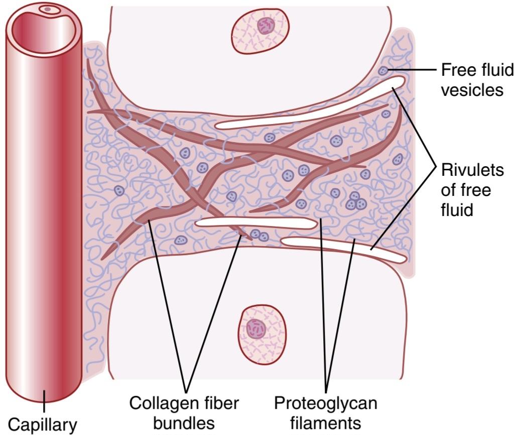
Figure 16-4. Structure of the interstitium. Proteoglycan filaments are everywhere in the spaces between the collagen fiber bundles. Free fluid vesicles and small amounts of free fluid in the form of rivulets occasionally also occur.
شکل 16-4. ساختار بینابینی. رشته های پروتئوگلیکان در همه جا در فضاهای بین بسته های فیبر کلاژن قرار دارند. گاهی اوقات وزیکول های مایع آزاد و مقادیر کمی مایع آزاد به شکل ریوولت نیز ایجاد می شود.
Gel in the Interstitium. The fluid in the interstitium is derived by filtration and diffusion from the capillaries. It contains almost the same constituents as plasma except for much lower concentrations of proteins because proteins do not easily pass outward through the pores of the capillaries. The interstitial fluid is entrapped mainly in the minute spaces among the proteoglycan filaments. This combination of proteoglycan filaments and fluid en- trapped within them has the characteristics of a gel; it is therefore called tissue gel.
ژل در بینابینی. مایع موجود در بینابینی از فیلتراسیون و انتشار از مویرگ ها به دست می آید. حاوی تقریباً همان مواد تشکیل دهنده پلاسما است، به جز غلظت بسیار کمتر پروتئین، زیرا پروتئین ها به راحتی از منافذ مویرگ ها به بیرون عبور نمی کنند. مایع بینابینی عمدتاً در فضاهای ریز بین رشته های پروتئوگلیکان به دام افتاده است. این ترکیب از رشتههای پروتئوگلیکان و مایع محبوس شده در درون آنها ویژگیهای یک ژل را دارد. بنابراین ژل بافت نامیده می شود.
Because of the large number of proteoglycan filaments, it is difficult for fluid to flow easily through the tissue gel. Instead, fluid mainly diffuses through the gel; that is, it moves molecule by molecule from one place to another by kinetic thermal motion rather than by large numbers of molecules moving together.
به دلیل تعداد زیاد رشته های پروتئوگلیکان، جریان آسان مایع در ژل بافت دشوار است. در عوض، مایع عمدتاً از طریق ژل پخش می شود. یعنی مولکول به مولکول از یک مکان به مکان دیگر با حرکت حرارتی جنبشی به جای تعداد زیادی مولکول که با هم حرکت می کنند حرکت می کند.
Diffusion through the gel occurs about 95% to 99% as rapidly as it does through free fluid. For the short distances between the capillaries and tissue cells, this diffusion allows for rapid transport through the interstitium, not only of water molecules but also of substances such as electrolytes, low-molecular-weight nutrients, cellular excreta, oxygen, and carbon dioxide.
انتشار از طریق ژل حدود 95% تا 99% به همان سرعتی که از طریق مایع آزاد انجام می شود، رخ می دهد. برای فواصل کوتاه بین مویرگ ها و سلول های بافتی، این انتشار امکان انتقال سریع از طریق بینابینی را نه تنها مولکول های آب بلکه همچنین موادی مانند الکترولیت ها، مواد مغذی با وزن مولکولی کم، مدفوع سلولی، اکسیژن و دی اکسید کربن را فراهم می کند.
Free Fluid in the Interstitium. Although almost all the fluid in the interstitium normally is entrapped within the tissue gel, occasionally small rivulets of free fluid and small free fluid vesicles are also present, which means fluid that is free of the proteoglycan molecules and therefore can flow freely. When a dye is injected into the circulating blood, it often can be seen to flow through the interstitium in the small rivulets, usually coursing along the surfaces of collagen fibers or surfaces of cells.
مایعات رایگان در بینابینی. اگرچه تقریباً تمام مایع بینابینی به طور معمول در داخل ژل بافت به دام میافتد، گاهی اوقات جریانهای کوچکی از مایع آزاد و وزیکولهای کوچک مایع آزاد نیز وجود دارند، که به معنای مایعی است که عاری از مولکولهای پروتئوگلیکان است و بنابراین میتواند آزادانه جریان یابد. هنگامی که یک رنگ به خون در گردش تزریق می شود، اغلب می توان مشاهده کرد که از طریق بینابینی در نهرهای کوچک جریان می یابد، که معمولاً در امتداد سطوح رشته های کلاژن یا سطوح سلول ها جریان دارند.
The amount of free fluid present in most normal tissues is slight, usually less than 1%. Conversely, when the tissues develop edema, these small pockets and rivulets of free fluid expand tremendously until one half or more of the edema fluid becomes free-flowing fluid, independent of the proteoglycan filaments.
مقدار مایع آزاد موجود در اکثر بافت های طبیعی اندک است، معمولاً کمتر از 1٪. برعکس، وقتی بافتها دچار ادم میشوند، این حفرههای کوچک و جویبارهای مایع آزاد بهشدت منبسط میشوند تا جایی که نیمی یا بیشتر از مایع ادم مستقل از رشتههای پروتئوگلیکان به مایع آزاد تبدیل میشود.
FLUID FILTRATION ACROSS CAPILLARIES
The hydrostatic pressure in the capillaries tends to force fluid and its dissolved substances through the capillary pores into the interstitial spaces. Conversely, osmotic pressure caused by the plasma proteins (called colloid osmotic pressure) tends to cause fluid movement by osmosis from the interstitial spaces into the blood. This osmotic pressure exerted by the plasma proteins normally prevents significant loss of fluid volume from the blood into the interstitial spaces.
فیلتراسیون سیالات در سراسر مویرگها
فشار هیدرواستاتیک در مویرگ ها تمایل دارد مایع و مواد محلول آن را از طریق منافذ مویرگی به فضاهای بینابینی وادار کند. برعکس، فشار اسمزی ناشی از پروتئینهای پلاسما (که فشار اسمزی کلوئیدی نامیده میشود) تمایل به حرکت مایع با اسمز از فضاهای بین بافتی به داخل خون دارد. این فشار اسمزی که توسط پروتئین های پلاسما اعمال می شود به طور معمول از کاهش قابل توجه حجم مایع از خون به فضاهای بینابینی جلوگیری می کند.
Also important is the lymphatic system, which returns to the circulation the small amounts of excess protein and fluid that leak from the blood into the interstitial spaces. In the remainder of this chapter, we discuss the mechanisms that control capillary filtration and lymph flow function together to regulate the respective volumes of the plasma and interstitial fluid.
همچنین سیستم لنفاوی مهم است که مقادیر کمی پروتئین و مایع اضافی را که از خون به فضاهای بینابینی نشت می کند به گردش خون باز می گرداند. در ادامه این فصل، مکانیسمهایی را مورد بحث قرار میدهیم که فیلتراسیون مویرگی و عملکرد جریان لنفاوی را با هم کنترل میکنند تا حجم مربوط به پلاسما و مایع بینابینی را تنظیم کنند.
Hydrostatic and Colloid Osmotic Forces Determine Fluid Movement Through the Capillary Membrane. Figure 16-5 shows the four primary forces that determine whether fluid will move out of the blood into the interstitial fluid or in the opposite direction. These forces, called Starling forces, were named after the physiologist Ernest Starling who first demonstrated their importance:
نیروهای اسمزی هیدرواستاتیک و کلوئیدی حرکت سیال را از طریق غشای مویرگی تعیین می کنند. شکل 16-5 چهار نیروی اولیه را نشان می دهد که تعیین می کند مایع از خون به داخل مایع بینابینی حرکت کند یا در جهت مخالف. این نیروها که نیروهای استارلینگ نامیده می شوند، به نام فیزیولوژیست ارنست استارلینگ که برای اولین بار اهمیت آنها را نشان داد، نامگذاری شدند:
1. The capillary hydrostatic pressure (Pc), which tends to force fluid outward through the capillary membrane.
۱. فشار هیدرواستاتیک مویرگی (Pc)، که تمایل دارد سیال را از طریق غشای مویرگی به بیرون فشار دهد.
2. The interstitial fluid hydrostatic pressure (Pif), which tends to force fluid inward through the cap- illary membrane when Pif is positive but outward when Pif is negative.
2. فشار هیدرواستاتیک سیال بینابینی (Pif)، که تمایل دارد سیال را به داخل غشای مویرگی در زمانی که Pif مثبت است، به سمت خارج و زمانی که Pif منفی است، به سمت بیرون هدایت می کند.
3. The capillary plasma colloid osmotic pressure (Ip), which tends to cause osmosis of fluid inward through the capillary membrane
3. فشار اسمزی کلوئیدی پلاسمای مویرگی (Ip)، که تمایل دارد باعث اسمز مایع به داخل از طریق غشای مویرگی شود.
4. The interstitial fluid colloid osmotic pressure (Пif), which tends to cause osmosis of fluid outward through the capillary membrane
4. فشار اسمزی کلوئیدی مایع بینابینی (Пif)، که تمایل دارد باعث اسمز مایع به بیرون از طریق غشای مویرگی شود.

Figure 16-5. Fluid pressure and colloid osmotic pressure forces operate at the capillary membrane and tend to move fluid outward or inward through the membrane pores.
شکل 16-5. فشار سیال و نیروهای فشار اسمزی کلوئیدی در غشای مویرگی اعمال می شوند و تمایل دارند مایع را از طریق منافذ غشا به سمت بیرون یا داخل حرکت دهند.
If the sum of these forces-the net filtration pressure- is positive, there will be a net fluid filtration across the capillaries. If the sum of the Starling forces is negative, there will be a net fluid absorption from the interstitial spaces into the capillaries. The net filtration pressure (NFP) is calculated as follows:
اگر مجموع این نیروها – فشار خالص فیلتراسیون – مثبت باشد، یک فیلتر خالص مایع در سراسر مویرگ ها وجود خواهد داشت. اگر مجموع نیروهای استارلینگ منفی باشد، جذب خالص مایع از فضاهای بین بافتی به داخل مویرگ ها وجود خواهد داشت. فشار خالص فیلتراسیون (NFP) به صورت زیر محاسبه می شود:

As discussed later, the NFP is slightly positive under normal conditions, resulting in a net filtration of fluid across the capillaries into the interstitial space in most organs. The rate of fluid filtration in a tissue is also determined by the number and size of the pores in each capillary, as well as the number of capillaries in which blood is flowing. These factors are usually expressed together as the capillary filtration coefficient (K). The Kf is therefore a measure of the capacity of the capillary membranes to filter water for a given NFP and is usually expressed as ml/ min per mm Hg NFP.
همانطور که بعداً بحث شد، NFP در شرایط عادی کمی مثبت است، که منجر به فیلتر خالص مایع در سراسر مویرگ ها به فضای بین بافتی در اکثر اندام ها می شود. سرعت فیلتراسیون مایع در بافت نیز با تعداد و اندازه منافذ در هر مویرگ و همچنین تعداد مویرگ هایی که خون در آنها جریان دارد تعیین می شود. این عوامل معمولاً با هم به عنوان ضریب فیلتراسیون مویرگی (K) بیان می شوند. بنابراین Kf اندازهگیری ظرفیت غشاهای مویرگی برای فیلتر کردن آب برای یک NFP معین است و معمولاً به صورت ml/min بر میلیمتر جیوه NFP بیان میشود.
The rate of capillary fluid filtration is therefore determined as follows:
بنابراین نرخ فیلتراسیون مایع مویرگی به صورت زیر تعیین می شود:

In the following sections, we discuss each of the forces that determine the rate of capillary fluid filtration.
در بخشهای بعدی، در مورد هر یک از نیروهایی که میزان فیلتراسیون سیال مویرگی را تعیین میکنند صحبت میکنیم.
CAPILLARY HYDROSTATIC PRESSURE
Various methods have been used to estimate the capillary hydrostatic pressure: (1) direct micropipette cannulation of the capillaries, which gives an average capillary pressure of about 25 mm Hg in some tissues, such as the skeletal muscle and gut, and (2) indirect functional measurement of the capillary pressure, which gives a capillary pressure averaging about 17 mm Hg in these tissues.
فشار هیدرواستاتیک مویرگی
روشهای مختلفی برای تخمین فشار هیدرواستاتیک مویرگی استفاده شده است: (1) کانولاسیون مستقیم مویرگها با میکروپیپت، که فشار مویرگی متوسطی در حدود 25 میلیمتر جیوه در برخی بافتها، مانند ماهیچههای اسکلتی و روده ایجاد میکند، و (2) اندازهگیری غیرمستقیم عملکردی مویرگی، که فشار H را در حدود 1 میلیمتر در بافت مویرگی افزایش میدهد.
Micropipette Method for Measuring Capillary Pressure. To measure pressure in a capillary by cannulation, a microscopic glass pipette is thrust directly into the capillary, and the pressure is measured by an appropriate micromanometer system. Using this method, capillary pressures have been measured in exposed tissues of animals and in large capillary loops of the eponychium at the base of the fingernail in humans. These measurements have given pressures of 30 to 40 mm Hg in the arterial ends of the capillaries, 10 to 15 mm Hg in the venous ends, and about 25 mm Hg in the middle.
روش میکروپیپت برای اندازه گیری فشار مویرگی. برای اندازهگیری فشار در یک مویرگ با کانولاسیون، یک پیپت شیشهای میکروسکوپی مستقیماً به داخل مویرگ رانده میشود و فشار توسط یک سیستم میکرومانومتر مناسب اندازهگیری میشود. با استفاده از این روش، فشار مویرگی در بافت های در معرض حیوانات و در حلقه های مویرگی بزرگ اپونیشیوم در پایه ناخن در انسان اندازه گیری شده است. این اندازهگیریها فشاری بین 30 تا 40 میلیمتر جیوه در انتهای شریانی مویرگها، 10 تا 15 میلیمتر جیوه در انتهای وریدی و حدود 25 میلیمتر جیوه در وسط نشان میدهد.
In some capillaries, such as the glomerular capillaries of the kidneys, the pressures measured by the micropipette method are much higher, averaging about 60 mm Hg. The peritubular capillaries of the kidneys, in contrast, have a hydrostatic pressure that averages only about 13 mm Hg. Thus, the capillary hydrostatic pressures in different tissues are highly variable, depending on the particular tissue and the physiological condition.
در برخی از مویرگ ها مانند مویرگ های گلومرولی کلیه، فشارهای اندازه گیری شده با روش میکروپیپت بسیار بیشتر و به طور متوسط حدود 60 میلی متر جیوه است. در مقابل، مویرگ های اطراف لوله کلیه ها دارای فشار هیدرواستاتیکی هستند که به طور متوسط تنها حدود 13 میلی متر جیوه است. بنابراین، فشارهای هیدرواستاتیک مویرگی در بافت های مختلف بسته به بافت خاص و شرایط فیزیولوژیکی بسیار متغیر است.
INTERSTITIAL FLUID HYDROSTATIC PRESSURE
There are several methods for measuring interstitial fluid hydrostatic pressure, each of which gives slightly different values, depending on the method used and the tissue in which the pressure is measured. In loose subcutaneous tissue, interstitial fluid pressure measured by the different methods is usually a few millimeters of mercury less than atmospheric pressure; that is, the values are called negative interstitial fluid pressure. In other tissues that are surrounded by capsules, such as the kidneys, the interstitial pressure is generally positive (i.e., greater than atmospheric pressure). The methods most widely used have been: (1) measurement of the pressure with a micropipette inserted into the tissues; (2) measurement of the pressure from implanted perforated capsules; and (3) measurement of the pressure from a cotton wick inserted into the tissue. These different methods provide different values for interstitial hydrostatic pressure, even in the same tissues.
فشار هیدرواستاتیک سیالات بینابینی
روشهای مختلفی برای اندازهگیری فشار هیدرواستاتیک مایع بینابینی وجود دارد که هر کدام بسته به روش مورد استفاده و بافتی که فشار در آن اندازهگیری میشود، مقادیر کمی متفاوت میدهد. در بافت شل زیر جلدی، فشار مایع بینابینی که با روشهای مختلف اندازهگیری میشود، معمولاً چند میلیمتر جیوه کمتر از فشار اتمسفر است. یعنی مقادیر را فشار سیال بینابینی منفی می نامند. در بافت های دیگری که توسط کپسول احاطه شده اند، مانند کلیه ها، فشار بینابینی به طور کلی مثبت است (یعنی بیشتر از فشار اتمسفر). روش هایی که به طور گسترده مورد استفاده قرار می گیرند عبارتند از: (1) اندازه گیری فشار با میکروپیپت وارد شده به بافت ها. (2) اندازه گیری فشار از کپسول های سوراخ شده کاشته شده. و (3) اندازه گیری فشار از فتیله پنبه ای وارد شده به بافت. این روشهای مختلف مقادیر متفاوتی را برای فشار هیدرواستاتیک بینابینی، حتی در همان بافتها، ارائه میکنند.
Measurement of Interstitial Fluid Pressure Using Micropipettes. The same type of micropipette used for measuring capillary pressure can also be used in some tissues for measuring interstitial fluid pressure. The tip of the micropipette is about 1 micrometer in diameter, but even this is 20 or more times larger than the sizes of the spaces between the proteoglycan filaments of the interstitium. Therefore, the pressure that is measured is probably the pressure in a free fluid pocket.
اندازه گیری فشار سیال بینابینی با استفاده از میکروپیپت. همان نوع میکروپیپت مورد استفاده برای اندازه گیری فشار مویرگی نیز می تواند در برخی از بافت ها برای اندازه گیری فشار مایع بینابینی استفاده شود. قطر نوک میکروپیپت حدود 1 میکرومتر است، اما حتی این قطر 20 بار یا بیشتر از اندازه فضاهای بین رشته های پروتئوگلیکان بینابینی است. بنابراین، فشاری که اندازهگیری میشود احتمالاً فشار در یک پاکت سیال آزاد است.
Pressures measured using the micropipette method range from -2 to +2 mm Hg in loose tissues, such as skin but, in most cases, they average slightly less than atmospheric pressure.
فشارهای اندازه گیری شده با استفاده از روش میکروپیپت از 2- تا 2+ میلی متر جیوه در بافت های شل مانند پوست متغیر است، اما در بیشتر موارد به طور متوسط کمی کمتر از فشار اتمسفر است.
Measurement of Interstitial Free Fluid Pressure in Implanted Perforated Hollow Capsules. Interstitial free fluid pressure measured when using 2-centimeter diameter capsules in normal loose subcutaneous tissue averages about -6 mm Hg but, with smaller capsules, the values are not greatly different from the -2 mm Hg measured by the micropipette.
اندازه گیری فشار مایع آزاد بینابینی در کپسول های توخالی سوراخ شده کاشته شده. فشار مایع آزاد بینابینی که هنگام استفاده از کپسولهای با قطر 2 سانتیمتر در بافت نرمال شل زیر جلدی اندازهگیری میشود، به طور متوسط حدود 6- میلیمتر جیوه است، اما در کپسولهای کوچکتر، این مقادیر تفاوت زیادی با 2- میلیمتر جیوه اندازهگیری شده توسط میکروپیپت ندارند.
Interstitial Fluid Pressures in Tightly Encased Tissues. Some tissues of the body are surrounded by tight en- casements, such as the cranial vault around the brain, the strong fibrous capsule around the kidney, the fibrous sheaths around the muscles, and the sclera around the eye. In most of these tissues, regardless of the method used for measurement, the interstitial fluid pressures are positive. However, these interstitial fluid pressures almost invariably are still less than the pressures exerted on the outsides of the tissues by their encasements. For example, the cerebrospinal fluid pressure surrounding the brain of an animal lying on its side averages about +10 mm Hg, whereas the brain interstitial fluid pressure averages about +4 to +6 mm Hg. In the kidneys, the capsular pressure surrounding the kidney averages about +13 mm Hg, whereas the reported renal interstitial fluid pressures have averaged about +6 mm Hg. Thus, if one remembers that the pressure exerted on the skin is atmospheric pressure, which is considered to be zero pressure, one might formulate a general rule that the normal interstitial fluid pressure is usually several millimeters of mercury negative with respect to the pressure that surrounds each tissue.
فشار مایعات بینابینی در بافت های محصور شده محکم. برخی از بافت های بدن با محفظه های محکم احاطه شده اند، مانند طاق جمجمه در اطراف مغز، کپسول فیبری قوی اطراف کلیه، غلاف های فیبری اطراف ماهیچه ها و صلبیه اطراف چشم. در اکثر این بافت ها، صرف نظر از روشی که برای اندازه گیری استفاده می شود، فشار مایع بین بافتی مثبت است. با این حال، این فشارهای مایع میان بافتی تقریباً همیشه کمتر از فشارهایی است که بر روی قسمتهای بیرونی بافتها توسط پوشش آنها اعمال میشود. به عنوان مثال، فشار مایع مغزی نخاعی احاطه کننده مغز حیوانی که به پهلو خوابیده است به طور متوسط حدود 10+ میلی متر جیوه است، در حالی که فشار مایع بین بافتی مغز به طور متوسط حدود 4+ تا 6+ میلی متر جیوه است. در کلیه ها، فشار کپسولی اطراف کلیه به طور متوسط حدود 13+ میلی متر جیوه است، در حالی که فشار مایع بین بافتی کلیه گزارش شده به طور متوسط حدود 6+ میلی متر جیوه است. بنابراین، اگر کسی به خاطر داشته باشد که فشار وارد شده بر روی پوست، فشار اتمسفر است که فشار صفر در نظر گرفته میشود، میتوان یک قانون کلی را فرموله کرد که فشار طبیعی مایع میان بافتی معمولاً چندین میلیمتر جیوه منفی با توجه به فشاری است که هر بافت را احاطه میکند.
In most natural cavities of the body, where there is free fluid in dynamic equilibrium with the surrounding interstitial fluids, the pressures that have been measured have been negative. Some of these cavities and pressure measurements are as follows:
در اکثر حفره های طبیعی بدن، جایی که مایع آزاد در تعادل دینامیکی با مایعات بینابینی اطراف وجود دارد، فشارهای اندازه گیری شده منفی بوده است. برخی از این حفره ها و اندازه گیری فشار به شرح زیر است:
• Intrapleural space: -8 mm Hg
• Joint synovial spaces: -4 to -6 mm Hg.
• Epidural space: -4 to -6 mm Hg
•فضای داخل پلورال: -8 میلی متر جیوه
•فضاهای سینوویال مفصلی: -4 تا -6 میلی متر جیوه.
•فضای اپیدورال: -4 تا -6 میلی متر جیوه
Summary: Interstitial Fluid Pressure in Loose Subcutaneous Tissue Usually Subatmospheric. Although the aforementioned different methods give slightly different values for interstitial fluid pressure, most physiologists believe that the interstitial fluid pressure in loose subcutaneous tissue is, in normal conditions, slightly less subatmospheric, averaging about -3 mm Hg.
خلاصه: فشار سیال بینابینی در بافت شل زیر پوستی معمولاً زیر جوی است. اگرچه روشهای مختلف فوقالذکر مقادیر کمی متفاوت برای فشار مایع میان بافتی میدهند، اکثر فیزیولوژیستها معتقدند که فشار مایع بینابینی در بافت شل زیرپوستی، در شرایط عادی، اندکی کمتر از سطح زیر اتمسفر است و به طور متوسط حدود 3- میلیمتر جیوه است.
Pumping by the Lymphatic System-Basic Cause of the Negative Interstitial Fluid Pressure. The lymphatic system is discussed later in the chapter, but first we need to understand the basic role that this system plays in determining interstitial fluid pressure. The lymphatic system is a kind of scavenger system that removes excess fluid, excess protein molecules, debris, and other matter from the tissue spaces. Normally, when fluid enters the terminal lymphatic capillaries, the lymph vessel walls automatically contract for a few seconds and pump the fluid into the blood circulation. This overall process creates the slight negative pressure that has been measured for fluid in the interstitial spaces.
پمپاژ توسط سیستم لنفاوی – علت اصلی فشار منفی مایع بینابینی. سیستم لنفاوی بعداً در این فصل مورد بحث قرار می گیرد، اما ابتدا باید نقش اساسی این سیستم در تعیین فشار مایع بین بافتی را درک کنیم. سیستم لنفاوی نوعی سیستم رفتگر است که مایع اضافی، مولکول های پروتئین اضافی، باقی مانده ها و سایر مواد را از فضاهای بافتی خارج می کند. به طور معمول، هنگامی که مایع وارد مویرگ های لنفاوی انتهایی می شود، دیواره های عروق لنفاوی به طور خودکار برای چند ثانیه منقبض می شوند و مایع را به گردش خون پمپ می کنند. این فرآیند کلی باعث ایجاد فشار منفی خفیفی می شود که برای مایع در فضاهای بینابینی اندازه گیری شده است.
PLASMA COLLOID OSMOTIC PRESSURE
فشار اسموتیک کلوئیدی پلاسما
Plasma Proteins Cause Colloid Osmotic Pressure. As discussed in Chapter 4, only the molecules or ions that fail to pass through the pores of a semipermeable membrane exert osmotic pressure. Because the proteins are the only dissolved constituents in the plasma and interstitial fluids that do not readily pass through the capillary pores, it is the proteins of the plasma and interstitial fluids that are responsible for the osmotic pressures on the two sides of the capillary membrane. To distinguish this osmotic pres- sure from that which occurs at the cell membrane, it is called colloid osmotic pressure or oncotic pressure. The term colloid osmotic pressure is derived from the fact that a protein solution resembles a colloidal solution, despite the fact that it is actually a true molecular solution.
پروتئین های پلاسما باعث فشار اسمزی کلوئیدی می شوند. همانطور که در فصل 4 بحث شد، تنها مولکول ها یا یون هایی که از منافذ یک غشای نیمه تراوا عبور نمی کنند، فشار اسمزی اعمال می کنند. از آنجایی که پروتئین ها تنها اجزای حل شده در پلاسما و مایعات بین بافتی هستند که به راحتی از منافذ مویرگی عبور نمی کنند، این پروتئین های پلاسما و مایعات بینابینی هستند که مسئول فشار اسمزی در دو طرف غشای مویرگی هستند. برای تشخیص این فشار اسمزی از فشاری که در غشای سلولی ایجاد می شود، فشار اسمزی کلوئیدی یا فشار انکوتیک نامیده می شود. اصطلاح فشار اسمزی کلوئیدی از این واقعیت ناشی می شود که یک محلول پروتئینی شبیه یک محلول کلوئیدی است، علی رغم این واقعیت که در واقع یک محلول مولکولی واقعی است.
Normal Values for Plasma Colloid Osmotic Pressure. The colloid osmotic pressure of normal human plasma averages about 28 mm Hg; 19 mm of this pressure is caused by molecular effects of the dissolved protein, and 9 mm is caused by the Donnan effect—that is, extra osmotic pressure caused by sodium, potassium, and the other cations bound to the plasma proteins.
مقادیر طبیعی فشار اسمزی کلوئیدی پلاسما. فشار اسمزی کلوئیدی پلاسمای طبیعی انسان به طور متوسط حدود 28 میلی متر جیوه است. 19 میلی متر از این فشار ناشی از اثرات مولکولی پروتئین محلول و 9 میلی متر ناشی از اثر دانان است – یعنی فشار اسمزی اضافی ناشی از سدیم، پتاسیم و سایر کاتیون های متصل به پروتئین های پلاسما.
Effect of the Different Plasma Proteins on Colloid Osmotic Pressure. The plasma proteins are a mixture that contains albumin, globulins, and fibrinogen, with an average molecular weight of 69,000, 140,000, and 400,000, respectively. Thus, 1 gram of globulin contains only half as many molecules as 1 gram of albumin, and 1 gram of fibrinogen contains only one sixth as many molecules as 1 gram of albumin. It should be recalled from the discussion of osmotic pressure in Chapter 4 that osmotic pressure is determined by the number of molecules dissolved in a fluid rather than by the mass of these molecules. The following chart gives both the relative mass concentrations (g/dl) of the different types of proteins in normal plasma and their respective contributions to the total plasma colloid osmotic pressure (Ip). These values include the Don- nan effect of ions bound to the plasma proteins:
اثر پروتئین های مختلف پلاسما بر فشار اسمزی کلوئیدی. پروتئین های پلاسما مخلوطی هستند که حاوی آلبومین، گلوبولین ها و فیبرینوژن با وزن مولکولی متوسط به ترتیب 69000، 140000 و 400000 هستند. بنابراین، 1 گرم گلوبولین حاوی تنها نیمی از مولکول های 1 گرم آلبومین است و 1 گرم فیبرینوژن دارای تنها یک ششم مولکول های 1 گرم آلبومین است. از بحث فشار اسمزی در فصل 4 باید به خاطر داشت که فشار اسمزی با تعداد مولکول های حل شده در یک سیال به جای جرم این مولکول ها تعیین می شود. نمودار زیر هم غلظت جرمی نسبی (g/dl) انواع مختلف پروتئین ها در پلاسمای نرمال و هم سهم مربوطه آنها به کل فشار اسمزی کلوئیدی پلاسما (IP) را نشان می دهد. این مقادیر شامل اثر Donnan یون های متصل به پروتئین های پلاسما است:

Thus, about 80% of the total colloid osmotic pressure of the plasma results from the albumin, 20% from the globulins, and almost none from fibrinogen. Therefore, from the point of view of capillary and tissue fluid dynamics, it is mainly albumin that is important.
بنابراین، حدود 80٪ از کل فشار اسمزی کلوئیدی پلاسما از آلبومین، 20٪ از گلوبولین ها، و تقریباً هیچ یک از فیبرینوژن ناشی نمی شود. بنابراین، از نظر دینامیک مایع مویرگی و بافت، عمدتاً آلبومین است که مهم است.
INTERSTITIAL FLUID COLLOID OSMOTIC PRESSURE
Although the size of the usual capillary pore is smaller than the molecular sizes of the plasma proteins, this is not true of all the pores. Therefore, small amounts of plasma proteins do leak into the interstitial spaces through pores and by transcytosis in small vesicles.
فشار اسموتیک کلوئیدی بینابینی
اگرچه اندازه منافذ مویرگی معمولی کوچکتر از اندازه های مولکولی پروتئین های پلاسما است، اما این در مورد همه منافذ صادق نیست. بنابراین، مقادیر کمی از پروتئین های پلاسما از طریق منافذ و با ترانس سیتوز در وزیکول های کوچک به فضاهای بینابینی نشت می کنند.
The total quantity of protein in the entire 12 liters of interstitial fluid of the body is slightly greater than the total quantity of protein in the plasma but, because this volume is four times the volume of plasma, the average protein concentration of the interstitial fluid of most tissues is usually only 40% of that in plasma, or about 3 g/ dl. Quantitatively, the average interstitial fluid colloid osmotic pressure for this concentration of proteins is about 8 mm Hg.
مقدار کل پروتئین در کل 12 لیتر مایع بین بافتی بدن کمی بیشتر از مقدار کل پروتئین در پلاسما است، اما به دلیل اینکه این حجم چهار برابر حجم پلاسما است، میانگین غلظت پروتئین مایع بین بافتی اغلب بافت ها معمولاً تنها 40 درصد آن در پلاسما یا حدود 3 گرم در لیتر است. از نظر کمی، متوسط فشار اسمزی کلوئیدی مایع بینابینی برای این غلظت از پروتئین ها حدود 8 میلی متر جیوه است.
FLUID VOLUME EXCHANGE THROUGH THE CAPILLARY MEMBRANE
The different factors affecting fluid movement through the capillary membrane have been discussed, so we can put all these factors together to see how the capillary system maintains normal fluid volume distribution between the plasma and interstitial fluid.
تبادل حجم مایع از طریق غشای مویرگی
عوامل مختلف مؤثر بر حرکت مایع از طریق غشای مویرگی مورد بحث قرار گرفتهاند، بنابراین میتوانیم همه این عوامل را کنار هم بگذاریم تا ببینیم چگونه سیستم مویرگی توزیع حجم مایع طبیعی را بین پلاسما و مایع بینابینی حفظ میکند.
The average capillary pressure at the arterial ends of the capillaries is 15 to 25 mm Hg greater than at the venous ends. Because of this difference, fluid filters out of the capillaries at their arterial ends but, at their venous ends, fluid is reabsorbed back into the capillaries (see Figure 16-3). Thus, a small amount of fluid actually “flows” through the tissues from the arterial ends of the capillaries to the venous ends. The dynamics of this flow are as follows.
میانگین فشار مویرگی در انتهای شریانی مویرگ ها 15 تا 25 میلی متر جیوه بیشتر از انتهای وریدی است. به دلیل این تفاوت، مایع از مویرگ ها در انتهای شریانی آنها فیلتر می شود، اما در انتهای وریدی آنها، مایع دوباره به داخل مویرگ ها جذب می شود (شکل 16-3 را ببینید). بنابراین، مقدار کمی مایع در واقع از طریق بافت ها از انتهای شریانی مویرگ ها به انتهای وریدی “جریان می یابد”. پویایی این جریان به شرح زیر است.
Analysis of the Forces Causing Filtration at the Arterial End of the Capillary. The approximate average forces operative at the arterial end of the capillary that cause movement through the capillary membrane are shown as follows:
تجزیه و تحلیل نیروهای ایجاد کننده فیلتراسیون در انتهای شریانی مویرگ. میانگین تقریبی نیروهای اعمال شده در انتهای شریانی مویرگ که باعث حرکت در غشای مویرگی می شود به شرح زیر نشان داده شده است:

Thus, the summation of forces at the arterial end of the capillary shows a net filtration pressure of 13 mm Hg, tending to move fluid outward through the capillary pores. This 13 mm Hg filtration pressure causes, on average, about 1/200 of the plasma in the flowing blood to filter out of the arterial ends of the capillaries into the interstitial spaces each time the blood passes through the capillaries.
بنابراین، مجموع نیروها در انتهای شریانی مویرگ، فشار خالص فیلتراسیون 13 میلی متر جیوه را نشان می دهد، که تمایل دارد مایع را از طریق منافذ مویرگی به سمت خارج حرکت دهد. این فشار فیلتراسیون 13 میلیمتر جیوه باعث میشود که هر بار که خون از مویرگها عبور میکند، بهطور میانگین حدود 1/200 پلاسمای خون جاری از انتهای شریانی مویرگها به فضاهای بینابینی فیلتر میشود.
Analysis of Reabsorption at the Venous End of the Capillary. The low blood pressure at the venous end of the capillary changes the balance of forces in favor of absorption as follows:
تجزیه و تحلیل بازجذب در انتهای وریدی مویرگ. فشار خون پایین در انتهای وریدی مویرگی تعادل نیروها را به نفع جذب به شرح زیر تغییر می دهد:
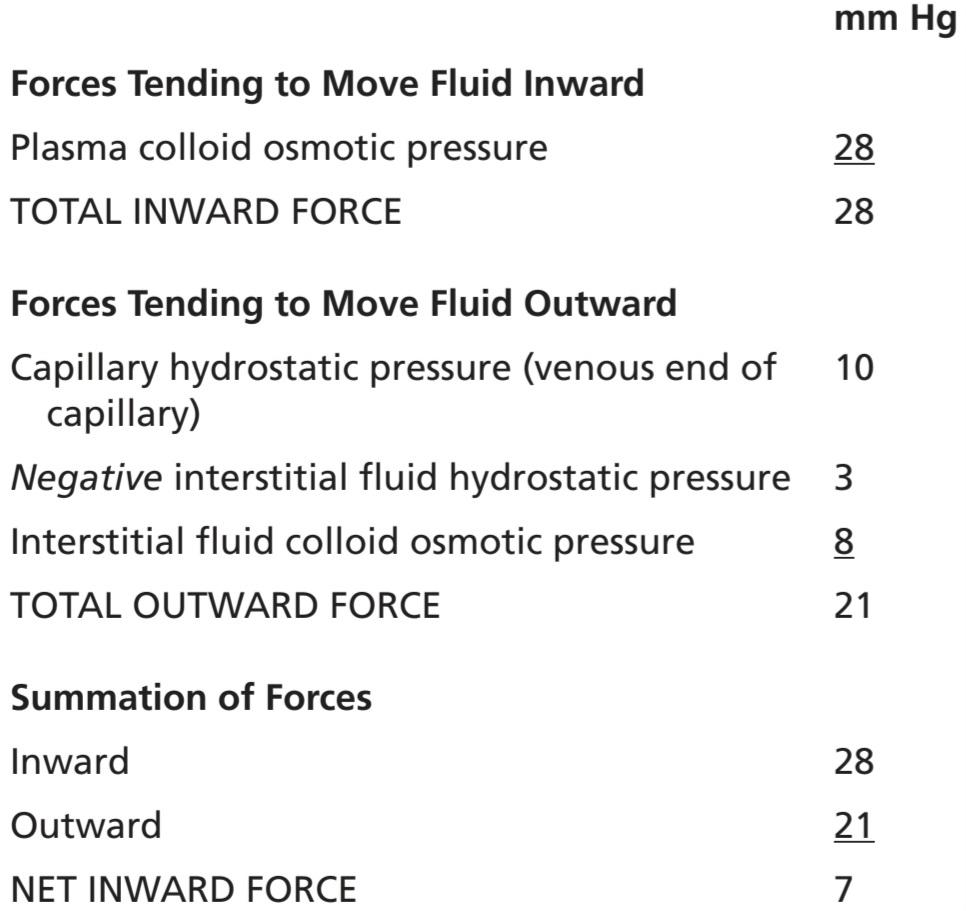
Thus, there is a net reabsorption pressure of 7 mm Hg at the venous ends of the capillaries. This reabsorption pressure is considerably less than the filtration pressure at the capillary arterial ends, but remember that the venous capillaries are more numerous and more permeable than the arterial capillaries. Thus less reabsorption pressure is required to cause inward movement of fluid.
بنابراین، فشار خالص بازجذب 7 میلی متر جیوه در انتهای وریدی مویرگ ها وجود دارد. این فشار بازجذب به طور قابل توجهی کمتر از فشار فیلتراسیون در انتهای شریان های مویرگی است، اما به یاد داشته باشید که مویرگ های وریدی از مویرگ های شریانی پرتعدادتر و نفوذپذیرتر هستند. بنابراین فشار بازجذب کمتری برای ایجاد حرکت مایع به داخل لازم است.
The reabsorption pressure causes about nine tenths of the fluid that has filtered out of the arterial ends of the capillaries to be reabsorbed at the venous ends. The remaining one tenth flows into the lymph vessels and returns to the circulating blood.
فشار بازجذب باعث می شود حدود نه دهم مایعی که از انتهای شریانی مویرگ ها فیلتر شده است در انتهای وریدی دوباره جذب شود. یک دهم باقی مانده به رگ های لنفاوی جریان می یابد و به خون در گردش باز می گردد.
STARLING EQUILIBRIUM FOR CAPILLARY EXCHANGE
Ernest Starling pointed out more than a century ago that under normal conditions, a state of near-equilibrium exists in most capillaries. That is, the amount of fluid filtering outward from the arterial ends of capillaries almost exactly equals the fluid returned to the circulation by absorption. The slight disequilibrium that does occur accounts for the fluid that is eventually returned to the circulation by way of the lymphatics.
تعادل خیره شدن برای تبادل مویرگی
ارنست استارلینگ بیش از یک قرن پیش اشاره کرد که در شرایط عادی، وضعیت تقریباً تعادل در اکثر مویرگ ها وجود دارد. یعنی مقدار مایعی که از انتهای شریانی مویرگ ها به سمت خارج فیلتر می شود تقریباً دقیقاً برابر با مایعی است که با جذب به گردش خون باز می گردد. عدم تعادل خفیفی که اتفاق می افتد به دلیل مایعی است که در نهایت از طریق غدد لنفاوی به گردش خون باز می گردد.
The following chart shows the principles of the Starling equilibrium. For this chart, the pressures in the arterial and venous capillaries are averaged to calculate mean the functional capillary pressure for the entire length of the capillary. This mean functional capillary pressure is calculated to be 17.3 mm Hg.
نمودار زیر اصول تعادل استارلینگ را نشان می دهد. برای این نمودار، فشارها در مویرگ های شریانی و وریدی به طور متوسط محاسبه می شود تا میانگین فشار مویرگی عملکردی برای کل طول مویرگ محاسبه شود. این فشار عملکردی متوسط مویرگی 17.3 میلی متر جیوه محاسبه می شود.


Thus, for the total capillary circulation, we find a nearequilibrium between the total outward forces, 28.3 mm Hg, and the total inward force, 28.0 mm Hg. This slight imbalance of forces, 0.3 mm Hg, causes slightly more filtration of fluid into the interstitial spaces than reabsorption. This slight excess of filtration is called net filtration, and it is the fluid that must be returned to the circulation through the lymphatics. The normal rate of net filtration in the entire body, not including the kidneys, is only about 2 ml/min.
بنابراین، برای گردش کل مویرگی، ما یک تعادل تقریباً بین کل نیروهای بیرونی، 28.3 میلی متر جیوه، و کل نیروی داخل، 28.0 میلی متر جیوه پیدا می کنیم. این عدم تعادل جزئی نیروها، 0.3 میلیمتر جیوه، کمی بیشتر از جذب مجدد مایع به داخل فضاهای بین بافتی فیلتر میشود. این مقدار جزئی فیلتراسیون، فیلتراسیون خالص نامیده می شود و این مایعی است که باید از طریق لنفاوی به گردش خون بازگردانده شود. سرعت طبیعی فیلتراسیون خالص در کل بدن، بدون احتساب کلیه ها، تنها حدود 2 میلی لیتر در دقیقه است.
CAPILLARY FILTRATION COEFFICIENT
In the previous example, an average net imbalance of forces at the capillary membranes of 0.3 mm Hg causes net fluid filtration in the entire body of 2 ml/min. Expressing the net fluid filtration rate for each mm Hg imbalance, one finds a net filtration rate of 6.67 ml/min of fluid per mm Hg for the entire body. This value is called the whole body capillary filtration coefficient.
ضریب فیلتراسیون مویرگی
در مثال قبلی، عدم تعادل خالص نیروها در غشاهای مویرگی 0.3 میلی متر جیوه باعث فیلتراسیون خالص مایع در کل بدن 2 میلی لیتر در دقیقه می شود. با بیان نرخ خالص فیلتراسیون مایع برای هر عدم تعادل میلیمتر جیوه، نرخ تصفیه خالص 6.67 میلیلیتر در دقیقه مایع در هر میلیمتر جیوه برای کل بدن وجود دارد. این مقدار ضریب فیلتراسیون مویرگی کل بدن نامیده می شود.
The filtration coefficient can also be expressed for separate parts of the body in terms of the rate of filtration per minute per mm Hg per 100 grams of tissue. On this basis, the capillary filtration coefficient of the average tissue is about 0.01 ml/min per mm Hg per 100 g of tissue. How- ever, because of extreme differences in permeabilities and surface areas of the capillary systems in different tissues, this coefficient varies more than 100-fold among the different tissues. It is very small in brain and muscle, moderately large in subcutaneous tissue, large in the intestine, and extremely large in the liver and glomerulus of the kidney, where the capillary surface is large, and the pores are numerous or wide open. By the same token, the permeation of proteins through the capillary membranes also varies greatly. The concentration of protein in the interstitial fluid of muscles is about 1.5 g/dl; in subcutaneous tissue, it is 2 g/dl; in the intestine, it is 4 g/dl; and, in the liver, it is 6 g/dl.
همچنین ضریب فیلتراسیون را می توان برای قسمت های جداگانه بدن بر حسب نرخ فیلتراسیون در دقیقه بر میلی متر جیوه در هر 100 گرم بافت بیان کرد. بر این اساس، ضریب فیلتراسیون مویرگی بافت متوسط حدود 0.01 میلی لیتر در دقیقه در هر میلی متر جیوه به ازای هر 100 گرم بافت است. با این حال، به دلیل تفاوت های شدید در نفوذپذیری و سطح سیستم های مویرگی در بافت های مختلف، این ضریب بیش از 100 برابر در بین بافت های مختلف متفاوت است. از نظر مغز و ماهیچه بسیار کوچک، در بافت زیر جلدی نسبتاً بزرگ، در روده بزرگ و در کبد و گلومرول کلیه بسیار بزرگ است، جایی که سطح مویرگها بزرگ است و منافذ متعدد یا کاملاً باز هستند. به همین ترتیب، نفوذ پروتئین ها از طریق غشاهای مویرگی نیز بسیار متفاوت است. غلظت پروتئین در مایع بین بافتی عضلات حدود 1.5 گرم در دسی لیتر است. در بافت زیر جلدی 2 گرم در دسی لیتر است. در روده 4 گرم در دسی لیتر است. و در کبد 6 گرم در دسی لیتر است.
Effect of Abnormal Imbalance of Forces at the Capillary Membrane. If the mean capillary pressure rises significantly above the average value of 17 mm Hg, the net force tending to cause filtration of fluid into the tissue spaces rises. Thus, a 20-mm Hg rise in mean capillary pressure causes an increase in net filtration pressure from 0.3 to 20.3 mm Hg, which results in 68 times as much net filtration of fluid into the interstitial spaces as normally occurs. To prevent accumulation of excess fluid in these spaces would require 68 times the normal flow of fluid into the lymphatic system, an amount that is 2 to 5 times too much for the lymphatics to carry away. As a result, fluid will begin to accumulate in the interstitial spaces and edema will result.
اثر عدم تعادل غیرعادی نیروها در غشای مویرگی. اگر فشار متوسط مویرگی به طور قابل توجهی بالاتر از مقدار متوسط 17 میلی متر جیوه باشد، نیروی خالصی که تمایل به فیلتر کردن مایع به داخل فضاهای بافت دارد افزایش می یابد. بنابراین، افزایش 20 میلیمتر جیوه در فشار متوسط مویرگی باعث افزایش فشار خالص فیلتراسیون از 0.3 به 20.3 میلیمتر جیوه میشود که منجر به 68 برابر فیلتراسیون خالص مایع به فضاهای بینابینی نسبت به حالت عادی میشود. برای جلوگیری از تجمع مایع اضافی در این فضاها، نیاز به 68 برابر جریان طبیعی مایع به سیستم لنفاوی است، مقداری که 2 تا 5 برابر بیش از حد برای انتقال لنفاوی است. در نتیجه، مایع در فضاهای بینابینی شروع به تجمع می کند و ادم ایجاد می شود.
Conversely, if the capillary pressure falls very low, net reabsorption of fluid into the capillaries will occur instead of net filtration, and the blood volume will increase at the expense of the interstitial fluid volume. These effects of imbalance at the capillary membrane in relationship to the development of the different types of edema are discussed in Chapter 25.
برعکس، اگر فشار مویرگی بسیار کم شود، به جای فیلتراسیون خالص، بازجذب خالص مایع به داخل مویرگها اتفاق میافتد و حجم خون به هزینه حجم مایع بینابینی افزایش مییابد. این اثرات عدم تعادل در غشای مویرگی در رابطه با ایجاد انواع مختلف ادم در فصل 25 مورد بحث قرار گرفته است.
LYMPHATIC SYSTEM
The lymphatic system represents an accessory route through which fluid can flow from the interstitial spaces into the blood. Most importantly, the lymphatics can carry proteins and large particulate matter away from the tissue spaces, neither of which can be removed by absorption directly into the blood capillaries. This return of proteins to the blood from the interstitial spaces is an essential function, without which we would die within about 24 hours.
سیستم لنفاوی
سیستم لنفاوی یک مسیر کمکی را نشان می دهد که از طریق آن مایع می تواند از فضاهای بینابینی به خون جریان یابد. مهمتر از همه، غدد لنفاوی میتوانند پروتئینها و ذرات بزرگ را از فضاهای بافتی دور کنند، که هیچکدام نمیتوانند با جذب مستقیم به مویرگهای خونی خارج شوند. این بازگشت پروتئین ها به خون از فضاهای بینابینی یک عملکرد ضروری است که بدون آن ما در عرض 24 ساعت می میریم.
LYMPH CHANNELS OF THE BODY
Almost all tissues of the body have special lymph channels that drain excess fluid directly from the interstitial spaces. The exceptions include the superficial portions of the skin, central nervous system, endomysium of muscles, and bones. However, even these tissues have minute interstitial channels called prelymphatics through which interstitial fluid can flow; this fluid eventually empties into lymphatic vessels or, in the case of the brain, into the cerebrospinal fluid and then directly back into the blood.
کانال های لنفاوی بدن
تقریباً تمام بافتهای بدن دارای کانالهای لنفاوی خاصی هستند که مایع اضافی را مستقیماً از فضاهای بینابینی تخلیه میکنند. استثناها شامل بخش های سطحی پوست، سیستم عصبی مرکزی، اندومیزیوم ماهیچه ها و استخوان ها هستند. با این حال، حتی این بافتها دارای کانالهای بینابینی کوچکی به نام پیشلنفاتیک هستند که مایع میان بافتی میتواند از طریق آنها جریان یابد. این مایع در نهایت به عروق لنفاوی یا در مورد مغز به مایع مغزی نخاعی تخلیه می شود و سپس مستقیماً به خون باز می گردد.
Essentially all the lymph vessels from the lower part of the body eventually empty into the thoracic duct, which in turn empties into the blood venous system at the juncture of the left internal jugular vein and left subclavian vein, as shown in Figure 16-6.
اساساً تمام عروق لنفاوی از قسمت تحتانی بدن در نهایت به داخل مجرای قفسه سینه تخلیه می شوند، که به نوبه خود در محل اتصال ورید ژوگولار داخلی چپ و ورید ساب کلاوین چپ به سیستم وریدی خون تخلیه می شود، همانطور که در شکل 16-6 نشان داده شده است.
Lymph from the left side of the head, left arm, and parts of the chest region also enters the thoracic duct before it empties into the veins.
لنف سمت چپ سر، بازوی چپ و قسمتهایی از قفسه سینه نیز قبل از تخلیه به داخل سیاهرگها وارد مجرای قفسه سینه میشود.
Lymph from the right side of the neck and head, right arm, and parts of the right thorax enters the right lymph duct (much smaller than the thoracic duct), which empties into the blood venous system at the juncture of the right subclavian vein and internal jugular vein.
لنف سمت راست گردن و سر، بازوی راست و قسمتهایی از قفسه سینه راست وارد مجرای لنفاوی راست (بسیار کوچکتر از مجرای سینهای) میشود که در محل اتصال ورید ساب کلاوین راست و ورید ژوگولار داخلی به سیستم وریدی خون میریزد.

Figure 16-6. The lymphatic system.
شکل 16-6. سیستم لنفاوی.
Terminal Lymphatic Capillaries and Their Permeability. Most of the fluid filtering from the arterial ends of blood capillaries flows among the cells and finally is reabsorbed back into the venous ends of the blood capillaries but, on average, about one tenth of the fluid instead enters the lymphatic capillaries and returns to the blood through the lymphatic system rather than through the venous capillaries. The total quantity of all this lymph is normally only 2 to 3 L/day.
مویرگهای لنفاوی انتهایی و نفوذپذیری آنها. بیشتر مایع فیلتر شده از انتهای شریانی مویرگهای خون در میان سلولها جریان مییابد و در نهایت دوباره به انتهای وریدی مویرگهای خون بازجذب میشود، اما به طور متوسط، حدود یک دهم مایع در عوض وارد مویرگهای لنفاوی میشود و از طریق سیستم لنفاوی به جای مویرگهای وریدی به خون باز میگردد. مقدار کل این لنف معمولاً فقط 2 تا 3 لیتر در روز است.
The fluid that returns to the circulation by way of the lymphatics is extremely important because substances of high molecular weight, such as proteins, cannot be absorbed from the tissues in any other way, although they can enter the lymphatic capillaries almost unimpeded. The reason for this mechanism is a special structure of the lymphatic capillaries, demonstrated in Figure 16-7. This figure shows the endothelial cells of the lymphatic capillary attached by anchoring filaments to the surrounding connective tissue. At the junctions of adjacent endothelial cells, the edge of one endothelial cell overlaps the edge of the adjacent cell in such a way that the overlapping edge is free to flap inward, thus forming a minute valve that opens to the interior of the lymphatic capillary. Interstitial fluid, along with its suspended particles, can push the valve open and flow directly into the lymphatic capillary. However, this fluid has difficulty leaving the capillary once it has entered because any backflow closes the flap valve. Thus, the lymphatics have valves at the very tips of the terminal lymphatic capillaries, as well as valves along their larger vessels, up to the point where they empty into the blood circulation.
مایعی که از طریق غدد لنفاوی به گردش خون باز می گردد بسیار مهم است زیرا مواد با وزن مولکولی بالا مانند پروتئین ها به هیچ وجه نمی توانند از بافت ها جذب شوند، اگرچه می توانند تقریباً بدون مانع وارد مویرگ های لنفاوی شوند. دلیل این مکانیسم ساختار خاصی از مویرگ های لنفاوی است که در شکل 16-7 نشان داده شده است. این شکل سلول های اندوتلیال مویرگ لنفاوی را نشان می دهد که توسط رشته های لنگر به بافت همبند اطراف متصل شده اند. در محل اتصال سلول های اندوتلیال مجاور، لبه یک سلول اندوتلیال روی لبه سلول مجاور همپوشانی دارد، به گونه ای که لبه همپوشانی آزاد است تا به سمت داخل فلپ شود، بنابراین یک دریچه دقیقه ای تشکیل می شود که به سمت داخل مویرگ لنفاوی باز می شود. مایع بینابینی، همراه با ذرات معلق آن، می تواند دریچه را باز کند و مستقیماً به مویرگ لنفاوی جریان یابد. با این حال، این مایع پس از ورود به مویرگی با مشکل خارج می شود زیرا هر جریان برگشتی دریچه فلپ را می بندد. بنابراین، غدد لنفاوی دارای دریچههایی در انتهای مویرگهای لنفاوی انتهایی و همچنین دریچههایی در امتداد عروق بزرگتر خود تا جایی که به گردش خون تخلیه میشوند، دارند.

Figure 16-7. Special structure of the lymphatic capillaries that permits passage of substances of high molecular weight into the lymph.
شکل 16-7. ساختار ویژه مویرگ های لنفاوی که اجازه عبور مواد با وزن مولکولی بالا را به داخل لنف می دهد.
FORMATION OF LYMPH
Lymph is derived from interstitial fluid that flows into the lymphatics. Therefore, lymph as it first enters the terminal lymphatics has almost the same composition as the interstitial fluid.
تشکیل لنف
لنف از مایع بینابینی که به غدد لنفاوی می ریزد مشتق می شود. بنابراین، لنف زمانی که برای اولین بار وارد غدد لنفاوی انتهایی می شود، تقریباً ترکیبی مشابه مایع بینابینی دارد.
The protein concentration in the interstitial fluid of most tissues averages about 2 g/dl, and the protein concentration of lymph flowing from these tissues is near this value. Lymph formed in the liver has a protein concentration as high as 6 g/dl, and lymph formed in the intestines has a protein concentration as high as 3 to 4 g/dl. Because about two thirds of all lymph normally is derived from the liver and intestines, the thoracic duct lymph, which is a mixture of lymph from all areas of the body, usually has a protein concentration of 3 to 5 g/dl.
غلظت پروتئین در مایع بینابینی اکثر بافت ها به طور متوسط حدود 2 گرم در دسی لیتر است و غلظت پروتئین لنفاوی جاری از این بافت ها نزدیک به این مقدار است. لنف تشکیل شده در کبد دارای غلظت پروتئین به اندازه 6 گرم در دسی لیتر است و لنف تشکیل شده در روده دارای غلظت پروتئینی به میزان 3 تا 4 گرم در دسی لیتر است. از آنجایی که حدود دو سوم کل لنف به طور معمول از کبد و روده مشتق می شود، لنف مجرای قفسه سینه، که مخلوطی از لنف از تمام نواحی بدن است، معمولاً دارای غلظت پروتئین 3 تا 5 گرم در دسی لیتر است.
The lymphatic system is also one of the major routes for absorption of nutrients from the gastrointestinal tract, especially for absorption of virtually all fats in food, as discussed in Chapter 66. After a fatty meal, thoracic duct lymph sometimes contains as much as 1% to 2% fat.
سیستم لنفاوی همچنین یکی از راه های اصلی برای جذب مواد مغذی از دستگاه گوارش است، به ویژه برای جذب تقریباً تمام چربی های موجود در غذا، همانطور که در فصل 66 بحث شد.
Finally, even large particles, such as bacteria, can push their way between the endothelial cells of the lymphatic capillaries and in this way enter the lymph. As the lymph passes through the lymph nodes, these particles are almost entirely removed and destroyed, as discussed in Chapter 34.
در نهایت، حتی ذرات بزرگ، مانند باکتری، می توانند راه خود را بین سلول های اندوتلیال مویرگ های لنفاوی فشار داده و از این طریق وارد لنف شوند. همانطور که در فصل 34 بحث شد، همانطور که لنف از غدد لنفاوی عبور می کند، این ذرات تقریباً به طور کامل حذف و از بین می روند.
RATE OF LYMPH FLOW
About 100 ml/hr of lymph flows through the thoracic duct of a resting human, and approximately another 20 ml flows into the circulation each hour through other channels, making a total estimated lymph flow of about 120 ml/hr or 2 to 3 L/day.
سرعت جریان لنفاوی
حدود 100 میلی لیتر در ساعت لنف از طریق مجرای قفسه سینه یک انسان در حال استراحت جریان دارد و تقریباً 20 میلی لیتر دیگر در هر ساعت از طریق کانال های دیگر وارد گردش خون می شود که مجموع جریان لنفاوی تخمینی حدود 120 میلی لیتر در ساعت یا 2 تا 3 لیتر در روز است.
Effect of Interstitial Fluid Pressure on Lymph Flow. Figure 16-8 shows the effect of different levels of interstitial fluid hydrostatic pressure on lymph flow, as measured in animals. Note that normal lymph flow is very little at interstitial fluid pressures more negative than the normal value of -6 mm Hg. Then, as the pressure rises to 0 mm Hg (atmospheric pressure), flow increases more than 20- fold. Therefore, any factor that increases interstitial fluid pressure also increases lymph flow if the lymph vessels are functioning normally. Such factors include the following:
تأثیر فشار مایع بینابینی بر جریان لنفاوی. شکل 16-8 تأثیر سطوح مختلف فشار هیدرواستاتیک مایع بینابینی را بر جریان لنفاوی نشان می دهد که در حیوانات اندازه گیری شده است. توجه داشته باشید که جریان لنفاوی طبیعی در فشارهای مایع بینابینی منفی تر از مقدار طبیعی -6 میلی متر جیوه است. سپس، با افزایش فشار به 0 میلی متر جیوه (فشار اتمسفر)، جریان بیش از 20 برابر افزایش می یابد. بنابراین، هر عاملی که فشار مایع بینابینی را افزایش دهد، اگر عروق لنفاوی به طور طبیعی کار کنند، جریان لنفاوی را نیز افزایش می دهد. چنین عواملی شامل موارد زیر است:
• Elevated capillary hydrostatic pressure
• Decreased plasma colloid osmotic pressure
• Increased interstitial fluid colloid osmotic pressure
• Increased permeability of the capillaries
• افزایش فشار هیدرواستاتیک مویرگی
• کاهش فشار اسمزی کلوئیدی پلاسما
• افزایش فشار اسمزی کلوئیدی مایع بینابینی
• افزایش نفوذپذیری مویرگها
All these factors favor net fluid movement into the interstitium, thus increasing interstitial fluid volume, interstitial fluid pressure, and lymph flow all at the same time.
همه این عوامل به حرکت خالص مایع به داخل بینابینی کمک می کنند، بنابراین حجم مایع بین بافتی، فشار مایع بین بافتی و جریان لنف را به طور همزمان افزایش می دهند.
However, note in Figure 16-8 that when the interstitial fluid hydrostatic pressure becomes 1 or 2 mm Hg greater than atmospheric pressure (>0 mm Hg), lymph flow fails to rise any further at still higher pressures. This results from the fact that the increasing tissue pressure not only increases entry of fluid into the lymphatic capillaries, but also compresses the outside surfaces of the larger lymphatics, thus impeding lymph flow. At the higher pressures, these two factors balance each other, so lymph flow reaches a maximum flow rate. This maxi- mum flow rate is illustrated by the upper level plateau in Figure 16-8.
با این حال، در شکل 16-8 توجه داشته باشید که وقتی فشار هیدرواستاتیک مایع بینابینی 1 یا 2 میلیمتر جیوه بیشتر از فشار اتمسفر (بیش از 0 میلیمتر جیوه) میشود، جریان لنفاوی در فشارهای باالتر بیشتر نمیشود. این نتیجه از این واقعیت است که افزایش فشار بافت نه تنها باعث افزایش ورود مایع به مویرگهای لنفاوی میشود، بلکه سطوح خارجی غدد لنفاوی بزرگتر را نیز فشرده میکند و در نتیجه جریان لنفاوی را مختل میکند. در فشارهای بالاتر، این دو عامل یکدیگر را متعادل می کنند، بنابراین جریان لنفاوی به حداکثر سرعت جریان می رسد. این حداکثر سرعت جریان توسط فلات سطح بالایی در شکل 16-8 نشان داده شده است.

Figure 16-8. Relationship between interstitial fluid pressure and lymph flow in the leg of a dog. Note that lymph flow reaches a maximum when the interstitial pressure (Pi) rises slightly above atmospheric pressure (0 mm Hg). (Courtesy Dr. Harry Gibson and Dr. Aubrey Taylor.)
شکل 16-8. رابطه بین فشار مایع بینابینی و جریان لنفاوی در پای سگ توجه داشته باشید که جریان لنفاوی زمانی به حداکثر می رسد که فشار میان بافتی (Pi) کمی بالاتر از فشار اتمسفر (0 میلی متر جیوه) باشد. (با احترام از دکتر هری گیبسون و دکتر اوبری تیلور.)
Lymphatic Pump Increases Lymph Flow. Valves exist in all lymph channels. Figure 16-9 shows typical valves for collecting lymphatics into which the lymphatic capillaries empty.
پمپ لنفاوی جریان لنفاوی را افزایش می دهد. دریچه ها در تمام کانال های لنفاوی وجود دارند. شکل 16-9 دریچه های معمولی را برای جمع آوری غدد لنفاوی نشان می دهد که مویرگ های لنفاوی داخل آن خالی می شوند.
Videos of exposed lymph vessels in animals and in humans have shown that when a collecting lymphatic or larger lymph vessel becomes stretched with fluid, the smooth muscle in the wall of the vessel automatically contracts. Furthermore, each segment of the lymph vessel between successive valves functions as a separate automatic pump. That is, even slight filling of a segment causes it to contract, and the fluid is pumped through the next valve into the next lymphatic segment. This fluid fills the subsequent segment and a few seconds later it also contracts, with the process continuing all along the lymph vessel until the fluid is finally emptied into the blood circulation. In a very large lymph vessel, such as the thoracic duct, this lymphatic pump can generate pressure as high as 50 to 100 mm Hg.
ویدئوهای رگ های لنفاوی در معرض در حیوانات و انسان ها نشان داده است که وقتی یک رگ لنفاوی جمع کننده یا بزرگتر با مایع کشیده می شود، ماهیچه صاف در دیواره رگ به طور خودکار منقبض می شود. علاوه بر این، هر بخش از رگ لنفاوی بین دریچه های متوالی به عنوان یک پمپ خودکار جداگانه عمل می کند. یعنی حتی پر شدن جزئی یک قطعه باعث انقباض آن می شود و مایع از طریق دریچه بعدی به بخش لنفاوی بعدی پمپ می شود. این مایع قسمت بعدی را پر می کند و چند ثانیه بعد نیز منقبض می شود و این روند در تمام طول رگ لنفاوی ادامه می یابد تا در نهایت مایع به گردش خون تخلیه شود. در یک رگ لنفاوی بسیار بزرگ، مانند مجرای قفسه سینه، این پمپ لنفاوی می تواند فشاری بین 50 تا 100 میلی متر جیوه ایجاد کند.

Figure 16-9. Structure of lymphatic capillaries and a collecting lymphatic, with the lymphatic valves also shown.
شکل 16-9. ساختار مویرگ های لنفاوی و یک لنفاوی جمع کننده با دریچه های لنفاوی نیز نشان داده شده است.
Pumping Caused by External Intermittent Compression of the Lymphatics. In addition to the pumping caused by intrinsic intermittent contraction of the lymph vessel walls, any external factor that intermittently compresses the lymph vessel can also cause pumping. In order of their importance, such factors are as follows:
پمپاژ ناشی از فشرده سازی متناوب خارجی غدد لنفاوی. علاوه بر پمپاژ ناشی از انقباض متناوب ذاتی دیواره عروق لنفاوی، هر عامل خارجی که به طور متناوب رگ لنفاوی را تحت فشار قرار دهد نیز می تواند باعث پمپاژ شود. این عوامل به ترتیب اهمیت به شرح زیر است:
• Contraction of surrounding skeletal muscles
• Movement of the parts of the body
• Pulsations of arteries adjacent to the lymphatics
• Compression of the tissues by objects outside the body
• انقباض عضلات اسکلتی اطراف
• حرکت اعضای بدن
• نبضهای شریانهای مجاور مجرای لنفاوی
• فشرده سازی بافت ها توسط اجسام خارج از بدن
The lymphatic pump becomes very active during exercise, often increasing lymph flow 10- to 30-fold. Conversely, during periods of rest, lymph flow is sluggish (almost zero).
پمپ لنفاوی در طول ورزش بسیار فعال می شود و اغلب جریان لنفاوی را 10 تا 30 برابر افزایش می دهد. برعکس، در طول دوره های استراحت، جریان لنفاوی کند است (تقریباً صفر).
Lymphatic Capillary Pump. The terminal lymphatic capillary is also capable of pumping lymph, in addition to the pumping by the larger lymph vessels. As explained earlier in the chapter, the anchoring filaments on the walls of the lymphatic capillaries tightly adhere to the surrounding tissue cells. Therefore, each time excess fluid enters the tissue and causes the tissue to swell, the anchoring filaments pull on the wall of the lymphatic capillary, and fluid flows into the terminal lymphatic capillary through the junctions between the endothelial cells. Then, when the tissue is compressed, the pressure inside the capillary increases and causes the overlapping edges of the endothelial cells to close like valves. Therefore, the pressure pushes the lymph forward into the collecting lymphatic instead of backward through the cell junctions.
پمپ مویرگی لنفاوی. مویرگ لنفاوی انتهایی همچنین قادر به پمپاژ لنف، علاوه بر پمپاژ توسط عروق لنفاوی بزرگتر است. همانطور که قبلا در این فصل توضیح داده شد، رشته های لنگر بر روی دیواره های مویرگ های لنفاوی محکم به سلول های بافت اطراف می چسبند. بنابراین، هر بار که مایع اضافی وارد بافت می شود و باعث متورم شدن بافت می شود، رشته های لنگر بر روی دیواره مویرگ لنفاوی می کشند و مایع از طریق اتصالات بین سلول های اندوتلیال به مویرگ لنفاوی انتهایی جریان می یابد. سپس، هنگامی که بافت فشرده می شود، فشار داخل مویرگ افزایش می یابد و باعث می شود که لبه های روی هم افتاده سلول های اندوتلیال مانند دریچه ها بسته شوند. بنابراین، فشار، لنف را به سمت جلو به سمت لنفاوی جمعآوریکننده هل میدهد، نه اینکه از طریق اتصالات سلولی به عقب برود.
The lymphatic capillary endothelial cells also contain a few contractile actomyosin filaments. In some animal tis- sues (e.g., a bat wing), these filaments have been observed to cause rhythmical contraction of the lymphatic capillaries in the same rhythmic way that many of the small blood vessels and larger lymphatic vessels contract. Therefore, it is probable that at least part of lymph pumping results from lymph capillary endothelial cell contraction in addition to contraction of the larger muscular lymphatics.
سلول های اندوتلیال مویرگی لنفاوی همچنین حاوی چند رشته اکتومیوزین انقباضی هستند. در برخی بافتهای حیوانی (مانند بال خفاش)، مشاهده شده است که این رشتهها باعث انقباض ریتمیک مویرگهای لنفاوی میشوند به همان شیوه ریتمیک که بسیاری از رگهای خونی کوچک و عروق لنفاوی بزرگتر منقبض میشوند. بنابراین، این احتمال وجود دارد که حداقل بخشی از پمپاژ لنف ناشی از انقباض سلول های اندوتلیال مویرگی لنفاوی علاوه بر انقباض لنفاوی عضلانی بزرگتر باشد.
Summary of Factors That Determine Lymph Flow. From the previous discussion, one can see that the two primary factors that determine lymph flow are (1) the interstitial fluid pressure and (2) the activity of the lymphatic pump. Therefore, one can state that, roughly, the rate of lymph flow is determined by the product of interstitial fluid pressure times the activity of the lymphatic pump.
خلاصه عواملی که جریان لنف را تعیین می کنند. از بحث قبلی، می توان دریافت که دو عامل اصلی که جریان لنف را تعیین می کنند عبارتند از (1) فشار مایع بینابینی و (2) فعالیت پمپ لنفاوی. بنابراین، می توان گفت که تقریباً میزان جریان لنفاوی توسط حاصل ضرب فشار مایع بینابینی ضربدر فعالیت پمپ لنفاوی تعیین می شود.
Lymphatic System Plays a Key Role in Controlling Interstitial Fluid Protein Concentration, Volume, and Pressure
It is already clear that the lymphatic system functions as an overflow mechanism to return excess proteins and excess fluid volume from the tissue spaces to the circulation. Therefore, the lymphatic system also plays a central role in controlling the following: (1) concentration of proteins in the interstitial fluids; (2) volume of interstitial fluid; and (3) interstitial fluid pressure. Here is an explanation of how these factors interact.
سیستم لنفاوی نقش کلیدی در کنترل غلظت، حجم و فشار پروتئین مایع بینابینی ایفا می کند.
در حال حاضر واضح است که سیستم لنفاوی به عنوان یک مکانیسم سرریز برای بازگرداندن پروتئین های اضافی و حجم مایع اضافی از فضاهای بافتی به گردش خون عمل می کند. بنابراین، سیستم لنفاوی نیز نقش اصلی را در کنترل موارد زیر ایفا می کند: (1) غلظت پروتئین ها در مایعات بینابینی. (2) حجم مایع بینابینی؛ و (3) فشار مایع بینابینی. در اینجا توضیحی در مورد نحوه تعامل این عوامل ارائه شده است.
1.Remember that small amounts of proteins leak continuously out of the blood capillaries into the interstitium. Only minute amounts, if any, of the leaked proteins return to the circulation by way of the venous ends of the blood capillaries. Therefore, these proteins tend to accumulate in the interstitial fluid, which in turn increases the colloid osmotic pressure of the interstitial fluids.
1. به یاد داشته باشید که مقادیر کمی از پروتئین ها به طور مداوم از مویرگ های خون به داخل بینابینی نشت می کند. فقط مقادیر بسیار کمی از پروتئین های نشت شده از طریق انتهای وریدی مویرگ های خون به گردش خون باز می گردند. بنابراین، این پروتئین ها تمایل به تجمع در مایع بینابینی دارند که به نوبه خود فشار اسمزی کلوئیدی مایعات بینابینی را افزایش می دهد.
2. The increasing colloid osmotic pressure in the interstitial fluid shifts the balance of forces at the blood capillary membranes in favor of fluid filtration into the interstitium. Therefore, in effect, fluid is trans- located osmotically outward through the capillary wall by the proteins and into the interstitium, thus increasing both interstitial fluid volume and interstitial fluid pressure.
2. افزایش فشار اسمزی کلوئیدی در مایع بینابینی، توازن نیروها را در غشای مویرگی خون به نفع فیلتراسیون مایع به داخل بینابینی تغییر می دهد. بنابراین، در واقع، مایع به صورت اسمزی از طریق دیواره مویرگی توسط پروتئین ها و به داخل بینابینی منتقل می شود، بنابراین حجم مایع بین بافتی و فشار مایع بین بافتی افزایش می یابد.
3. The increasing interstitial fluid pressure greatly increases the rate of lymph flow, which carries away the excess interstitial fluid volume and excess protein that has accumulated in the spaces.
3. افزایش فشار مایع بین بافتی، سرعت جریان لنفاوی را به شدت افزایش می دهد، که حجم اضافی مایع بین بافتی و پروتئین اضافی را که در فضاها انباشته شده است، از بین می برد.
Thus, once the interstitial fluid protein concentration reaches a certain level and causes comparable increases in interstitial fluid volume and pressure, the return of protein and fluid by way of the lymphatic system becomes great enough to balance the rate of leakage of these into the interstitium from the blood capillaries. Therefore, the quantitative values of all these factors reach a steady state, and they remain balanced at these steady state levels until some factor changes the rate of leakage of proteins and fluid from the blood capillaries.
بنابراین، هنگامی که غلظت پروتئین مایع بین بافتی به سطح معینی می رسد و باعث افزایش قابل مقایسه در حجم و فشار مایع بین بافتی می شود، بازگشت پروتئین و مایع از طریق سیستم لنفاوی به اندازه کافی بزرگ می شود تا میزان نشت این پروتئین ها از مویرگ های خون به داخل بینابینی را متعادل کند. بنابراین، مقادیر کمی همه این عوامل به یک حالت ثابت میرسند و تا زمانی که عاملی سرعت نشت پروتئینها و مایعات از مویرگهای خون را تغییر دهد، در این سطوح حالت پایدار متعادل میمانند.
Significance of Negative Interstitial Fluid Pressure for Holding Body Tissues Together
Traditionally, it has been assumed that the different tissues of the body are held together entirely by connective tissue fibers. However, connective tissue fibers are very weak or even absent at many places in the body, particularly at points where tissues slide over one another (e.g., skin sliding over the back of the hand or over the face). Yet, even at these places, the tissues are held together by the negative interstitial fluid pressure, which is actually a partial vacuum. When the tissues lose their negative pressure, fluid accumulates in the spaces, and the condition known as edema occurs. This condition is discussed in Chapter 25.
اهمیت فشار منفی مایع میان بافتی برای نگه داشتن بافت های بدن در کنار هم
به طور سنتی، فرض بر این است که بافت های مختلف بدن به طور کامل توسط الیاف بافت همبند در کنار هم قرار می گیرند. با این حال، الیاف بافت همبند در بسیاری از نقاط بدن بسیار ضعیف یا حتی وجود ندارند، به ویژه در نقاطی که بافت ها روی یکدیگر می لغزند (مانند لغزش پوست روی پشت دست یا روی صورت). با این حال، حتی در این مکانها، بافتها توسط فشار منفی مایع میان بافتی که در واقع یک خلاء جزئی است، در کنار هم نگه داشته میشوند. هنگامی که بافت ها فشار منفی خود را از دست می دهند، مایع در فضاها جمع می شود و وضعیتی به نام ادم ایجاد می شود. این شرایط در فصل 25 مورد بحث قرار گرفته است.
کلیک کنید «Bibliography: فهرست کتب مربوطه»
Alitalo K: The lymphatic vasculature in disease. Nat Med 17:1371, 2011.
Chidlow JH Jr, Sessa WC: Caveolae, caveolins, and cavins: complex control of cellular signalling and inflammation. Cardiovasc Res 86:219, 2010.
Dejana E: Endothelial cell-cell junctions: happy together. Nat Rev Mol Cell Biol 5:261, 2004.
Gutterman DD, Chabowski DS, Kadlec AO, et. al: The human micro-
circulation: regulation of flow and beyond. Circ Res 118:157, 2016.
Guyton AC: Interstitial fluid pressure: II. Pressure-volume curves of interstitial space. Circ Res 16:452, 1965.
Guyton AC, Granger HJ, Taylor AE: Interstitial fluid pressure. Physiol Rev 51:527, 1971.
Jourde-Chiche N, Fakhouri F, Dou L, Bellien J, et al: Endothelium structure and function in kidney health and disease. Nat Rev Neph- rol 15:87, 2019.
Komarova YA, Kruse K, Mehta D, Malik AB: Protein interactions at endothelial junctions and signaling mechanisms regulating en- dothelial permeability. Circ Res 120:179, 2017.
Mehta D, Malik AB: Signaling mechanisms regulating endothelial per- meability. Physiol Rev 86:279, 2006.
Michel CC, Curry FE: Microvascular permeability. Physiol Rev 79:703, 1999.
Oliver G: Lymphatic vasculature development. Nat Rev Immunol 4:35, 2004.
Parker JC: Hydraulic conductance of lung endothelial phenotypes and Starling safety factors against edema. Am J Physiol Lung Cell Mol Physiol 292:L378, 2007.
Potente M, Mäkinen T: Vascular heterogeneity and specialization in development and disease. Nat Rev Mol Cell Biol 18:477, 2017.
Predescu SA, Predescu DN, Malik AB: Molecular determinants of en- dothelial transcytosis and their role in endothelial permeability. Am J Physiol Lung Cell Mol Physiol 293:1823, 2007.
Townsley MI: Structure and composition of pulmonary arteries, capil- laries, and veins. Compr Physiol 2:675, 2012
Wiig H, Swartz MA: Interstitial fluid and lymph formation and trans- port: physiological regulation and roles in inflammation and can- cer. Physiol Rev 92:1005, 2012.
🚀 با ما همراه شوید!
تازهترین مطالب و آموزشهای مغز و اعصاب را از دست ندهید. با فالو کردن کانال تلگرام آیندهنگاران مغز، از ما حمایت کنید!



