اعصاب جمجمهای؛ عصب جمجمهای VIII: عصب دهلیزی-حلزونی


دعای مطالعه [ نمایش ]
بِسْمِ الله الرَّحْمنِ الرَّحیمِ
اَللّهُمَّ اَخْرِجْنى مِنْ ظُلُماتِ الْوَهْمِ
خدايا مرا بيرون آور از تاريكىهاى وهم،
وَ اَكْرِمْنى بِنُورِ الْفَهْمِ
و به نور فهم گرامى ام بدار،
اَللّهُمَّ افْتَحْ عَلَيْنا اَبْوابَ رَحْمَتِكَ
خدايا درهاى رحمتت را به روى ما بگشا،
وَانْشُرْ عَلَيْنا خَزائِنَ عُلُومِكَ بِرَحْمَتِكَ يا اَرْحَمَ الرّاحِمينَ
و خزانههاى علومت را بر ما باز كن به امید رحمتت اى مهربانترين مهربانان.
آیندهنگاران مغز: «ما مغز را میشناسیم، تا آینده را بسازیم.»
کتاب «نوروآناتومی بالینی اسنل» اثر دکتر ریچارد اس. اسنل، با ویرایش جدید دکتر رایان اسپلیتگربر، همچنان بهعنوان مرجع طلایی این حوزه شناخته میشود. این اثر با تلفیق تازهترین پژوهشها و کاربردهای بالینی، پلی استوار میان دانش پایه و عمل پزشکی بنا میکند.
تلاشهای علمی و سالها تدریس ارزشمند استاد گرانقدر دکتر محمدتقی جغتایی الهامبخش ترجمه و گسترش این دانش در ایران بوده است.
ترجمه دقیق این کتاب توسط برند علمی آیندهنگاران مغز به مدیریت داریوش طاهری، امکان دسترسی فارسیزبانان به مرزهای نوین علوم اعصاب را فراهم کرده و رسالتی علمی در جهت شناخت عمیقتر مغز و ترسیم آیندهای روشنتر بر عهده گرفته است.
»» فصل ۱۱: هستههای اعصاب جمجمهای؛ بخش هشتم مبانی و اصول
»» Chapter 11: Cranial Nerve Nuclei
VESTIBULOCOCHLEAR NERVE (CRANIAL NERVE VIII)
This nerve consists of two distinct parts, the vestibular nerve and the cochlear nerve, which are concerned with the transmission of afferent information from the internal ear to the central nervous system (Figs. 11-13 and 11-14).
عصب دهلیزی-حلزونی (عصب جمجمهای هشتم)
این عصب از دو بخش مجزا تشکیل شده است، عصب دهلیزی و عصب حلزونی که مربوط به انتقال اطلاعات آوران از گوش داخلی به سیستم عصبی مرکزی هستند (شکلهای 11-13 و 11-14).
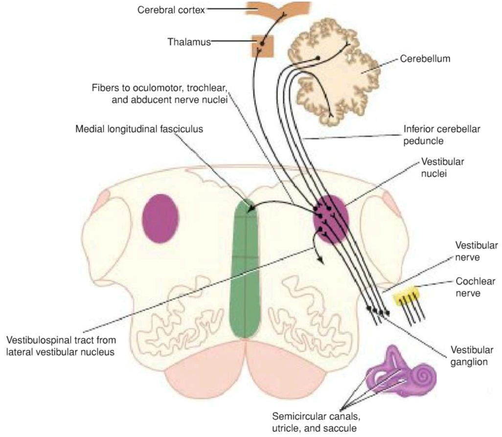
Figure 11-13 Vestibular nerve nuclei and their central connections.
شکل ۱۱-۱۳ هستههای عصب دهلیزی و اتصالات مرکزی آنها.
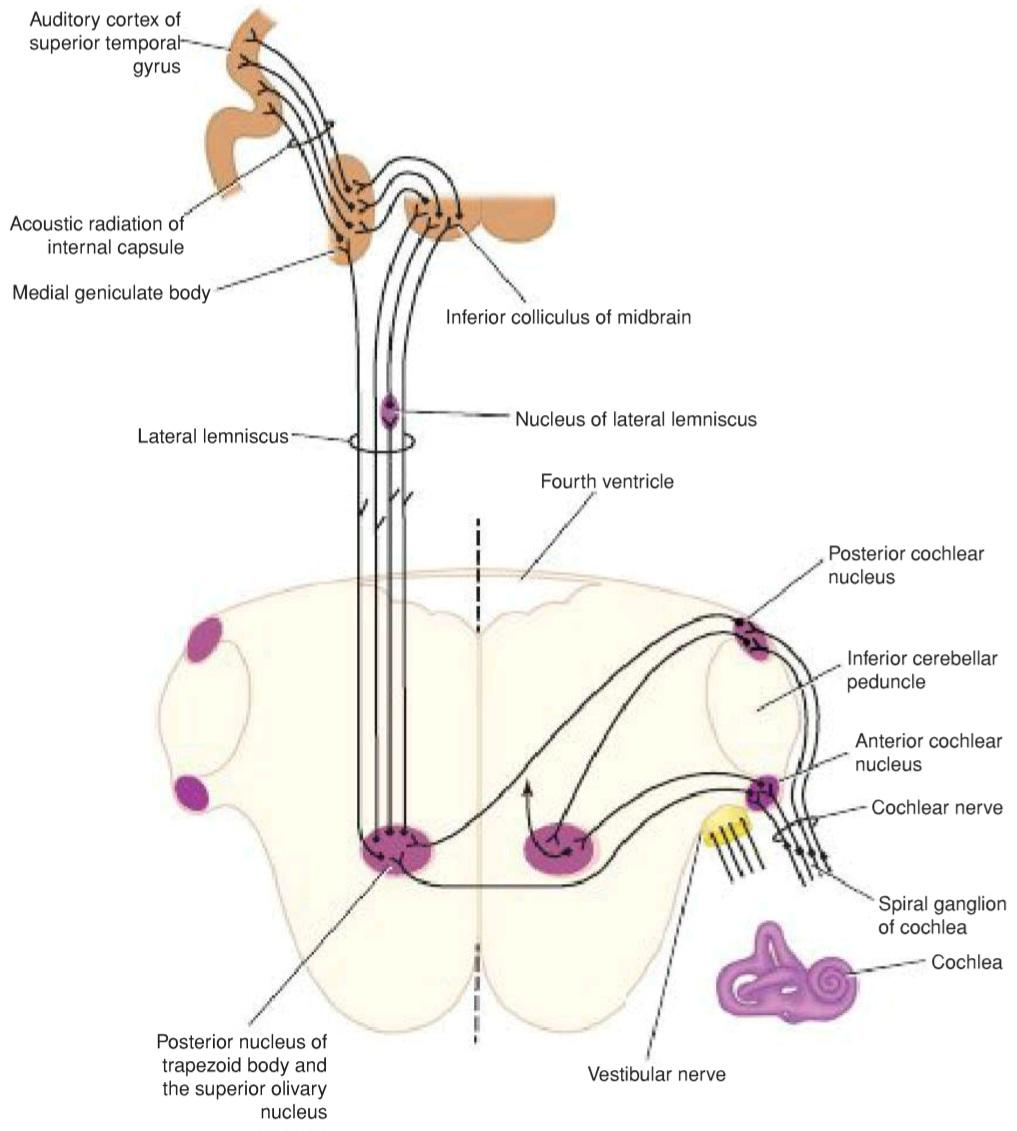
Figure 11-14 Cochlear nerve nuclei and their central connections. The descending pathways have been omitted.
شکل ۱۱-۱۴ هستههای عصب حلزونی و اتصالات مرکزی آنها. مسیرهای نزولی حذف شدهاند.
Vestibular Nerve
The vestibular nerve conducts nerve impulses from the utricle and saccule that provide information concerning
Vestibulocochlear Nerve (Cranial Nerve VIII) the position of the head; the nerve also conducts impulses from the semicircular canals that provide information concerning movements of the head.
عصب دهلیزی
عصب دهلیزی تکانههای عصبی را از اتریکول و ساکول هدایت میکند که اطلاعاتی در مورد موقعیت سر ارائه میدهند. عصب دهلیزی حلزونی (عصب جمجمهای VIII) همچنین تکانههایی را از مجاری نیمدایرهای هدایت میکند که اطلاعاتی در مورد حرکات سر ارائه میدهند.
The nerve fibers of the vestibular nerve are the central processes of nerve cells located in the vestibular ganglion, which is situated in the internal acoustic meatus. They enter the anterior surface of the brainstem in a groove between the lower border of the pons and the upper part of the medulla oblongata (see Fig. 11-13). When they enter the vestibular nuclear complex, the fibers divide into short ascending and long descending fibers; a small number of fibers pass directly to the cerebellum through the inferior cerebellar peduncle, bypassing the vestibular nuclei.
فیبرهای عصبی عصب دهلیزی، زوائد مرکزی سلولهای عصبی واقع در گانگلیون دهلیزی هستند که در مجرای شنوایی داخلی قرار دارد. آنها در شیاری بین لبه پایینی پل مغزی و قسمت بالایی بصلالنخاع وارد سطح قدامی ساقه مغز میشوند (شکل 11-13 را ببینید). هنگامی که آنها وارد کمپلکس هستهای دهلیزی میشوند، به الیاف کوتاه صعودی و بلند نزولی تقسیم میشوند. تعداد کمی از الیاف مستقیماً از طریق پایه مخچه تحتانی عبور میکنند و هستههای دهلیزی را دور میزنند.
Vestibular Nuclear Complex
This complex consists of a group of nuclei situated beneath the floor of the fourth ventricle (see Fig. 11-13). Four nuclei may be recognized: (1) the lateral vestibular nucleus, (2) the superior vestibular nucleus, (3) the medial vestibular nucleus, and (4) the inferior vestibular nucleus (see Fig. 5-14).
مجتمع هستهای دهلیزی
این مجتمع شامل گروهی از هستهها است که در زیر کف بطن چهارم قرار دارند (شکل 11-13 را ببینید). چهار هسته را میتوان تشخیص داد: (1) هسته دهلیزی جانبی، (2) هسته دهلیزی فوقانی، (3) هسته دهلیزی میانی و (4) هسته دهلیزی تحتانی (شکل 5-14 را ببینید).
The vestibular nuclei receive afferent fibers from the utricle and saccule and the semicircular canals through the vestibular nerve and fibers from the cerebellum through the inferior cerebellar peduncle (see Fig. 11-13). Efferent fibers from the nuclei pass to the cerebellum through the inferior cerebellar peduncle. Efferent fibers also descend uncrossed to the spinal cord from the lateral vestibular nucleus and form the vestibulospinal tract. In addition, efferent fibers pass to the nuclei of the oculomotor, trochlear, and abducens nerves through the medial longitudinal fasciculus.
هستههای دهلیزی، فیبرهای آوران را از اتریکول و ساکول و مجاری نیمدایرهای از طریق عصب دهلیزی و فیبرهای مخچه را از طریق پایه مخچهای تحتانی دریافت میکنند (شکل 11-13 را ببینید). فیبرهای وابران از هستهها از طریق پایه مخچهای تحتانی به مخچه میروند. فیبرهای وابران همچنین از هسته دهلیزی جانبی به صورت غیر متقاطع به نخاع نزول میکنند و راه دهلیزی-نخاعی را تشکیل میدهند. علاوه بر این، فیبرهای وابران از طریق فاسیکول طولی داخلی به هستههای اعصاب حرکتی چشمی، قرقرهای و ابدوسنس میروند.
These connections enable the movements of the head and the eyes to be coordinated so that visual fixation on an object can be maintained. In addition, information received from the internal ear can assist in maintaining balance by influencing the muscle tone of the limbs and trunk.
این ارتباطات باعث میشوند حرکات سر و چشمها هماهنگ شوند تا تثبیت بینایی روی یک جسم حفظ شود. علاوه بر این، اطلاعات دریافتی از گوش داخلی میتواند با تأثیر بر تون عضلانی اندامها و تنه به حفظ تعادل کمک کند.
Ascending fibers also pass upward from the vestibular nuclei to the cerebral cortex, to the vestibular area in the postcentral gyrus just above the lateral fissure. These fibers are thought to relay in the ventral posterior nuclei of the thalamus. The cerebral cortex probably serves to orient the individual consciously in space.
فیبرهای صعودی همچنین از هستههای دهلیزی به سمت قشر مغز، به ناحیه دهلیزی در شکنج پس مرکزی درست بالای شیار جانبی، به سمت بالا میروند. تصور میشود که این فیبرها در هستههای خلفی شکمی تالاموس رله میشوند. قشر مغز احتمالاً برای جهتیابی آگاهانه فرد در فضا عمل میکند.
Cochlear Nerve
The cochlear nerve conducts nerve impulses concerned with sound from the organ of Corti in the cochlea. The fibers of the cochlear nerve are the central processes of nerve cells located in the spiral ganglion of the cochlea (Fig. 11-15). They enter the anterior surface of the brainstem at the lower border of the pons on the lateral side of the emerging facial nerve by the vestibular nerve (see Fig. 11-14). On entering the pons, the nerve fibers divide, with one branch entering the posterior cochlear nucleus and the other branch entering the anterior cochlear nucleus.
عصب حلزونی
عصب حلزونی، تکانههای عصبی مربوط به صدا را از اندام کورتی در حلزون گوش هدایت میکند. فیبرهای عصب حلزونی، زوائد مرکزی سلولهای عصبی واقع در گانگلیون مارپیچی حلزون گوش هستند (شکل 11-15). آنها توسط عصب دهلیزی (شکل 11-14 را ببینید) وارد سطح قدامی ساقه مغز در مرز پایینی پل مغزی در سمت جانبی عصب صورتی در حال ظهور میشوند. هنگام ورود به پل مغزی، فیبرهای عصبی تقسیم میشوند و یک شاخه وارد هسته حلزونی خلفی و شاخه دیگر وارد هسته حلزونی قدامی میشود.
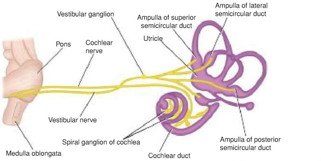
Figure 11-15 Distribution of the vestibulocochlear nerve.
شکل ۱۱-۱۵ توزیع عصب دهلیزی-حلزونی.
Cochlear Nuclei
The anterior and posterior cochlear nuclei are situated on the surface of the inferior cerebellar peduncle (see Fig. 11-14). They receive afferent fibers from the cochlea through the cochlear nerve. The cochlear nuclei send axons (second-order neuron fibers) that run medially through the pons to end in the trapezoid body and the olivary nucleus. Here, they are relayed in the posterior nucleus of the trapezoid body and the superior olivary nucleus on the same or the opposite side. The axons now ascend through the posterior part of the pons and midbrain and form a tract known as the lateral lemniscus. Each lateral lemniscus, therefore, consists of third-order neurons from both sides. As these fibers ascend, some of them relay in small groups of nerve cells, collectively known as the nucleus of the lateral lemniscus.
هستههای حلزونی
هستههای حلزونی قدامی و خلفی بر روی سطح پایه مخچه تحتانی قرار دارند (شکل 11-14 را ببینید). آنها از طریق عصب حلزونی، فیبرهای آوران را از حلزون دریافت میکنند. هستههای حلزونی، آکسونها (فیبرهای نورونی مرتبه دوم) را میفرستند که از طریق پل مغزی به سمت داخل امتداد مییابند تا در جسم ذوزنقهای و هسته زیتونی ختم شوند. در اینجا، آنها در هسته خلفی جسم ذوزنقهای و هسته زیتونی فوقانی در همان طرف یا طرف مقابل رله میشوند. آکسونها اکنون از طریق قسمت خلفی پل مغزی و مغز میانی بالا میروند و مسیری را تشکیل میدهند که به عنوان لمنیسکوس جانبی شناخته میشود. بنابراین، هر لمنیسکوس جانبی از نورونهای مرتبه سوم از هر دو طرف تشکیل شده است. با بالا رفتن این فیبرها، برخی از آنها در گروههای کوچکی از سلولهای عصبی رله میشوند که در مجموع به عنوان هسته لمنیسکوس جانبی شناخته میشوند.
On reaching the midbrain, the fibers of the lateral lemniscus either terminate in the nucleus of the inferior colliculus or are relayed in the medial geniculate body and pass to the auditory cortex of the cerebral hemisphere through the acoustic radiation of the internal capsule.
الیاف لمنیسکوس جانبی با رسیدن به مغز میانی، یا در هسته کولیکولوس تحتانی خاتمه مییابند یا در جسم زانویی داخلی رله میشوند و از طریق تابش صوتی کپسول داخلی به قشر شنوایی نیمکره مغزی منتقل میشوند.
The primary auditory cortex (areas 41 and 42) includes the gyrus of Heschl on the upper surface of the superior temporal gyrus. The recognition and interpretation of sounds, on the basis of past experience, take place in the secondary auditory area.
قشر شنوایی اولیه (نواحی ۴۱ و ۴۲) شامل شکنج هشل در سطح بالایی شکنج گیجگاهی فوقانی است. تشخیص و تفسیر صداها، بر اساس تجربیات گذشته، در ناحیه شنوایی ثانویه انجام میشود.
Nerve impulses from the ear are transmitted along auditory pathways on both sides of the brainstem, with more being projected along the contralateral pathway. Many collateral branches are given off to the reticular activating system of the brainstem (see p. 301). The tonotopic organization present in the organ of Corti is preserved within the cochlear nuclei, the inferior colliculi, and the primary auditory area.
پیامهای عصبی از گوش در امتداد مسیرهای شنوایی در دو طرف ساقه مغز منتقل میشوند و بیشتر آنها در امتداد مسیر مقابل قرار میگیرند. بسیاری از شاخههای جانبی به سیستم فعالکننده مشبک ساقه مغز منتقل میشوند (به صفحه ۳۰۱ مراجعه کنید). سازمان تونوتوپیک موجود در اندام کورتی در هستههای حلزونی، کولیکولوس تحتانی و ناحیه شنوایی اولیه حفظ میشود.
Descending Auditory Pathways
Descending fibers originating in the auditory cortex and in other nuclei in the auditory pathway accompany the ascending pathway. These fibers are bilateral and end on nerve cells at different levels of the auditory pathway and on the hair cells of the organ of Corti. These fibers are believed to serve as a feedback mechanism and inhibit the reception of sound. They may also have a role in the process of auditory sharpening, suppressing some signals and enhancing others.
مسیرهای شنوایی نزولی
فیبرهای نزولی که از قشر شنوایی و سایر هستههای مسیر شنوایی منشأ میگیرند، مسیر صعودی را همراهی میکنند. این فیبرها دو طرفه هستند و به سلولهای عصبی در سطوح مختلف مسیر شنوایی و سلولهای مویی اندام کورتی ختم میشوند. اعتقاد بر این است که این فیبرها به عنوان یک مکانیسم بازخورد عمل میکنند و مانع از دریافت صدا میشوند. آنها همچنین ممکن است در فرآیند تیز کردن شنوایی، سرکوب برخی سیگنالها و تقویت برخی دیگر نقش داشته باشند.
Vestibulocochlear Nerve Course
The vestibular and cochlear parts of the nerve leave the anterior surface of the brain between the lower border of the pons and the medulla oblongata (see Fig. 11-15). They run laterally in the posterior cranial fossa and enter the internal acoustic meatus with the facial nerve. The fibers are then distributed to the different parts of the internal ear.
مسیر عصب دهلیزی-حلزونی
بخشهای دهلیزی و حلزونی عصب، سطح قدامی مغز را بین لبه پایینی پل مغزی و بصل النخاع ترک میکنند (شکل 11-15 را ببینید). آنها به صورت جانبی در حفره جمجمهای خلفی امتداد یافته و همراه با عصب صورتی وارد مجرای شنوایی داخلی میشوند. سپس فیبرها در قسمتهای مختلف گوش داخلی توزیع میشوند.
«فصل یازدهم کتاب نورواناتومی بالینی اسنل»
🚀 با ما همراه شوید!
تازهترین مطالب و آموزشهای مغز و اعصاب را از دست ندهید. با فالو کردن کانال تلگرام آیندهنگاران مغز، از ما حمایت کنید!