
مخ؛ دیانسفالون: تالاموس، ساب تالاموس، اپیتالاموس و هیپوتالاموس؛ فصل هفتم نوروآناتومی بالینی اسنل


دعای مطالعه [ نمایش ]
بِسْمِ الله الرَّحْمنِ الرَّحیمِ
اَللّهُمَّ اَخْرِجْنى مِنْ ظُلُماتِ الْوَهْمِ
خدايا مرا بيرون آور از تاريكىهاى وهم،
وَ اَكْرِمْنى بِنُورِ الْفَهْمِ
و به نور فهم گرامى ام بدار،
اَللّهُمَّ افْتَحْ عَلَيْنا اَبْوابَ رَحْمَتِكَ
خدايا درهاى رحمتت را به روى ما بگشا،
وَانْشُرْ عَلَيْنا خَزائِنَ عُلُومِكَ بِرَحْمَتِكَ يا اَرْحَمَ الرّاحِمينَ
و خزانههاى علومت را بر ما باز كن به امید رحمتت اى مهربانترين مهربانان.
آیندهنگاران مغز: «ما مغز را میشناسیم، تا آینده را بسازیم.»
کتاب «نوروآناتومی بالینی اسنل» اثر دکتر ریچارد اس. اسنل، با ویرایش جدید دکتر رایان اسپلیتگربر، همچنان بهعنوان مرجع طلایی این حوزه شناخته میشود. این اثر با تلفیق تازهترین پژوهشها و کاربردهای بالینی، پلی استوار میان دانش پایه و عمل پزشکی بنا میکند.
تلاشهای علمی و سالها تدریس ارزشمند استاد گرانقدر آقای دکتر محمدتقی جغتایی الهامبخش ترجمه و گسترش این دانش در ایران بوده است.
ترجمه دقیق این کتاب توسط برند علمی آیندهنگاران مغز به مدیریت داریوش طاهری، امکان دسترسی فارسیزبانان به مرزهای نوین علوم اعصاب را فراهم کرده و رسالتی علمی در جهت شناخت عمیقتر مغز و ترسیم آیندهای روشنتر بر عهده گرفته است.
»» فصل ۷: مخ؛ بخش اول مبانی و اصول
»» Chapter 7: Cerebrum
CHAPTER OBJECTIVES
• To introduce the student to the complexities of the forebrain
• To understand the definition of the diencephalon and accurately localize the thalamus and hypothalamus by studying the sagittal, coronal, and horizontal sections of the brain
• To understand the exact position of the main conduit of the ascending and descending tracts, namely the internal capsule, which is so often the site of pathologic lesions
اهداف فصل
• آشنایی دانشجو با پیچیدگیهای مغز پیشین
• درک تعریف دیانسفالون و تعیین دقیق محل تالاموس و هیپوتالاموس با مطالعه بخشهای ساژیتال، کرونال و افقی مغز
• درک موقعیت دقیق مجرای اصلی مسیرهای صعودی و نزولی، یعنی کپسول داخلی، که اغلب محل ضایعات پاتولوژیک است
A 23-year-old man is referred to a neurologist because of intermittent attacks of headaches, dizziness, and weakness and numbness of the left leg. On close questioning, the patient admits that the headache is made worse by changing the position of his head. A computed tomography (CT) scan reveals a small white opaque ball at the anterior end of the third ventricle. A diagnosis of a colloid cyst of the third ventricle is made.
مردی ۲۳ ساله به دلیل حملات متناوب سردرد، سرگیجه و ضعف و بیحسی پای چپ به متخصص مغز و اعصاب ارجاع داده میشود. در بازجویی دقیق، بیمار اذعان میکند که سردرد با تغییر وضعیت سر بدتر میشود. سیتیاسکن یک گوی کوچک سفید مات را در انتهای قدامی بطن سوم نشان میدهد. تشخیص کیست کلوئیدی بطن سوم داده میشود.
The aggravation of the headache caused by changing the position of the head can be explained by the fact that the cyst is mobile and suspended from the choroid plexus. When the head is moved into certain positions, the ball-like cyst blocks the foramen of Monro on the right side, further raising the intracerebral pressure and increasing the hydrocephalus. The weakness and numbness of the left leg are due to pressure on the right thalamus and the tracts in the right internal capsule, produced by the slowly expanding tumor. The patient makes a complete recovery after surgical excision of the tumor.
تشدید سردرد ناشی از تغییر موقعیت سر را میتوان با این واقعیت توضیح داد که کیست متحرک است و از شبکه کوروئید آویزان است. هنگامی که سر در موقعیتهای خاصی حرکت داده میشود، کیست توپ مانند، سوراخ مونرو را در سمت راست مسدود میکند و فشار داخل مغزی را بیشتر افزایش میدهد و هیدروسفالی را تشدید میکند. ضعف و بیحسی پای چپ به دلیل فشار بر تالاموس راست و مسیرهای موجود در کپسول داخلی راست است که توسط تومور به آرامی در حال گسترش ایجاد میشود. بیمار پس از برداشتن جراحی تومور، بهبودی کامل مییابد.
The cerebral hemispheres are developed from the telencephalon and form the largest part of the brain. Each hemisphere has a covering of gray matter, the cortex and internal masses of gray matter, the basal nuclei, and a lateral ventricle. The basic anatomical structure of this area is described so that the student can be prepared for the complexities associated with functional localization.
نیمکرههای مغزی از تلانسفالون توسعه یافتهاند و بزرگترین بخش مغز را تشکیل میدهند. هر نیمکره دارای پوششی از ماده خاکستری، قشر و تودههای داخلی ماده خاکستری، هستههای قاعدهای و یک بطن جانبی است. ساختار آناتومیکی اساسی این ناحیه به گونهای شرح داده شده است که دانشجو بتواند برای پیچیدگیهای مرتبط با مکانیابی عملکردی آماده شود.

SUBDIVISIONS
The cerebrum is the largest part of the brain, situated in the anterior and middle cranial fossae of the skull and occupying the whole concavity of the vault of the skull. It may be divided into two parts: the diencephalon, which forms the central core, and the telencephalon, which forms the cerebral hemispheres.
زیربخشها
مخ بزرگترین بخش مغز است که در حفرههای جمجمهای قدامی و میانی جمجمه قرار دارد و تمام فرورفتگی طاق جمجمه را اشغال میکند. میتوان آن را به دو بخش تقسیم کرد: دیانسفالون، که هسته مرکزی را تشکیل میدهد، و تلانسفالون، که نیمکرههای مغزی را تشکیل میدهد.
DIENCEPHALON
The diencephalon consists of the third ventricle and the structures that form its boundaries (Figs. 7-1 and 7-2). It extends posteriorly to the point where the third ventricle becomes continuous with the cerebral aqueduct and anteriorly as far as the interventricular foramina (Fig. 7-3). Thus, the diencephalon is a midline structure with symmetrical right and left halves. Obviously, these subdivisions of the brain are made for convenience, and from a functional point of view, nerve fibers freely cross the boundaries.
دیانسفالون
دیانسفالون از بطن سوم و ساختارهایی که مرزهای آن را تشکیل میدهند، تشکیل شده است (شکلهای 7-1 و 7-2). این بخش از عقب تا نقطهای که بطن سوم با مجرای مغزی در امتداد قرار میگیرد و از جلو تا سوراخهای بین بطنی امتداد مییابد (شکل 7-3). بنابراین، دیانسفالون یک ساختار خط وسط با نیمههای راست و چپ متقارن است. بدیهی است که این زیربخشهای مغز برای راحتی ساخته شدهاند و از دیدگاه عملکردی، فیبرهای عصبی آزادانه از مرزها عبور میکنند.
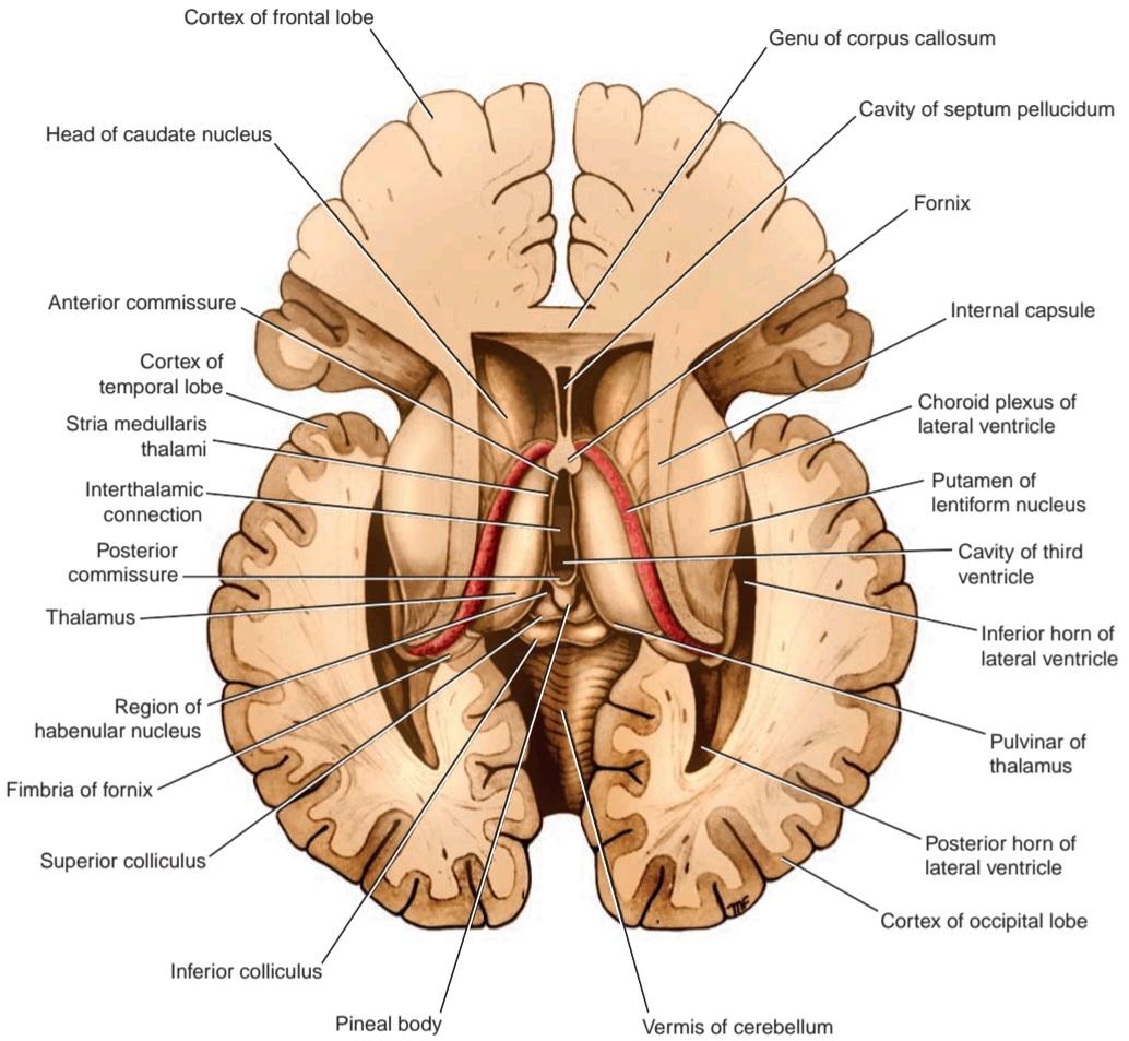
Figure 7.1 Horizontal section of the brain showing the third and lateral ventricles exposed by dissection from above.
شکل ۱.۷ برش افقی مغز که بطنهای سوم و جانبی را نشان میدهد که با تشریح از بالا نمایان شدهاند.

Figure 7-2 Inferior surface of the brain showing parts of the diencephalon.
شکل ۷-۲ سطح تحتانی مغز که بخشهایی از دیانسفالون را نشان میدهد.

Figure 7-3 Sagittal section of the brain showing the medial surface of the diencephalon.
شکل ۷-۳ برش ساژیتال مغز که سطح داخلی دیانسفالون را نشان میدهد.
Gross Features
The inferior surface of the diencephalon is the only area exposed to the surface in the intact brain (see Fig. 7-2; see also Atlas Plate 1). It is formed by hypothalamic and other structures, which include, from anterior to posterior, the optic chiasma, with the optic tract on either side; the infundibulum, with the tuber cinereum; and the mammillary bodies.
ویژگیهای کلی
سطح تحتانی دیانسفالون تنها ناحیهای است که در مغز سالم در معرض سطح قرار دارد (شکل 7-2 را ببینید؛ همچنین به صفحه اطلس 1 مراجعه کنید). این سطح توسط هیپوتالاموس و سایر ساختارها تشکیل شده است که از جلو به عقب شامل کیاسمای بینایی، با مجرای بینایی در دو طرف آن؛ اینفاندیبولوم، با غده سینروم؛ و اجسام پستانی میشود.
The superior surface of the diencephalon is concealed by the fornix, which is a thick bundle of fibers that originates in the hippocampus of the temporal lobe and arches posteriorly over the thalamus (see Fig. 7-3; see also Atlas Plate 8) to join the mammillary body.
سطح فوقانی دیانسفالون توسط فورنیکس پنهان شده است، که دستهای ضخیم از الیاف است که از هیپوکامپ لوب گیجگاهی منشأ میگیرد و به صورت قوسی در خلف تالاموس قرار میگیرد (شکل 7-3 را ببینید؛ همچنین به صفحه 8 اطلس مراجعه کنید) تا به جسم پستانی متصل شود.
The actual superior wall of the diencephalon is formed by the roof of the third ventricle. This consists of a layer of ependyma, which is continuous with the rest of the ependymal lining of the third ventricle. It is covered superiorly by a vascular fold of pia mater, called the tela choroidea of the third ventricle. From the roof of the third ventricle, a pair of vascular processes, the choroid plexuses of the third ventricle, project downward from the midline into the cavity of the third ventricle.
دیواره فوقانی واقعی دیانسفالون توسط سقف بطن سوم تشکیل شده است. این دیواره از یک لایه اپاندیم تشکیل شده است که در امتداد بقیه پوشش اپاندیمی بطن سوم قرار دارد. این دیواره در بالا توسط یک چین عروقی از نرم شامه به نام تلا مشیمیه بطن سوم پوشیده شده است. از سقف بطن سوم، یک جفت زائده عروقی، شبکههای مشیمیه بطن سوم، از خط میانی به سمت پایین و به داخل حفره بطن سوم امتداد یافتهاند.
The lateral surface of the diencephalon is bounded by the internal capsule of white matter and consists of nerve fibers that connect the cerebral cortex with other parts of the brainstem and spinal cord (see Fig. 7-1).
سطح جانبی دیانسفالون توسط کپسول داخلی ماده سفید محدود شده است و از الیاف عصبی تشکیل شده است که قشر مغز را با سایر قسمتهای ساقه مغز و نخاع متصل میکند (شکل 7-1 را ببینید).
Because the diencephalon is divided into symmetrical halves by the slitlike third ventricle, it also has a medial surface. The medial surface of the diencephalon (i.e., the lateral wall of the third ventricle) is formed in its superior part by the medial surface of the thalamus and in its inferior part by the hypothalamus (see Fig. 7-3; see also Atlas Plate 8). These two areas are separated from one another by a shallow sulcus, the hypothalamic sulcus. A bundle of nerve fibers, which are afferent fibers to the habenular nucleus, forms a ridge along the superior margin of the medial surface of the diencephalon and is called the stria medullaris thalami (see Fig. 7-1).
از آنجا که دیانسفالون توسط بطن سوم شکاف مانند به نیمههای متقارن تقسیم میشود، دارای یک سطح داخلی نیز میباشد. سطح داخلی دیانسفالون (یعنی دیواره جانبی بطن سوم) در قسمت فوقانی توسط سطح داخلی تالاموس و در قسمت تحتانی توسط هیپوتالاموس تشکیل میشود (شکل 7-3 را ببینید؛ همچنین به صفحه اطلس 8 مراجعه کنید). این دو ناحیه توسط یک شیار کم عمق، شیار هیپوتالاموس، از یکدیگر جدا میشوند. دستهای از الیاف عصبی، که الیاف آوران به هسته هابنولار هستند، یک برآمدگی در امتداد لبه فوقانی سطح داخلی دیانسفالون تشکیل میدهند و به آن استریا مدولا تالامی میگویند (شکل 7-1 را ببینید).
The diencephalon can be divided into four major parts: (1) the thalamus, (2) the subthalamus, (3) the epithalamus, and (4) the hypothalamus.
دیانسفالون را میتوان به چهار بخش اصلی تقسیم کرد: (1) تالاموس، (2) ساب تالاموس، (3) اپی تالاموس و (4) هیپوتالاموس.
Thalamus
The thalamus is a large ovoid mass of gray matter that forms the major part of the diencephalon. It is a region of great functional importance and serves as a cell station to all the main sensory systems (except the olfactory pathway). The activities of the thalamus are closely related to that of the cerebral cortex and damage to the thalamus causes great loss of cerebral function.
تالاموس
تالاموس یک توده بزرگ تخممرغی شکل از ماده خاکستری است که بخش عمده دیانسفالون را تشکیل میدهد. این ناحیه از اهمیت عملکردی بالایی برخوردار است و به عنوان یک ایستگاه سلولی برای تمام سیستمهای حسی اصلی (به جز مسیر بویایی) عمل میکند. فعالیتهای تالاموس ارتباط نزدیکی با فعالیتهای قشر مغز دارد و آسیب به تالاموس باعث از دست رفتن بخش زیادی از عملکرد مغز میشود.
The thalamus is situated on each side of the third ventricle (see Fig. 7-3; see also Atlas Plate 5). The anterior end of the thalamus is narrow and rounded and forms the posterior boundary of the interventricular foramen. The posterior end (Fig. 7-4) is expanded to form the pulvinar, which overhangs the superior colliculus and the superior brachium. The lateral geniculate body forms a small elevation on the under aspect of the lateral portion of the pulvinar.
تالاموس در هر دو طرف بطن سوم قرار دارد (شکل 7-3 را ببینید؛ همچنین به صفحه اطلس 5 مراجعه کنید). انتهای قدامی تالاموس باریک و گرد است و مرز خلفی سوراخ بین بطنی را تشکیل میدهد. انتهای خلفی (شکل 7-4) منبسط شده و پولوینار را تشکیل میدهد که بر روی کولیکولوس فوقانی و براکیوم فوقانی قرار دارد. جسم زانویی خارجی، برآمدگی کوچکی را در قسمت زیرین قسمت جانبی پولوینار تشکیل میدهد.
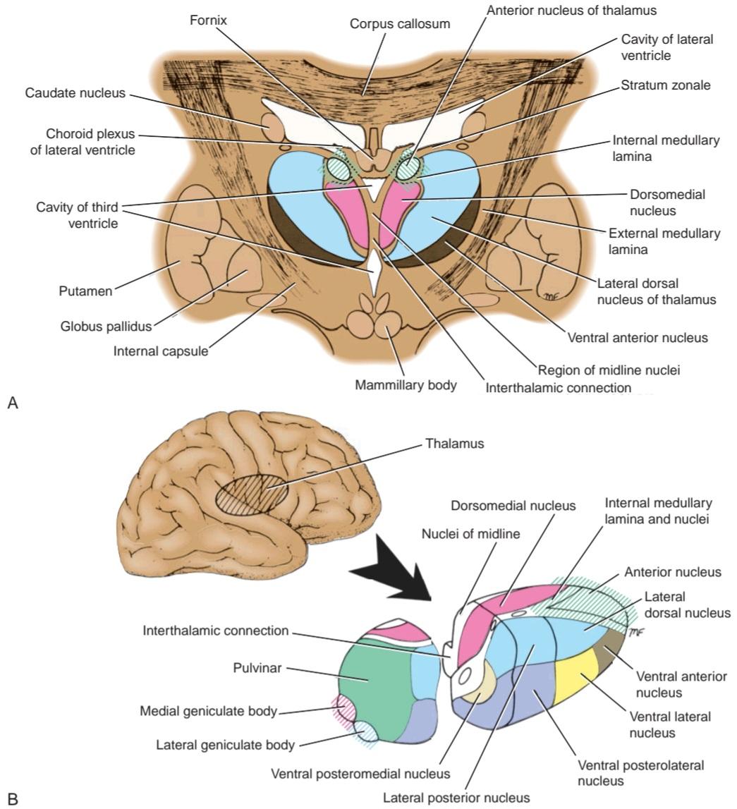
Figure 7-4 Nuclei of the thalamus. A: Transverse section through the anterior end of the thalamus. B: Diagram showing the position of the thalamus within the right cerebral hemisphere and the relative position of the thalamic nuclei to one another.
شکل ۷-۴ هستههای تالاموس. الف: برش عرضی از انتهای قدامی تالاموس. ب: نموداری که موقعیت تالاموس را در نیمکره مغزی راست و موقعیت نسبی هستههای تالاموس نسبت به یکدیگر نشان میدهد.
The superior surface of the thalamus is covered medially by the tela choroidea and the fornix, and laterally, it is covered by ependyma and forms part of the floor of the lateral ventricle; the lateral part is partially hidden by the choroid plexus of the lateral ventricle (see Fig. 7-1). The inferior surface is continuous with the tegmentum of the midbrain (see Fig. 7-3).
سطح فوقانی تالاموس از سمت داخل توسط تلا مشیمیه و فورنیکس و از سمت خارج توسط اپاندیم پوشیده شده و بخشی از کف بطن جانبی را تشکیل میدهد؛ بخش جانبی تا حدی توسط شبکه مشیمیه بطن جانبی پنهان است (شکل 7-1 را ببینید). سطح تحتانی با تگمنتوم مغز میانی در امتداد است (شکل 7-3 را ببینید).
The medial surface of the thalamus forms the superior part of the lateral wall of the third ventricle and is usually connected to the opposite thalamus by a band of gray matter, the interthalamic connection (interthalamic adhesion) (see Fig. 7-3).
سطح داخلی تالاموس قسمت فوقانی دیواره جانبی بطن سوم را تشکیل میدهد و معمولاً توسط نواری از ماده خاکستری، اتصال بین تالاموسی (چسبندگی بین تالاموسی) به تالاموس مقابل متصل میشود (شکل 7-3 را ببینید).
The lateral surface of the thalamus is separated from the lentiform nucleus by the very important band of white matter called the internal capsule (see Fig. 7-1).
سطح جانبی تالاموس توسط نوار بسیار مهمی از ماده سفید به نام کپسول داخلی از هسته عدسی شکل جدا میشود (شکل 7-1 را ببینید).
The subdivisions of the thalamus (see Fig. 7-4) and the detailed description of the thalamic nuclei and their connections are given on first page of Chapter 12.
زیربخشهای تالاموس (شکل 7-4 را ببینید) و شرح مفصلی از هستههای تالاموس و اتصالات آنها در صفحه اول فصل 12 ارائه شده است.
The thalamus is a very important cell station that receives the main sensory tracts (except the olfactory pathway). It should be regarded as a station where much of the information is integrated and relayed to the cerebral cortex and many other subcortical regions. It also plays a key role in the integration of visceral and somatic functions. For more information on the function of the thalamus, see Chapter 12.
تالاموس یک ایستگاه سلولی بسیار مهم است که مسیرهای حسی اصلی (به جز مسیر بویایی) را دریافت میکند. باید آن را به عنوان ایستگاهی در نظر گرفت که بسیاری از اطلاعات در آن ادغام شده و به قشر مغز و بسیاری از مناطق زیرقشری دیگر منتقل میشوند. همچنین نقش کلیدی در ادغام عملکردهای احشایی و جسمی ایفا میکند. برای اطلاعات بیشتر در مورد عملکرد تالاموس، به فصل ۱۲ مراجعه کنید.
Subthalamus
The subthalamus lies inferior to the thalamus and, therefore, is situated between the thalamus and the tegmentum of the midbrain; craniomedially, it is related to the hypothalamus.
ساب تالاموس
ساب تالاموس در پایین تالاموس قرار دارد و بنابراین، بین تالاموس و تگمنتوم مغز میانی قرار دارد؛ از نظر جمجمهای-داخلی با هیپوتالاموس مرتبط است.
ساب تالاموس همچنین شامل بسیاری از مسیرهای مهم است که از تگمنتوم به هستههای تالاموس میروند؛ انتهای جمجمهای لمنیسهای میانی، نخاعی و سه قلو نمونههایی از آن هستند.
The structure of the subthalamus is extremely complex, and only a brief description is given here. Among the collections of nerve cells found in the subthalamus are the cranial ends of the red nuclei and the substantia nigra. The subthalamic nucleus has the shape of a biconvex lens. The nucleus has important connections with the corpus striatum (see p. 310); as a result, it is involved in the control of muscle activity.
ساختار ساب تالاموس بسیار پیچیده است و در اینجا فقط توضیح مختصری ارائه شده است. در میان مجموعه سلولهای عصبی موجود در ساب تالاموس، انتهای جمجمهای هستههای قرمز و جسم سیاه وجود دارد. هسته ساب تالاموس به شکل یک عدسی دو محدب است. این هسته ارتباطات مهمی با جسم مخطط دارد (به صفحه ۳۱۰ مراجعه کنید)؛ در نتیجه، در کنترل فعالیت عضلات نقش دارد.
The subthalamus also contains many important tracts that pass up from the tegmentum to the thalamic nuclei; the cranial ends of the medial, spinal, and trigeminal lemnisci are examples.
ساب تالاموس همچنین حاوی مسیرهای مهم بسیاری است که از تگمنتوم به هستههای تالاموس میروند؛ انتهای جمجمهای لمنیسهای میانی، نخاعی و سه قلو نمونههایی از این مسیرها هستند.
Epithalamus
The epithalamus consists of the habenular nuclei and their connections and the pineal gland.
اپیتالاموس
اپیتالاموس از هستههای هابنولار و اتصالات آنها و غده پینهآل تشکیل شده است.
Habenular Nucleus
The habenular nucleus is a small group of neurons situated just medial to the posterior surface of the thalamus. Afferent fibers are received from the amygdaloid nucleus in the temporal lobe (see p. 261) through the stria medullaris thalami; other fibers pass from the hippocampal formation through the fornix. Some of the fibers of the stria medullaris thalami cross the midline and reach the habenular nucleus of the opposite side; these latter fibers form the habenular commissure (see Fig. 7-3). Axons from the habenular nucleus pass to the interpeduncular nucleus in the roof of the interpeduncular fossa, the tectum of the midbrain, the thalamus, and the reticular formation of the midbrain. The habenular nucleus is believed to be a center for integration of olfactory, visceral, and somatic afferent pathways.
هسته هابنولار
هسته هابنولار گروه کوچکی از نورونها است که درست در قسمت داخلی سطح خلفی تالاموس قرار دارد. فیبرهای آوران از هسته آمیگدالوئید در لوب گیجگاهی (به صفحه 261 مراجعه کنید) از طریق استریا بصل النخاع دریافت میشوند؛ فیبرهای دیگر از طریق فورنیکس از تشکیلات هیپوکامپ عبور میکنند. برخی از فیبرهای استریا بصل النخاع از خط میانی عبور کرده و به هسته هابنولار طرف مقابل میرسند؛ این فیبرهای اخیر رابط هابنولار را تشکیل میدهند (به شکل 7-3 مراجعه کنید). آکسونهای هسته هابنولار به هسته بین پایهای در سقف حفره بین پایهای، تکتوم مغز میانی، تالاموس و تشکیلات مشبک مغز میانی منتقل میشوند. اعتقاد بر این است که هسته هابنولار مرکزی برای ادغام مسیرهای آوران بویایی، احشایی و سوماتیک است.
Pineal Gland (Body)
The pineal gland is a small, conical structure that is attached by the pineal stalk to the diencephalon. It projects backward so that it lies posterior to the midbrain (see Fig. 7-3; see also Atlas Plate 8). The base of the pineal stalk possesses a recess that is continuous with the cavity of the third ventricle. The superior part of the base of the stalk contains the habenular commissure; the inferior part of the base of the stalk contains the posterior commissure.
غده پینه آل (بدنه)
غده پینه آل یک ساختار مخروطی کوچک است که توسط ساقه پینه آل به دیانسفالون متصل است. این غده به سمت عقب برجسته شده است به طوری که در خلف مغز میانی قرار دارد (شکل 7-3 را ببینید؛ همچنین به صفحه اطلس 8 مراجعه کنید). پایه ساقه پینه آل دارای یک فرورفتگی است که با حفره بطن سوم پیوسته است. قسمت فوقانی پایه ساقه حاوی رابط هابنولار است؛ قسمت تحتانی پایه ساقه حاوی رابط خلفی است.
On microscopic section, the pineal gland is seen to be incompletely divided into lobules by connective tissue septa that extend into the substance of the gland from the capsule. Two types of cells are found in the gland, the pinealocytes and the glial cells. Concretions of calcified material called brain sand progressively accumulate within the pineal gland with age (Fig. 7-5).
در برش میکروسکوپی، غده پینهآل به طور ناقص توسط دیوارههای بافت همبند که از کپسول به داخل ماده غده امتداد مییابند، به لوبولها تقسیم شده است. دو نوع سلول در غده یافت میشود، پینهآلوسیتها و سلولهای گلیال. با افزایش سن، تودههایی از مواد کلسیفیه به نام شن مغزی به تدریج در غده پینهآل تجمع مییابند (شکل 7-5).
The pineal gland possesses no nerve cells, but adrenergic sympathetic fibers derived from the superior cervical sympathetic ganglia enter the gland and run in association with the blood vessels and the pinealocytes.
غده پینهآل هیچ سلول عصبی ندارد، اما فیبرهای سمپاتیک آدرنرژیک مشتق شده از گانگلیونهای سمپاتیک فوقانی گردنی وارد غده شده و در ارتباط با رگهای خونی و پینهآلوسیتها حرکت میکنند.
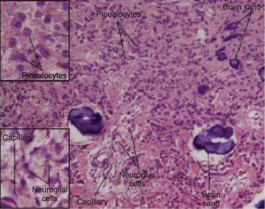
Figure 7-5 Photomicrograph of a section of the pineal gland stained with hematoxylin and eosin. (From Cui, D., Daley, W., Fratkin, J. D., Haines, D. E., Lynch, J. C., Naftel, J. P., Yang, G. Atlas of Histology with Functional and Clinical Correlations. Baltimore, MD: Wolters Kluwer, 2011.)
شکل ۷-۵ عکس میکروسکوپی از بخشی از غده پینهآل که با هماتوکسیلین و ائوزین رنگآمیزی شده است. (از Cui, D., Daley, W., Fratkin, J. D., Haines, D. E., Lynch, J. C., Naftel, J. P., Yang, G. Atlas of Histology with Functional and Clinical Correlations. Baltimore, MD: Wolters Kluwer, 2011.)
Pineal Gland Functions
The pineal gland, once thought to be of little significance, is now recognized as an important endocrine gland capable of influencing the activities of the pituitary gland, the islets of Langerhans of the pancreas, the parathyroids, the adrenal cortex and the adrenal medulla, and the gonads. The pineal secretions, produced by the pinealocytes, reach their target organs via the bloodstream or through the cerebrospinal fluid (CSF). Their actions are mainly inhibitory and either directly inhibit the production of hormones or indirectly inhibit the secretion of releasing factors by the hypothalamus. Notably, the pineal gland does not possess a blood-brain barrier.
عملکردهای غده پینه آل
غده پینه آل که زمانی اهمیت کمی داشت، اکنون به عنوان یک غده درون ریز مهم شناخته میشود که قادر به تأثیرگذاری بر فعالیتهای غده هیپوفیز، جزایر لانگرهانس پانکراس، پاراتیروئیدها، قشر آدرنال و مدولا آدرنال و غدد جنسی است. ترشحات پینه آل که توسط پینه آلوسیتها تولید میشوند، از طریق جریان خون یا از طریق مایع مغزی نخاعی (CSF) به اندامهای هدف خود میرسند. عملکرد آنها عمدتاً مهاری است و یا مستقیماً تولید هورمونها را مهار میکنند یا به طور غیرمستقیم ترشح عوامل آزادکننده توسط هیپوتالاموس را مهار میکنند. نکته قابل توجه این است که غده پینه آل فاقد سد خونی-مغزی است.
Animal experiments have shown that pineal activity exhibits a circadian rhythm that is influenced by light. The gland has been found to be most active during darkness. The probable nervous pathway from the retina runs to the suprachiasmatic nucleus of the hypothalamus, then to the tegmentum of the midbrain, and then to the pineal gland to stimulate its secretions. The latter part of this pathway may include the reticulospinal tract, the sympathetic outflow of the thoracic part of the spinal cord, and the superior cervical sympathetic ganglion and postganglionic nerve fibers that travel to the pineal gland on blood vessels.
آزمایشهای حیوانی نشان دادهاند که فعالیت غده پینهآل ریتم شبانهروزی دارد که تحت تأثیر نور قرار میگیرد. مشخص شده است که این غده در تاریکی بیشترین فعالیت را دارد. مسیر عصبی احتمالی از شبکیه به هسته سوپراکیاسماتیک هیپوتالاموس، سپس به تگمنتوم مغز میانی و سپس به غده پینهآل میرود تا ترشحات آن را تحریک کند. بخش دوم این مسیر ممکن است شامل مسیر رتیکولواسپینال، خروجی سمپاتیک قسمت سینهای نخاع و گانگلیون سمپاتیک گردنی فوقانی و فیبرهای عصبی پسگانگلیونی باشد که از طریق رگهای خونی به غده پینهآل میروند.
Melatonin and the enzymes needed for its production are present in high concentrations within the pineal gland. Melatonin and other substances are released into the blood or into the CSF of the third ventricle where they pass to the anterior lobe of the pituitary gland and inhibit the release of the gonadotrophic hormone. In humans, as in animals, the plasma melatonin level rises in darkness and falls during the day. The pineal gland apparently plays an important role in the regulation of reproductive function.
ملاتونین و آنزیمهای مورد نیاز برای تولید آن در غلظتهای بالا در غده پینهآل وجود دارند. ملاتونین و سایر مواد به داخل خون یا به مایع مغزی نخاعی بطن سوم آزاد میشوند، جایی که به لوب قدامی غده هیپوفیز میروند و ترشح هورمون گنادوتروپیک را مهار میکنند. در انسان، مانند حیوانات، سطح ملاتونین پلاسما در تاریکی افزایش مییابد و در طول روز کاهش مییابد. ظاهراً غده پینه آل نقش مهمی در تنظیم عملکرد تولید مثلی دارد.
Hypothalamus
The hypothalamus is that part of the diencephalon that extends from the region of the optic chiasma to the caudal border of the mammillary bodies (see Fig. 7-2; see also Atlas Plate 8). It lies below the hypothalamic sulcus on the lateral wall of the third ventricle. Thus, anatomically, the hypothalamus is a relatively small area of the brain that is strategically well placed close to the limbic system, the thalamus, the ascending and descending tracts, and the hypophysis. Microscopically, the hypothalamus is composed of small nerve cells that are arranged in groups or nuclei. The arrangement of these nuclei and their connections are fully described in Chapter 13.
هیپوتالاموس
هیپوتالاموس بخشی از دیانسفالون است که از ناحیه کیاسمای بینایی تا مرز دمی اجسام پستانی امتداد دارد (شکل 7-2 را ببینید؛ همچنین به صفحه اطلس 8 مراجعه کنید). این بخش در زیر شیار هیپوتالاموس روی دیواره جانبی بطن سوم قرار دارد. بنابراین، از نظر آناتومیکی، هیپوتالاموس ناحیه نسبتاً کوچکی از مغز است که از نظر استراتژیک به خوبی در نزدیکی سیستم لیمبیک، تالاموس، مسیرهای صعودی و نزولی و هیپوفیز قرار دارد. از نظر میکروسکوپی، هیپوتالاموس از سلولهای عصبی کوچکی تشکیل شده است که به صورت گروهها یا هستهها قرار گرفتهاند. ترتیب این هستهها و اتصالات آنها به طور کامل در فصل 13 شرح داده شده است.
Physiologically, nearly all the activities in the body are influenced by the hypothalamus. The hypothalamus controls and integrates the functions of the autonomic nervous system and the endocrine systems and plays a vital role in maintaining body homeostasis. It is involved in such activities as regulation of body temperature, body fluids, drives to eat and drink, sexual behavior, and emotion.
از نظر فیزیولوژیکی، تقریباً تمام فعالیتهای بدن تحت تأثیر هیپوتالاموس قرار دارند. هیپوتالاموس عملکردهای سیستم عصبی خودکار و سیستمهای غدد درونریز را کنترل و ادغام میکند و نقش حیاتی در حفظ هموستاز بدن ایفا میکند. این غده در فعالیتهایی مانند تنظیم دمای بدن، مایعات بدن، میل به خوردن و آشامیدن، رفتارهای جنسی و احساسات نقش دارد.
Hypothalamic Relations
Anterior to the hypothalamus is an area that extends forward from the optic chiasma to the lamina terminalis and the anterior commissure; it is referred to as the preoptic area. Caudally, the hypothalamus merges into the tegmentum of the midbrain. The thalamus lies superior to the hypothalamus, and the subthalamic region lies inferolaterally to the hypothalamus.
روابط هیپوتالاموسی
در جلوی هیپوتالاموس ناحیهای وجود دارد که از کیاسمای بینایی به سمت جلو تا لامینا ترمینالیس و رابط قدامی امتداد مییابد؛ به آن ناحیه پرهاپتیک گفته میشود. در قسمت خلفی، هیپوتالاموس در تگمنتوم مغز میانی ادغام میشود. تالاموس در بالای هیپوتالاموس قرار دارد و ناحیه سابتالاموس در قسمت تحتانی و جانبی هیپوتالاموس قرار دارد.
When observed from below, the hypothalamus is seen to be related to the following structures, from anterior to posterior: (1) the optic chiasma, (2) the tuber cinereum and the infundibulum, and (3) the mammillary bodies.
هنگام مشاهده از پایین، هیپوتالاموس از جلو به عقب با ساختارهای زیر مرتبط است: (1) کیاسمای بینایی، (2) توبر سینروم و اینفاندیبولوم، و (3) اجسام پستانی.
Optic Chiasma
The optic chiasma is a flattened bundle of nerve fibers situated at the junction of the anterior wall and floor of the third ventricle (see Figs. 7-2 and 7-3; see also Atlas Plate 8). The superior surface is attached to the lamina terminalis, and inferiorly, it is related to the hypophysis cerebri, from which it is separated by the diaphragma sellae. The anterolateral corners of the chiasma are continuous with the optic nerves, and the posterolateral corners are continuous with the optic tracts. A small recess, the optic recess of the third ventricle, lies on its superior surface.
کیاسمای بینایی
کیاسمای بینایی یک دسته مسطح از الیاف عصبی است که در محل اتصال دیواره قدامی و کف بطن سوم قرار دارد (به شکلهای 7-2 و 7-3 مراجعه کنید؛ همچنین به صفحه اطلس 8 مراجعه کنید). سطح فوقانی به لایه انتهایی مغز متصل است و در پایین، به هیپوفیز مغزی مرتبط است که توسط دیافراگم سلا از آن جدا میشود. گوشههای قدامی-جانبی کیاسمای قدامی-جانبی در امتداد اعصاب بینایی و گوشههای خلفی-جانبی در امتداد مجاری بینایی قرار دارند. یک فرورفتگی کوچک، فرورفتگی بینایی بطن سوم، در سطح فوقانی آن قرار دارد.
Importantly, remember that the fibers originating from the nasal half of each retina cross the median plane at the chiasma to enter the optic tract of the opposite side.
نکته مهم این است که به یاد داشته باشید الیافی که از نیمه بینی هر شبکیه سرچشمه میگیرند، از صفحه میانی در کیاسم عبور میکنند تا وارد مجرای بینایی طرف مقابل شوند.
Tuber Cinereum
The tuber cinereum is a convex mass of gray matter, as seen from the inferior surface (see Figs. 7-2 and 7-3; see also Atlas Plate 8). It is continuous inferiorly with the infundibulum. The infundibulum is hollow and becomes continuous with the posterior lobe of the hypophysis cerebri. The median eminence is a raised part of the tuber cinereum to which is attached the infundibulum. The median eminence, the infundibulum, and the posterior lobe (pars nervosa) of the hypophysis cerebri together form the neurohypophysis.
غده سینروم
غده سینروم از سطح تحتانی، تودهای محدب از ماده خاکستری است (شکلهای 7-2 و 7-3 را ببینید؛ همچنین به صفحه اطلس 8 مراجعه کنید). این غده در امتداد تحتانی با اینفاندیبولوم قرار دارد. اینفاندیبولوم توخالی است و با لوب خلفی هیپوفیز مغزی امتداد مییابد. برجستگی میانی، قسمت برجستهای از غده سینروم است که اینفاندیبولوم به آن متصل است. برجستگی میانی، اینفاندیبولوم و لوب خلفی (پارس عصبی) هیپوفیز مغزی با هم نوروهیپوفیز را تشکیل میدهند.
Mammillary Bodies
The mammillary bodies are two small hemispherical bodies situated side by side posterior to the tuber cinereum (see Figs. 7-2 and 7-3; see also Atlas Plate 8). They possess a central core of gray matter invested by a capsule of myelinated nerve fibers. Posterior to the mammillary bodies lies an area of the brain that is pierced by a number of small apertures and is called the posterior perforated substance. These apertures transmit the central branches of the posterior cerebral arteries.
اجسام پستانی
اجسام پستانی دو جسم نیمکره کوچک هستند که در کنار هم و در پشت غده سینروم قرار گرفتهاند (شکلهای 7-2 و 7-3 را ببینید؛ همچنین به صفحه اطلس 8 مراجعه کنید). آنها دارای یک هسته مرکزی از ماده خاکستری هستند که توسط کپسولی از الیاف عصبی میلیندار پوشانده شده است. در پشت اجسام پستانی، ناحیهای از مغز قرار دارد که توسط تعدادی روزنه کوچک سوراخ شده است و ماده سوراخدار خلفی نامیده میشود. این روزنهها شاخههای مرکزی شریانهای مغزی خلفی را منتقل میکنند.
Third Ventricle
The third ventricle, which is derived from the forebrain vesicle, is a slitlike cleft between the two thalami (see Figs. 7-1 and 7-3; see also Atlas Plates 5 and 8). It communicates anteriorly with the lateral ventricles through the interventricular foramina (foramina of Monro), and it communicates posteriorly with the fourth ventricle through the cerebral aqueduct. The third ventricle has anterior, posterior, lateral, superior, and inferior walls and is lined with ependyma.
بطن سوم
بطن سوم که از وزیکول مغز قدامی مشتق شده است، شکافی شکاف مانند بین دو تالاموس است (به شکلهای 7-1 و 7-3 مراجعه کنید؛ همچنین به صفحات اطلس 5 و 8 مراجعه کنید). این بطن از جلو از طریق سوراخهای بین بطنی (سوراخهای مونرو) با بطنهای جانبی و از عقب از طریق مجرای مغزی با بطن چهارم ارتباط برقرار میکند. بطن سوم دارای دیوارههای قدامی، خلفی، جانبی، فوقانی و تحتانی است و با اپاندیم پوشیده شده است.
The anterior wall is formed by a thin sheet of gray matter, the lamina terminalis, across which runs the anterior commissure (see Fig. 7-3). The anterior commissure is a round bundle of nerve fibers that are situated anterior to the anterior columns of the fornix; they connect the right and left temporal lobes.
دیواره قدامی توسط یک لایه نازک از ماده خاکستری، لامینا ترمینالیس، تشکیل شده است که از روی آن رابط قدامی عبور میکند (شکل 7-3 را ببینید). رابط قدامی یک دسته گرد از الیاف عصبی است که در جلوی ستونهای قدامی فورنیکس قرار دارند؛ آنها لوبهای گیجگاهی راست و چپ را به هم متصل میکنند.
The posterior wall is formed by the opening into the cerebral aqueduct. Superior to this opening is the small posterior commissure. Superior to the commissure is the pineal recess, which projects into the stalk of the pineal body. Superior to the pineal recess is the small habenular commissure.
دیواره خلفی توسط دهانهای به داخل قنات مغزی تشکیل میشود. بالاتر از این دهانه، رابط خلفی کوچک قرار دارد. بالاتر از رابط، فرورفتگی پینهآل قرار دارد که به ساقه جسم پینهآل بیرون زده است. بالاتر از فرورفتگی پینهآل، رابط هابنولار کوچک قرار دارد.
The lateral wall is formed by the medial surface of the thalamus superiorly and the hypothalamus inferiorly. These two structures are separated by the hypothalamic sulcus. The lateral wall is limited superiorly by the stria medullaris thalami. The lateral walls are joined by the interthalamic connection.
دیواره جانبی توسط سطح داخلی تالاموس در بالا و هیپوتالاموس در پایین تشکیل شده است. این دو ساختار توسط شیار هیپوتالاموس از هم جدا میشوند. دیواره جانبی در بالا توسط استریا مدولا تالامی محدود میشود. دیوارههای جانبی توسط اتصال بین تالاموس به هم متصل میشوند.
The superior wall or roof is formed by a layer of ependyma that is continuous with the lining of the ventricle. Superior to this layer is a two-layered fold of pia mater called the tela choroidea of the third ventricle. The vascular tela choroidea projects downward on each side of the midline, invaginating the ependymal roof to form the choroid plexuses of the third ventricle. Within the tela choroidea lie the internal cerebral veins. Superiorly, the roof of the ventricle is related to the fornix and the corpus callosum.
دیواره یا سقف فوقانی توسط لایهای از اپاندیم تشکیل شده است که با پوشش بطن در امتداد است. در بالای این لایه، یک چین دو لایه از نرم شامه به نام تلا مشیمیه بطن سوم قرار دارد. تلا مشیمیه عروقی در هر طرف خط میانی به سمت پایین بیرون زده و سقف اپاندیم را به داخل فرو میبرد تا شبکههای مشیمیه بطن سوم را تشکیل دهد. در داخل تلا مشیمیه، رگهای مغزی داخلی قرار دارند. در بالا، سقف بطن با فورنیکس و جسم پینهای مرتبط است.
The inferior wall or floor is formed by the optic chiasma, the tuber cinereum, the infundibulum, with its funnel-shaped recess, and the mammillary bodies (see Figs. 7-2 and 7-3). The hypophysis is attached to the infundibulum. Posterior to these structures lies the tegmentum of the cerebral peduncles.
دیواره یا کف تحتانی توسط کیاسمای بینایی، غده سینروم، اینفاندیبولوم، با فرورفتگی قیفی شکل آن، و اجسام پستانی تشکیل شده است (شکلهای 7-2 و 7-3 را ببینید). هیپوفیز به اینفاندیبولوم متصل است. در خلف این ساختارها، تگمنتوم پایههای مغزی قرار دارد.
The ventricular system is fully described in Chapter 16.
سیستم بطنی به طور کامل در فصل ۱۶ شرح داده شده است.
«فصل هفتم کتاب نورواناتومی بالینی اسنل»
🚀 با ما همراه شوید!
تازهترین مطالب و آموزشهای مغز و اعصاب را از دست ندهید. با فالو کردن کانال تلگرام آیندهنگاران مغز، از ما حمایت کنید!



 ورود/ ثبت نام با جیمیل
