
یادداشتهای بالینی نوروآناتومی اسنل؛ هستههای قاعدهای (عقدههای قاعدهای)


دعای مطالعه [ نمایش ]
بِسْمِ الله الرَّحْمنِ الرَّحیمِ
اَللّهُمَّ اَخْرِجْنى مِنْ ظُلُماتِ الْوَهْمِ
خدايا مرا بيرون آور از تاريكىهاى وهم،
وَ اَكْرِمْنى بِنُورِ الْفَهْمِ
و به نور فهم گرامى ام بدار،
اَللّهُمَّ افْتَحْ عَلَيْنا اَبْوابَ رَحْمَتِكَ
خدايا درهاى رحمتت را به روى ما بگشا،
وَانْشُرْ عَلَيْنا خَزائِنَ عُلُومِكَ بِرَحْمَتِكَ يا اَرْحَمَ الرّاحِمينَ
و خزانههاى علومت را بر ما باز كن به امید رحمتت اى مهربانترين مهربانان.
آیندهنگاران مغز: «ما مغز را میشناسیم، تا آینده را بسازیم.»
کتاب «نوروآناتومی بالینی اسنل» اثر دکتر ریچارد اس. اسنل، با ویرایش جدید دکتر رایان اسپلیتگربر، همچنان بهعنوان مرجع طلایی این حوزه شناخته میشود. این اثر با تلفیق تازهترین پژوهشها و کاربردهای بالینی، پلی استوار میان دانش پایه و عمل پزشکی بنا میکند.
تلاشهای علمی و سالها تدریس ارزشمند استاد گرانقدر آقای دکتر محمدتقی جغتایی الهامبخش ترجمه و گسترش این دانش در ایران بوده است.
ترجمه دقیق این کتاب توسط برند علمی آیندهنگاران مغز به مدیریت داریوش طاهری، امکان دسترسی فارسیزبانان به مرزهای نوین علوم اعصاب را فراهم کرده و رسالتی علمی در جهت شناخت عمیقتر مغز و ترسیم آیندهای روشنتر بر عهده گرفته است.
»» فصل ۱۰: هستههای قاعدهای (عقدههای قاعدهای)؛ بخش یادداشتهای بالینی
»» Chapter 10: Basal Nuclei (Basal Ganglia)
Clinical Notes
Disorders of the basal nuclei are of two general types. Hyperkinetic disorders involve excessive and abnormal movements, such as seen with chorea, athetosis, and ballism. Hypokinetic disorders involve a lack or slowness of movement. Parkinson disease includes both types of motor disturbances.
یادداشتهای بالینی
اختلالات هستههای قاعدهای دو نوع کلی دارند. اختلالات هایپرکینتیک شامل حرکات بیش از حد و غیرطبیعی است، مانند آنچه در کره، آتتوز و بالیسم دیده میشود. اختلالات هایپوکینتیک شامل فقدان یا کندی حرکت است. بیماری پارکینسون شامل هر دو نوع اختلالات حرکتی است.
Chorea
In chorea, the patient exhibits involuntary, quick, jerky, irregular movements that are nonrepetitive. Swift grimaces and sudden movements of the head or limbs are good examples.
کره
در کره، بیمار حرکات غیرارادی، سریع، تند و نامنظمی را نشان میدهد که تکرار نمیشوند. شکلکهای سریع و حرکات ناگهانی سر یا اندامها نمونههای خوبی از این موارد هستند.
Huntington Disease
Huntington disease is an autosomal dominant inherited disease, with the onset occurring most often in adult life. Death occurs 15 to 20 years after onset. The disease has been traced to a single gene defect on chromosome 4. This gene encodes a protein, huntingtin, the function of which is not known. The codon (CAG) that encodes glutamine is repeated many more times than normal. The disease affects men and women with equal frequency and unfortunately often reveals itself only after they have had children.
بیماری هانتینگتون
بیماری هانتینگتون یک بیماری ارثی اتوزومال غالب است که شروع آن اغلب در بزرگسالی رخ میدهد. مرگ ۱۵ تا ۲۰ سال پس از شروع رخ میدهد. این بیماری به یک نقص ژنی واحد در کروموزوم ۴ نسبت داده شده است. این ژن پروتئینی به نام هانتینگتین را رمزگذاری میکند که عملکرد آن مشخص نیست. کدون (CAG) که گلوتامین را رمزگذاری میکند، بسیار بیشتر از حد معمول تکرار میشود. این بیماری مردان و زنان را با فراوانی یکسان تحت تأثیر قرار میدهد و متأسفانه اغلب تنها پس از بچهدار شدن آنها خود را نشان میدهد.
Patients have the following characteristic signs and symptoms:
بیماران علائم و نشانههای مشخصه زیر را دارند:
1. Choreiform movements first appear as involuntary movements of the extremities and twitching of the face (facial grimacing). Later, more muscle groups are involved, so the patient becomes immobile and unable to speak or swallow.
۱. حرکات کرهایشکل ابتدا به صورت حرکات غیرارادی اندامها و تکانهای صورت (شکلک درآوردن) ظاهر میشوند. بعداً، گروههای عضلانی بیشتری درگیر میشوند، بنابراین بیمار بیحرکت میشود و قادر به صحبت کردن یا بلعیدن نیست.
2. Progressive dementia occurs with loss of memory and intellectual capacity.
۲. زوال عقل پیشرونده با از دست دادن حافظه و ظرفیت فکری رخ میدهد.
In this disease, the GABA-secreting, substance P-secreting, and acetylcholine-secreting neurons of the striatonigralinhibiting pathway degenerate. This results in the dopasecreting neurons of the substantia nigra becoming overactive; thus, the nigrostriatal pathway inhibits the caudate nucleus and the putamen (Fig. 10-6). This inhibition produces the abnormal movements seen in this disease. Computed tomography scans show enlarged lateral ventricles due to degeneration of the caudate nuclei. Medical treatment of Huntington chorea has been disappointing.
در این بیماری، نورونهای ترشحکنندهی GABA، ترشحکنندهی مادهی P و ترشحکنندهی استیل کولین از مسیر مهارکنندهی استریاتونیگرا تخریب میشوند. این امر منجر به بیشفعال شدن نورونهای ترشحکنندهی دوپا در جسم سیاه میشود؛ بنابراین، مسیر نیگرواستریاتال، هستهی دمدار و پوتامن را مهار میکند (شکل 10-6). این مهار باعث ایجاد حرکات غیرطبیعی مشاهدهشده در این بیماری میشود. اسکنهای توموگرافی کامپیوتری، بطنهای جانبی بزرگشده را به دلیل تخریب هستههای دمدار نشان میدهند. درمان پزشکی کرهی هانتینگتون ناامیدکننده بوده است.

Figure 10-6 Diagram showing the degeneration of the inhibitory pathway between the corpus striatum and the substantia nigra seen in Huntington disease and the consequent reduction in the liberation of GABA, substance P, and acetylcholine in the substantia nigra.
شکل ۱۰-۶ نموداری که تخریب مسیر مهاری بین جسم مخطط و جسم سیاه را که در بیماری هانتینگتون مشاهده میشود و در نتیجه کاهش آزادسازی GABA، ماده P و استیل کولین در جسم سیاه را نشان میدهد.
Sydenham Chorea
Sydenham chorea (St. Vitus dance) is a disease of childhood in which rapid, irregular, involuntary movements of the limbs, face, and trunk occur. The condition is associated with rheumatic fever. The antigens of the streptococcal bacteria are similar in structure to the proteins present in the membranes of striatal neurons. The host’s antibodies not only combine with the bacterial antigens but also attack the membranes of the neurons of the basal ganglia. This results in the production of choreiform movements, which are fortunately transient, and full recovery is made.
کره سیدنهام
کره سیدنهام (رقص سنت ویتوس) یک بیماری دوران کودکی است که در آن حرکات سریع، نامنظم و غیرارادی اندامها، صورت و تنه رخ میدهد. این بیماری با تب روماتیسمی مرتبط است. آنتیژنهای باکتری استرپتوکوکی از نظر ساختاری مشابه پروتئینهای موجود در غشای نورونهای جسم مخطط هستند. آنتیبادیهای میزبان نه تنها با آنتیژنهای باکتریایی ترکیب میشوند، بلکه به غشای نورونهای عقدههای قاعدهای نیز حمله میکنند. این امر منجر به تولید حرکات کرهایشکل میشود که خوشبختانه گذرا هستند و بهبودی کامل حاصل میشود.
Hemiballismus
Hemiballismus is a form of involuntary movement confined to one side of the body. It usually involves the proximal extremity musculature, and the limb suddenly flies about out of control in all directions. The lesion, which is usually a small stroke, occurs in the opposite subthalamic nucleus or its connections; smooth movements of different parts of the body are integrated in the subthalamic nucleus.
همیبالیسموس
همیبالیسموس نوعی حرکت غیرارادی محدود به یک طرف بدن است. معمولاً عضلات پروگزیمال اندام را درگیر میکند و اندام ناگهان در تمام جهات از کنترل خارج میشود. ضایعه، که معمولاً یک ضربه کوچک است، در هسته سابتالاموس مقابل یا اتصالات آن رخ میدهد. حرکات روان قسمتهای مختلف بدن در هسته سابتالاموس ادغام میشوند.
Parkinson Disease
Parkinson disease is a progressive disease of unknown cause that commences between the ages of 45 and 55 years. It is associated with neuronal degeneration in the substantia nigra and, to a lesser extent, in the globus pallidus, putamen, and caudate nucleus. The disease affects about 1 million people in the United States.
بیماری پارکینسون
بیماری پارکینسون یک بیماری پیشرونده با علت ناشناخته است که بین سنین ۴۵ تا ۵۵ سالگی شروع میشود. این بیماری با تخریب نورونی در جسم سیاه و به میزان کمتر در گلوبوس پالیدوس، پوتامن و هسته دمی مرتبط است. این بیماری حدود ۱ میلیون نفر را در ایالات متحده تحت تأثیر قرار میدهد.
The degeneration of the neurons of the substantia nigra that send their axons to the corpus striatum results in a reduction in the release of the neurotransmitter dopamine within the corpus striatum (Figs. 10-7 and 10-8). This leads to hypersensitivity of the dopamine receptors in the postsynaptic neurons in the striatum.
تخریب نورونهای جسم سیاه که آکسونهای خود را به جسم مخطط میفرستند، منجر به کاهش آزادسازی انتقالدهنده عصبی دوپامین در جسم مخطط میشود (شکلهای 10-7 و 10-8). این امر منجر به حساسیت بیش از حد گیرندههای دوپامین در نورونهای پس سیناپسی در جسم مخطط میشود.
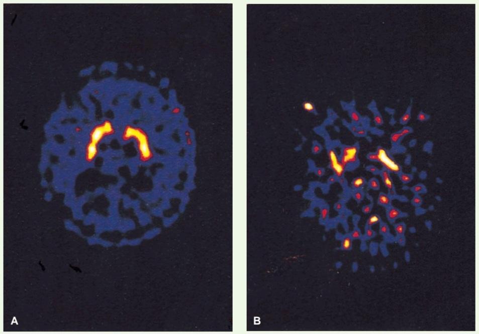
Figure 10-7 Axial (horizontal) positron emission tomography (PET) scans of a normal brain (A) and the brain of a patient with early Parkinson disease (B) following the injection of 18-F-fluorodopa. The normal brain image shows large amounts of the compound (yellow areas) distributed throughout the corpus striatum in both cerebral hemispheres. In the patient with Parkinson disease, the brain image shows that the total amount of the compound is low, and it is unevenly distributed in the corpus striatum. (Courtesy Dr. Holley Dey.)
شکل ۱۰-۷ اسکنهای توموگرافی گسیل پوزیترون (PET) محوری (افقی) از مغز طبیعی (A) و مغز بیمار مبتلا به مراحل اولیه بیماری پارکینسون (B) پس از تزریق 18-F-فلورودوپا. تصویر مغز طبیعی مقادیر زیادی از این ترکیب (نواحی زرد) را نشان میدهد که در سراسر جسم مخطط در هر دو نیمکره مغزی توزیع شده است. در بیمار مبتلا به پارکینسون، تصویر مغز نشان میدهد که مقدار کل این ترکیب کم است و به طور ناهموار در جسم مخطط توزیع شده است. (با احترام از دکتر هالی دی.)

Figure 10-8 Diagram showing the degeneration of the inhibitory pathway between the substantia nigra and the corpus striatum in Parkinson disease and the consequent reduction in the release of the neurotransmitter dopamine in the striatum.
شکل۱۰-۸ نموداری که تخریب مسیر مهاری بین جسم سیاه و جسم مخطط در بیماری پارکینسون و در نتیجه کاهش آزادسازی انتقالدهنده عصبی دوپامین در جسم مخطط را نشان میدهد.
Patients have the following characteristic signs and symptoms:
بیماران علائم و نشانههای مشخصه زیر را دارند:
1. Tremor. This is the result of the alternating contraction of agonists and antagonists. The tremor is slow and occurs most obviously when the limbs are at rest. It disappears during sleep. It should be distinguished from the intention tremor seen in cerebellar disease, which only occurs when purposeful active movement is attempted.
۱. لرزش. این لرزش نتیجه انقباض متناوب آگونیستها و آنتاگونیستها است. لرزش آهسته است و بیشتر در حالت استراحت اندامها دیده میشود. در هنگام خواب از بین میرود. باید آن را از لرزش عمدی که در بیماریهای مخچه دیده میشود، که فقط هنگام تلاش برای حرکت فعال هدفمند رخ میدهد، متمایز کرد.
2. Rigidity. This differs from the rigidity caused by lesions of the upper motor neurons in that it is present to an equal extent in opposing muscle groups. If the tremor is absent, the rigidity is felt as resistance to passive movement and is sometimes referred to as plastic rigidity. If the tremor is present, the muscle resistance is overcome as a series of jerks, called cogwheel rigidity.
۲. سفتی. این حالت با سفتی ناشی از ضایعات نورونهای حرکتی فوقانی متفاوت است، زیرا به میزان مساوی در گروههای عضلانی مخالف وجود دارد. اگر لرزش وجود نداشته باشد، سفتی به صورت مقاومت در برابر حرکت غیرفعال احساس میشود و گاهی اوقات به عنوان سفتی انعطافپذیر شناخته میشود. اگر لرزش وجود داشته باشد، مقاومت عضلانی به صورت یک سری حرکات ناگهانی، که سفتی چرخدنده نامیده میشود، برطرف میشود.
3. Bradykinesis. Initiating (akinesia) and performing new movements is difficult. The movements are slow, the face is expressionless, and the voice is slurred and unmodulated. Swinging of the arms in walking is lost.
۳. کندی حرکات. شروع (آکینزی) و انجام حرکات جدید دشوار است. حرکات کند، صورت بیحالت و صدا نامفهوم و نامنظم است. تاب دادن بازوها هنگام راه رفتن از بین میرود.
4. Postural disturbances. The patient stands with a stoop, and his or her arms are flexed. The patient walks by taking short steps and often is unable to stop. In fact, he or she may break into a shuffling run to maintain balance.
۴. اختلالات وضعیتی. بیمار با قوز ایستاده و بازوهایش خم شده است. بیمار با قدمهای کوتاه راه میرود و اغلب قادر به توقف نیست. در واقع، او ممکن است برای حفظ تعادل، شروع به دویدن کند.
5. Neither loss of muscle power nor loss of sensibility occurs. Since the corticospinal tracts are normal, the superficial abdominal reflexes are normal, and no Babinski response is seen. The deep tendon reflexes are normal.
۵. نه از دست دادن قدرت عضلانی و نه از دست دادن حس رخ نمیدهد. از آنجایی که راههای قشری-نخاعی طبیعی هستند، رفلکسهای شکمی سطحی طبیعی هستند و هیچ پاسخ بابنسکی مشاهده نمیشود. رفلکسهای تاندونی عمقی طبیعی هستند.
In a few types of Parkinson disease, the cause is known. Postencephalitic parkinsonism developed following the viral encephalitis outbreak of 1916 to 1917, in which damage occurred to the basal nuclei. Iatrogenic parkinsonism can be a side effect of antipsychotic drugs (e.g., phenothiazines). Meperidine analogues (used by drug addicts) and poisoning from carbon monoxide and manganese can also produce the symptoms of parkinsonism. Atherosclerotic parkinsonism can occur in elderly hypertensive patients.
در چند نوع بیماری پارکینسون، علت شناخته شده است. پارکینسونیسم پس از آنسفالیت پس از شیوع آنسفالیت ویروسی در سالهای ۱۹۱۶ تا ۱۹۱۷، که در آن آسیب به هستههای قاعدهای رخ داد، ایجاد شد. پارکینسونیسم یاتروژنیک میتواند یک عارضه جانبی داروهای ضد روانپریشی (مانند فنوتیازینها) باشد. آنالوگهای مپریدین (که توسط معتادان به مواد مخدر استفاده میشود) و مسمومیت با مونوکسید کربن و منگنز نیز میتوانند علائم پارکینسونیسم را ایجاد کنند. پارکینسونیسم آترواسکلروتیک میتواند در بیماران مسن مبتلا به فشار خون بالا رخ دهد.
Parkinson disease may be treated by elevating the brain dopamine level. Unfortunately, dopamine cannot cross the blood-brain barrier, but its immediate precursor L-dopa can and is used in its place. L-Dopa is taken up by the dopaminergic neurons in the basal nuclei and converted to dopamine. Selegiline, a drug that inhibits monoamine oxidase, which is responsible for destroying dopamine, is also of benefit in the treatment of the disease. Evidence shows that selegiline can slow the process of degeneration of the dopa-secreting neurons in the substantia nigra.
بیماری پارکینسون را میتوان با افزایش سطح دوپامین مغز درمان کرد. متأسفانه، دوپامین نمیتواند از سد خونی-مغزی عبور کند، اما پیشساز فوری آن، ال-دوپا، میتواند از سد خونی-مغزی عبور کند و به جای آن استفاده میشود. ال-دوپا توسط نورونهای دوپامینرژیک در هستههای قاعدهای جذب شده و به دوپامین تبدیل میشود. سلژیلین، دارویی که مونوآمین اکسیداز، مسئول تخریب دوپامین، را مهار میکند، نیز در درمان این بیماری مفید است. شواهد نشان میدهد که سلژیلین میتواند روند تخریب نورونهای ترشحکننده دوپا را در جسم سیاه کند کند.
Transplantation of human embryonic dopamine-producing neurons into the caudate nucleus and putamen has been shown to lead to improvement in motor function in Parkinson disease (Fig. 10-9). Evidence shows that the grafts can survive, and synaptic contacts are made. Unfortunately, many of the grafted neurons do not survive, and in many cases, the clinical improvement is counteracted by the continuing degeneration of the patient’s own dopa-producing neurons. Autotransplantation of suprarenal medullary cells can be a source of dopa-producing cells, but in the future, genetically engineered cells could be another source of dopa.
نشان داده شده است که پیوند نورونهای تولیدکننده دوپامین جنینی انسان به هسته دمی و پوتامن منجر به بهبود عملکرد حرکتی در بیماری پارکینسون میشود (شکل 10-9). شواهد نشان میدهد که پیوندها میتوانند زنده بمانند و تماسهای سیناپسی برقرار میشود. متأسفانه، بسیاری از نورونهای پیوند شده زنده نمیمانند و در بسیاری از موارد، بهبود بالینی با تخریب مداوم نورونهای تولیدکننده دوپا در خود بیمار خنثی میشود. پیوند خود به خودی سلولهای مدولاری فوق کلیوی میتواند منبعی از سلولهای تولیدکننده دوپا باشد، اما در آینده، سلولهای مهندسی ژنتیکی شده میتوانند منبع دیگری از دوپا باشند.

Figure 10-9 Change in 18-F-fluorodopa uptake in the brains of patients with Parkinson disease after transplantation, as shown in fluorodopa PET scans. In the panel on the far left, an axial (horizontal) section through the caudate nucleus and putamen of a normal subject shows intense uptake of 18-F-fluorodopa (red). On the right side, the upper panels show preoperative and 12-month postoperative scans in a patient in the transplantation group. Before surgery, the uptake of 18-F-fluorodopa was restricted to the region of the caudate nucleus. After transplantation, there was increased uptake of 18-F-fluorodopa in the putamen bilaterally. The lower panels show 18-F-fluorodopa scans in a patient in the sham-surgery group. There was no postoperative change in 18-F-fluorodopa uptake. (Courtesy of Freed, C. R., Greene, P. E., Breeze, R. E., et al. (2001). Transplantation of embryonic dopamine neurons for severe Parkinson’s disease. New England Journal of Medicine, 344(10), 710-719.)
شکل۱۰-۹ تغییر در جذب 18-F-فلورودوپا در مغز بیماران مبتلا به پارکینسون پس از پیوند، همانطور که در اسکنهای PET فلورودوپا نشان داده شده است. در پنل سمت چپ، یک برش محوری (افقی) از طریق هسته دمی و پوتامن یک فرد سالم، جذب شدید 18-F-فلورودوپا (قرمز) را نشان میدهد. در سمت راست، پنلهای بالایی اسکنهای قبل از عمل و 12 ماه پس از عمل را در یک بیمار در گروه پیوند نشان میدهند. قبل از جراحی، جذب 18-F-فلورودوپا به ناحیه هسته دمی محدود شده بود. پس از پیوند، جذب 18-F-فلورودوپا در پوتامن به صورت دو طرفه افزایش یافت. پنلهای پایینی اسکنهای 18-F-فلورودوپا را در یک بیمار در گروه جراحی ساختگی نشان میدهند. هیچ تغییری پس از عمل در جذب 18-F-فلورودوپا مشاهده نشد. (با تشکر از فرید، سی. آر.، گرین، پی. ای.، بریز، آر. ای. و همکاران (2001). پیوند نورونهای دوپامین جنینی برای بیماری پارکینسون شدید. مجله پزشکی نیوانگلند، 344(10)، 710-719.)
Since most of the symptoms of Parkinson disease are caused by an increased inhibitory output from the basal nuclei to the thalamus and the precentral motor cortex, surgical lesions in the globus pallidus (pallidotomy) have been shown to be effective in alleviating parkinsonian signs. At the present time, such procedures are restricted to patients who are no longer responding to medical treatment.
از آنجایی که بیشتر علائم بیماری پارکینسون ناشی از افزایش خروجی مهاری از هستههای پایه به تالاموس و قشر حرکتی پیشمرکزی است، نشان داده شده است که ایجاد ضایعات جراحی در گلوبوس پالیدوس (پالیدوتومی) در کاهش علائم پارکینسون مؤثر است. در حال حاضر، چنین روشهایی محدود به بیمارانی است که دیگر به درمان دارویی پاسخ نمیدهند.
Drug-Induced Parkinsonism
Although Parkinson disease (primary parkinsonism) is the most common type of parkinsonism found in clinical practice, drug-induced parkinsonism is becoming very prevalent. Drugs that block striatal dopamine receptors (D2) are often given for psychotic behavior (e.g., phenothiazines and butyrophenones). Other drugs may deplete striatal dopamine (e.g., tetrabenazines). Drug-induced parkinsonism disappears once the agent is withdrawn.
پارکینسونیسم ناشی از دارو
اگرچه بیماری پارکینسون (پارکینسونیسم اولیه) شایعترین نوع پارکینسونیسم است که در عمل بالینی یافت میشود، پارکینسونیسم ناشی از دارو بسیار شایع شده است. داروهایی که گیرندههای دوپامین جسم مخطط (D2) را مسدود میکنند، اغلب برای رفتارهای روانپریشی تجویز میشوند (به عنوان مثال، فنوتیازینها و بوتیروفنونها). سایر داروها ممکن است دوپامین جسم مخطط را کاهش دهند (به عنوان مثال، تترابنازینها). پارکینسونیسم ناشی از دارو پس از قطع دارو از بین میرود.
Athetosis
Athetosis consists of slow, sinuous, writhing movements that most commonly involve the distal segments of the limbs. Degeneration of the globus pallidus occurs with a breakdown of the circuitry involving the basal nuclei and the cerebral cortex.
آتتوز
آتتوز شامل حرکات آهسته، سینوسی و پیچشی است که معمولاً بخشهای انتهایی اندامها را درگیر میکند. تخریب گلوبوس پالیدوس با از کار افتادن مدارهای مربوط به هستههای قاعدهای و قشر مغز رخ میدهد.
Key Concepts
مفاهیم کلیدی
• The corpus striatum comprises gray matter that sits lateral to the thalamus and is divided by the internal capsule into the caudate nucleus and lentiform nucleus.
• جسم مخطط شامل ماده خاکستری است که در کنار تالاموس قرار گرفته و توسط کپسول داخلی به هسته دمدار و هسته عدسی شکل تقسیم میشود.
• The caudate nucleus is a large C-shaped structure, forming the lateral wall and floor of the lateral ventricle, and is divided into a head, body, and tail. It terminates anteriorly in the amygdaloid nucleus.
• هسته دمدار (caudate nucleus) یک ساختار بزرگ C شکل است که دیواره جانبی و کف بطن جانبی را تشکیل میدهد و به سر، بدنه و دم تقسیم میشود. این هسته در قسمت قدامی به هسته آمیگدالوئید ختم میشود.
• The lentiform nucleus consists of two nuclei, the putamen and globus pallidus. The paleness of the globus pallidus is due to the high concentration of myelinated nerve fibers.
• هسته عدسی شکل از دو هسته، پوتامن و گلوبوس پالیدوس تشکیل شده است. رنگ پریدگی گلوبوس پالیدوس به دلیل غلظت بالای فیبرهای عصبی میلیندار است.
• The corpus striatum, along with amygdaloid nucleus, substantia nigra, subthalamic nuclei, and claustrum, forms numerous complex afferent and efferent pathways.
• جسم مخطط، همراه با هسته آمیگدالوئید، جسم سیاه، هستههای ساب تالاموس و کلاستروم، مسیرهای آوران و وابران پیچیده متعددی را تشکیل میدهند.
• This circular process is initiated by motor information from the cortex, thalamus, and brainstem, processed by structures of the basal ganglia, and then channeled through the globus pallidus to influence muscular movements by returning and influencing the cerebral cortex.
• این فرآیند دایرهای توسط اطلاعات حرکتی از قشر مغز، تالاموس و ساقه مغز آغاز میشود، توسط ساختارهای عقدههای قاعدهای پردازش میشود و سپس از طریق گلوبوس پالیدوس هدایت میشود تا با بازگشت و تأثیرگذاری بر قشر مغز، بر حرکات عضلانی تأثیر بگذارد.
• The basal nuclei not only influence the execution of a particular movement but also help prepare for movements (i.e., placing the trunk in the appropriate position in preparation for the movement by the lower limbs).
• هستههای قاعدهای نه تنها بر اجرای یک حرکت خاص تأثیر میگذارند بلکه به آمادهسازی برای حرکات نیز کمک میکنند (یعنی قرار دادن تنه در موقعیت مناسب در آمادگی برای حرکت توسط اندامهای تحتانی).
«فصل دهم کتاب نورواناتومی بالینی اسنل»
🚀 با ما همراه شوید!
تازهترین مطالب و آموزشهای مغز و اعصاب را از دست ندهید. با فالو کردن کانال تلگرام آیندهنگاران مغز، از ما حمایت کنید!



