علوم اعصاب پروس؛ انتقال سیگنال در سیناپسهای الکتریکی و سیناپسهای شیمیایی


دعای مطالعه [ نمایش ]
بِسْمِ الله الرَّحْمنِ الرَّحیمِ
اَللّهُمَّ اَخْرِجْنى مِنْ ظُلُماتِ الْوَهْمِ
خدايا مرا بيرون آور از تاريكىهاى وهم،
وَ اَكْرِمْنى بِنُورِ الْفَهْمِ
و به نور فهم گرامى ام بدار،
اَللّهُمَّ افْتَحْ عَلَيْنا اَبْوابَ رَحْمَتِكَ
خدايا درهاى رحمتت را به روى ما بگشا،
وَانْشُرْ عَلَيْنا خَزائِنَ عُلُومِكَ بِرَحْمَتِكَ يا اَرْحَمَ الرّاحِمينَ
و خزانههاى علومت را بر ما باز كن به امید رحمتت اى مهربانترين مهربانان.
آیندهنگاران مغز: «ما مغز را میشناسیم، تا آینده را بسازیم.»
کتاب «علوم اعصاب» اثر پروس و همکاران بهعنوان یکی از جامعترین و معتبرترین منابع در حوزه علوم اعصاب، همچنان مرجع کلیدی برای درک پیچیدگیهای مغز و سیستم عصبی است. این اثر با بهرهگیری از تازهترین پژوهشها و توضیحات دقیق درباره سازوکارهای عصبی، پلی میان دانش پایه علوم اعصاب و کاربردهای بالینی ایجاد میکند و نقشی بیبدیل در آموزش، پژوهش و ارتقای دانش مغز و اعصاب ایفا مینماید.
ترجمه دقیق و علمی این شاهکار توسط برند علمی «آیندهنگاران مغز» به مدیریت داریوش طاهری، دسترسی فارسیزبانان به مرزهای نوین دانش علوم اعصاب را ممکن ساخته و رسالتی علمی برای ارتقای آموزش، فهم عمیقتر عملکرد مغز و سیستم عصبی و توسعه روشهای نوین در حوزه سلامت عصبی فراهم آورده است.
» کتاب علوم اعصاب پروس
» » فصل ۵: انتقال سیناپسی؛ قسمت دوم
» Neuroscience; Dale Purves, et al.
»» Chapter 5: Synaptic Transmission
در حال ویرایش
Signaling Transmission at Electrical Synapses
Figure 5.2A shows an electron micrograph of an electrical synapse from a mammalian brain. As in the diagrams in Figure 5.1A and B, it can be seen that the processes of the presynaptic and postsynaptic neurons are connected via a gap junction (Figure 5.2B). The connexons contained within gap junctions are key to understanding how electrical synapses work (Figure 5.2C). Connexons are composed of a unique family of ion channel proteins, the connexins, which serve as subunits to form connexon channels. There are twenty-one different types of human connexin genes (GJA–GJE) that are expressed in different cell types and yield connexons with diverse physiological properties. All connexins have four transmembrane domains, and all connexons consist of six connexins that come together to form a hemi-channel in both the preand postsynaptic neurons (Figure 5.2D). These hemi-channels are precisely aligned to form a pore that connects the two cells and permits electrical current to flow. The pore of a connexon channel is more than 1 nm in diameter, which is much larger than the pores of the voltage-gated ion channels described in Chapter 4. As a result, a variety of substances can simply diffuse between the cytoplasm of the preand postsynaptic neurons. In addition to ions, substances that diffuse through connexon pores include molecules with molecular weights as great as several hundred daltons. This permits important intracellular metabolites, such as ATP and second messengers (see Chapter 7), to be transferred between neurons.
انتقال سیگنال در سیناپسهای الکتریکی
شکل 5.2A یک ریزنگار الکترونی از یک سیناپس الکتریکی از مغز یک پستاندار را نشان میدهد. همانطور که در نمودارهای شکل 5.1A و B مشاهده میشود، میتوان مشاهده کرد که زوائد نورونهای پیشسیناپسی و پسسیناپسی از طریق یک اتصال شکافی به هم متصل میشوند (شکل 5.2B). کانکسونهای موجود در اتصالات شکافی، کلید درک نحوه عملکرد سیناپسهای الکتریکی هستند (شکل 5.2C). کانکسونها از یک خانواده منحصر به فرد از پروتئینهای کانال یونی، کانکسینها، تشکیل شدهاند که به عنوان زیر واحد برای تشکیل کانالهای کانکسونی عمل میکنند. بیست و یک نوع مختلف از ژنهای کانکسین انسانی (GJA-GJE) وجود دارد که در انواع مختلف سلول بیان میشوند و کانکسونهایی با خواص فیزیولوژیکی متنوع ایجاد میکنند. همه کانکسینها دارای چهار دامنه غشایی هستند و همه کانکسونها از شش کانکسین تشکیل شدهاند که برای تشکیل یک همی کانال در هر دو نورون پیشسیناپسی و پسسیناپسی گرد هم میآیند (شکل 5.2D). این همیکانالها دقیقاً تراز شدهاند تا منفذی را تشکیل دهند که دو سلول را به هم متصل میکند و اجازه جریان الکتریکی را میدهد. منفذ یک کانال کانکسون بیش از 1 نانومتر قطر دارد که بسیار بزرگتر از منافذ کانالهای یونی وابسته به ولتاژ است که در فصل 4 توضیح داده شده است. در نتیجه، انواع مواد میتوانند به سادگی بین سیتوپلاسم نورونهای پیشسیناپسی و پسسیناپسی منتشر شوند. علاوه بر یونها، موادی که از طریق منافذ کانکسون منتشر میشوند شامل مولکولهایی با وزن مولکولی به بزرگی چند صد دالتون هستند. این امر امکان انتقال متابولیتهای مهم درون سلولی، مانند ATP و پیامرسانهای ثانویه (به فصل 7 مراجعه کنید) را بین نورونها فراهم میکند.
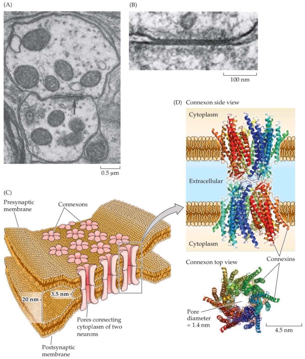
FIGURE 5.2 Structure of electrical synapses. (A) Electron micrograph of an electrical synapse (arrow) connecting two neurons within the inferior olive of a mammalian brain. (B) Higher-magnification electron micrograph of another electrical synapse, showing the gap junction structure characteristic of electrical synapses. (C) Gap junctions consist of connexons, hexameric complexes present in both the preand postsynaptic membranes. Channels assembled from connexons in these two membranes form pores that create electrical continuity between the two cells. (D) Crystallographic structure of connexons. Colors indicate individual connexins, integral membrane proteins that form the subunits of connexons. Side view shows the channels spanning the preand postsynaptic membranes; top view illustrates how six connexin subunits assemble in each membrane to form a channel with an exceptionally large pore. (A,B from Sotelo et al., 1974; D from Maeda et al., 2009.)
شکل ۵.۲ ساختار سیناپسهای الکتریکی. (الف) تصویر میکروسکوپ الکترونی از یک سیناپس الکتریکی (فلش) که دو نورون را در زیتون تحتانی مغز یک پستاندار به هم متصل میکند. (ب) تصویر میکروسکوپ الکترونی با بزرگنمایی بالاتر از یک سیناپس الکتریکی دیگر، که ساختار اتصال شکافی مشخصه سیناپسهای الکتریکی را نشان میدهد. (ج) اتصالات شکافی از کانکسونها، کمپلکسهای ششتایی موجود در هر دو غشای پیشسیناپسی و پسسیناپسی، تشکیل شدهاند. کانالهایی که از کانکسونها در این دو غشا تشکیل شدهاند، منافذی را تشکیل میدهند که پیوستگی الکتریکی بین دو سلول را ایجاد میکنند. (د) ساختار کریستالوگرافی کانکسونها. رنگها نشاندهنده کانکسینهای منفرد، پروتئینهای غشایی جداییناپذیری هستند که زیرواحدهای کانکسونها را تشکیل میدهند. نمای جانبی کانالهایی را نشان میدهد که غشاهای پیشسیناپسی و پسسیناپسی را در بر میگیرند. نمای بالا نشان میدهد که چگونه شش زیرواحد کانکسین در هر غشا جمع میشوند تا کانالی با یک منافذ فوقالعاده بزرگ تشکیل دهند. (الف، ب از Sotelo و همکاران، 1974؛ د از Maeda و همکاران، 2009.)
Although they are a distinct minority, electrical synapses have several functional advantages. One is that transmission is extraordinarily fast: Because passive current flow across connexons is virtually instantaneous, communication can occur without the delay that is characteristic of chemical synapses. The high speed of electrical synaptic transmission is apparent in the operation of the first electrical synapse to be discovered, which resides in the crayfish nervous system. A postsynaptic electrical signal is observed at this synapse within a fraction of a millisecond after the generation of a presynaptic action potential (Figure 5.3A). In fact, at least part of this brief synaptic delay is caused by propagation of the action potential into the presynaptic terminal, so there may be essentially no delay at all in the transmission of electrical signals across the synapse. Such synapses interconnect many of the neurons within the circuit that allows the crayfish to escape from its predators, thus minimizing the time between the presence of a threatening stimulus and a potentially lifesaving motor response.
اگرچه سیناپسهای الکتریکی اقلیت مشخصی هستند، اما چندین مزیت عملکردی دارند. یکی از آنها این است که انتقال فوقالعاده سریع است: از آنجا که جریان غیرفعال در سراسر کانکسونها عملاً آنی است، ارتباط میتواند بدون تأخیری که مشخصه سیناپسهای شیمیایی است، رخ دهد. سرعت بالای انتقال سیناپسی الکتریکی در عملکرد اولین سیناپس الکتریکی کشف شده، که در سیستم عصبی خرچنگ دریایی قرار دارد، آشکار است. یک سیگنال الکتریکی پسسیناپسی در کسری از میلیثانیه پس از تولید پتانسیل عمل پیشسیناپسی در این سیناپس مشاهده میشود (شکل 5.3A). در واقع، حداقل بخشی از این تأخیر سیناپسی کوتاه ناشی از انتشار پتانسیل عمل به ترمینال پیشسیناپسی است، بنابراین ممکن است اساساً هیچ تأخیری در انتقال سیگنالهای الکتریکی در سراسر سیناپس وجود نداشته باشد. چنین سیناپسهایی بسیاری از نورونهای درون مدار را به هم متصل میکنند که به خرچنگ دریایی اجازه میدهد از شکارچیان خود فرار کند، بنابراین زمان بین حضور یک محرک تهدیدکننده و یک پاسخ حرکتی بالقوه نجاتبخش را به حداقل میرسانند.
Another unique advantage of electrical synapses is that transmission can be bidirectional; although some connexons have special features for unidirectional transmission, in most cases current can flow in either direction, depending on which member of the coupled pair is invaded by an action potential. This allows electrical synapses to synchronize electrical activity among populations of neurons.
یکی دیگر از مزایای منحصر به فرد سیناپسهای الکتریکی این است که انتقال میتواند دو طرفه باشد؛ اگرچه برخی از کانکسونها ویژگیهای خاصی برای انتقال یک طرفه دارند، در بیشتر موارد جریان میتواند در هر دو جهت جریان یابد، بسته به اینکه کدام عضو از جفت جفت شده توسط پتانسیل عمل مورد حمله قرار گیرد. این امر به سیناپسهای الکتریکی اجازه میدهد تا فعالیت الکتریکی را در بین جمعیتهای نورونی همگامسازی کنند.
For example, the brainstem neurons that generate rhythmic electrical activity underlying breathing are synchronized by electrical synapses, as are populations of interneurons in the cerebral cortex, thalamus, cerebellum, and other brain regions (Figure 5.3B). Electrical transmission between vasopressinand oxytocin-secreting neurons in the hypothalamus ensures that all cells fire action potentials at about the same time, thus facilitating a synchronized burst of secretion of these hormones into the circulation (see Box 21A). The fact that connexon pores are large enough to allow second messengers to diffuse between cells also permits electrical synapses to synchronize the intracellular signaling of coupled cells. This feature may be particularly important for glial cells, which form large intracellular signaling networks via their gap junctions.
برای مثال، نورونهای ساقه مغز که فعالیت الکتریکی ریتمیک زیربنای تنفس را ایجاد میکنند، توسط سیناپسهای الکتریکی هماهنگ میشوند، همانطور که جمعیتهای نورونهای رابط در قشر مغز، تالاموس، مخچه و سایر نواحی مغز هماهنگ هستند (شکل 5.3B). انتقال الکتریکی بین نورونهای ترشحکننده وازوپرسین و اکسیتوسین در هیپوتالاموس تضمین میکند که همه سلولها تقریباً همزمان پتانسیل عمل ایجاد میکنند، بنابراین ترشح همزمان این هورمونها به گردش خون را تسهیل میکند (به کادر 21A مراجعه کنید). این واقعیت که منافذ کانکسون به اندازه کافی بزرگ هستند که به پیامرسانهای ثانویه اجازه انتشار بین سلولها را میدهند، به سیناپسهای الکتریکی نیز اجازه میدهد تا سیگنالدهی درون سلولی سلولهای جفتشده را هماهنگ کنند. این ویژگی ممکن است به ویژه برای سلولهای گلیال که از طریق اتصالات شکافدار خود شبکههای سیگنالدهی درون سلولی بزرگی را تشکیل میدهند، مهم باشد.


FIGURE 5.3 Function of gap junctions at electrical synapses. (A) Rapid transmission of signals at an electrical synapse in the crayfish. An action potential in the presynaptic neuron causes the postsynaptic neuron to be depolarized within a fraction of a millisecond. (B) Electrical synapses allow synchronization of electrical activity in hippocampal interneurons. In a pair of interneurons connected by electrical synapses, generation of an action potential in one neuron often results in the synchronized firing of an action potential in another neuron (asterisks). (A after Furshpan and Potter, 1959; B after Beierlein et al., 2000.)
شکل ۵.۳ عملکرد اتصالات شکافدار در سیناپسهای الکتریکی. (الف) انتقال سریع سیگنالها در یک سیناپس الکتریکی در خرچنگ دریایی. یک پتانسیل عمل در نورون پیشسیناپسی باعث میشود که نورون پسسیناپسی در کسری از میلیثانیه دپلاریزه شود. (ب) سیناپسهای الکتریکی امکان همگامسازی فعالیت الکتریکی در نورونهای رابط هیپوکامپ را فراهم میکنند. در یک جفت نورون رابط که توسط سیناپسهای الکتریکی به هم متصل شدهاند، تولید یک پتانسیل عمل در یک نورون اغلب منجر به شلیک همزمان یک پتانسیل عمل در نورون دیگر میشود (ستارهها). (الف برگرفته از Furshpan و Potter، 1959؛ ب برگرفته از Beierlein و همکاران، 2000.)
Signaling Transmission at Chemical Synapses
Figure 5.4A shows an electron micrograph of a chemical synapse in the cerebral cortex. This image illustrates the presynaptic terminal, with its abundance of synaptic vesicles, as well as the postsynaptic cell separated by a synaptic cleft. A three-dimensional rendering of this chemical synapse, constructed from many images including the one in Figure 5.4A, reveals these features as well as many more structures, including filamentous elements in both preand postsynaptic processes, as well as structures in the synaptic cleft (Figure 5.4B). In the presynaptic terminal, dense projections (dark blue) are associated with the active zone, the place where synaptic vesicles discharge their neurotransmitters into the synaptic cleft, while the blue structure on the postsynaptic side represents the postsynaptic density, a structure important for postsynaptic signaling at excitatory synapses (see Box 7B).
انتقال سیگنال در سیناپسهای شیمیایی
شکل 5.4A یک ریزنگار الکترونی از یک سیناپس شیمیایی در قشر مغز را نشان میدهد. این تصویر، پایانه پیشسیناپسی را با فراوانی وزیکولهای سیناپسی و همچنین سلول پسسیناپسی که توسط یک شکاف سیناپسی از هم جدا شدهاند، نشان میدهد. یک تصویر سهبعدی از این سیناپس شیمیایی، که از تصاویر زیادی از جمله تصویر موجود در شکل 5.4A ساخته شده است، این ویژگیها و همچنین ساختارهای بسیار بیشتری، از جمله عناصر رشتهای در هر دو فرآیند پیشسیناپسی و پسسیناپسی، و همچنین ساختارهای موجود در شکاف سیناپسی (شکل 5.4B) را نشان میدهد. در پایانه پیشسیناپسی، برآمدگیهای متراکم (آبی تیره) با منطقه فعال، مکانی که وزیکولهای سیناپسی انتقالدهندههای عصبی خود را به شکاف سیناپسی تخلیه میکنند، مرتبط هستند، در حالی که ساختار آبی در سمت پسسیناپسی نشان دهنده تراکم پسسیناپسی است، ساختاری که برای سیگنالدهی پسسیناپسی در سیناپسهای تحریکی مهم است (به کادر 7B مراجعه کنید).
Transmission at chemical synapses is based on the elaborate sequence of events depicted in Figure 5.4C. Prior to transmission, synaptic vesicles are formed and filled with neurotransmitter. Synaptic transmission is initiated when an action potential invades the terminal of the presynaptic neuron. The change in membrane potential caused by the arrival of the action potential leads to the opening of voltage-gated calcium channels in the presynaptic membrane. Because of the steep concentration gradient of Ca2+ across the presynaptic membrane (the external Ca2+ concentration is approximately 10–3 M, whereas the internal Ca2+ concentration is approximately 10–7 M), the opening of these channels causes a rapid influx of Ca2+ into the presynaptic terminal, with the result that the Ca2+ concentration of the cytoplasm in the terminal transiently rises to a much higher value. Elevation of the presynaptic Ca2+ concentration, in turn, allows synaptic vesicles to fuse with the plasma membrane of the presynaptic neuron. The Ca2+-dependent fusion of synaptic vesicles with the terminal membrane causes their contents, most importantly neurotransmitters, to be released into the synaptic cleft, a process called exocytosis.
انتقال در سیناپسهای شیمیایی بر اساس توالی پیچیده وقایع نشان داده شده در شکل 5.4C انجام میشود. قبل از انتقال، وزیکولهای سیناپسی تشکیل شده و با انتقالدهنده عصبی پر میشوند. انتقال سیناپسی زمانی آغاز میشود که یک پتانسیل عمل به ترمینال نورون پیشسیناپسی حمله میکند. تغییر در پتانسیل غشاء ناشی از ورود پتانسیل عمل منجر به باز شدن کانالهای کلسیم وابسته به ولتاژ در غشای پیشسیناپسی میشود. به دلیل شیب غلظت تند +Ca2 در سراسر غشای پیشسیناپسی (غلظت +Ca2 خارجی تقریباً 10-3 M است، در حالی که غلظت +Ca2 داخلی تقریباً 10-7 M است)، باز شدن این کانالها باعث هجوم سریع +Ca2 به ترمینال پیشسیناپسی میشود، در نتیجه غلظت +Ca2 سیتوپلاسم در ترمینال به طور موقت به مقدار بسیار بالاتری افزایش مییابد. افزایش غلظت +Ca2 پیشسیناپسی، به نوبه خود، به وزیکولهای سیناپسی اجازه میدهد تا با غشای پلاسمایی نورون پیشسیناپسی ادغام شوند. همجوشی وابسته به +Ca2 وزیکولهای سیناپسی با غشای انتهایی باعث میشود محتویات آنها، که مهمترین آنها انتقالدهندههای عصبی هستند، به داخل شکاف سیناپسی آزاد شوند، فرآیندی که اگزوسیتوز نامیده میشود.
Following exocytosis, transmitters diffuse across the synaptic cleft and bind to specific receptors on the membrane of the postsynaptic neuron. The binding of neurotransmitter to the receptors causes channels in the postsynaptic membrane to open (or sometimes to close), thus changing the ability of ions to flow across the postsynaptic membrane. The resulting neurotransmitter-induced current flow alters the conductance and (usually) the membrane potential of the postsynaptic neuron, increasing or decreasing the probability that the neuron will fire an action potential. Subsequent removal of the neurotransmitter from the synaptic cleft, by uptake into glial cells or by enzymatic degradation, terminates the action of the neurotransmitter. In this way, information is transmitted transiently from one neuron to another.
پس از اگزوسیتوز، انتقالدهندهها از طریق شکاف سیناپسی پخش میشوند و به گیرندههای خاصی روی غشای نورون پسسیناپسی متصل میشوند. اتصال انتقالدهنده عصبی به گیرندهها باعث باز شدن (یا گاهی بسته شدن) کانالها در غشای پسسیناپسی میشود و در نتیجه توانایی یونها برای عبور از غشای پسسیناپسی را تغییر میدهد. جریان القا شده توسط انتقالدهنده عصبی حاصل، رسانایی و (معمولاً) پتانسیل غشای نورون پسسیناپسی را تغییر میدهد و احتمال ایجاد پتانسیل عمل توسط نورون را افزایش یا کاهش میدهد. حذف بعدی انتقالدهنده عصبی از شکاف سیناپسی، با جذب در سلولهای گلیال یا با تخریب آنزیمی، عمل انتقالدهنده عصبی را خاتمه میدهد. به این ترتیب، اطلاعات به صورت گذرا از یک نورون به نورون دیگر منتقل میشوند.
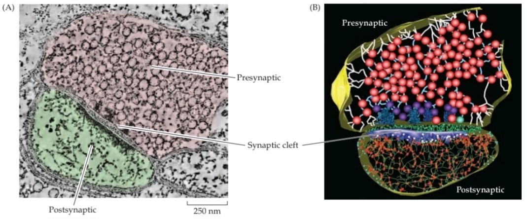
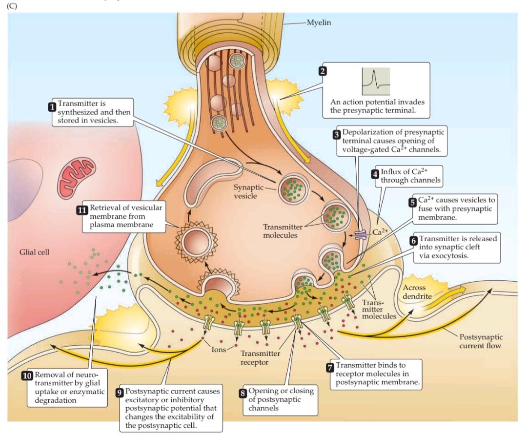
FIGURE 5.4 Structure and function of chemical synapses. (A) Structure of a chemical synapse in the cerebral cortex. A presynaptic terminal (pink) forms a synapse with a postsynaptic dendrite (green). (B) Three-dimensional reconstruction of the synapse shown in (A). Inside the presynaptic terminal, spheres indicate synaptic vesicles at various stages of their trafficking cycle, linear elements indicate intracellular filaments, and dark blue indicates dense projections associated with the active zone. Inside the postsynaptic neuron, the blue structure is the postsynaptic density, green structures represent filaments, red spheres indicate points where the filaments branch. Green material within the synaptic cleft indicates structures of unknown function. (C) Sequence of events involved in transmission at a typical chemical synapse. (A,B from Burette et al., 2012.)
شکل ۵.۴ ساختار و عملکرد سیناپسهای شیمیایی. (الف) ساختار یک سیناپس شیمیایی در قشر مغز. یک پایانه پیشسیناپسی (صورتی) با یک دندریت پسسیناپسی (سبز) یک سیناپس تشکیل میدهد. (ب) بازسازی سهبعدی سیناپس نشان داده شده در (الف). در داخل پایانه پیشسیناپسی، کرهها نشاندهنده وزیکولهای سیناپسی در مراحل مختلف چرخه انتقال آنها هستند، عناصر خطی نشاندهنده رشتههای درون سلولی هستند و آبی تیره نشاندهنده برآمدگیهای متراکم مرتبط با منطقه فعال است. در داخل نورون پسسیناپسی، ساختار آبی نشاندهنده تراکم پسسیناپسی است، ساختارهای سبز نشاندهنده رشتهها هستند، کرههای قرمز نقاطی را نشان میدهند که رشتهها در آنها منشعب میشوند. ماده سبز درون شکاف سیناپسی نشاندهنده ساختارهایی با عملکرد ناشناخته است. (ج) توالی رویدادهای دخیل در انتقال در یک سیناپس شیمیایی معمولی. (الف، ب از بورت و همکاران، 2012.)
Properties of Neurotransmitters
The notion that electrical information can be transferred from one neuron to the next by means of chemical signaling was the subject of intense debate through the first half of the twentieth century. In 1926, the German physiologist Otto Loewi performed a key experiment that supported this idea. Acting on an idea that allegedly came to him in the middle of the night, Loewi proved that electrical stimulation of the vagus nerve slows the heartbeat by releasing a chemical signal that was later shown to be acetylcholine (ACh). ACh is now known to be a neurotransmitter that acts not only in the heart but also at a variety of postsynaptic targets in the central and peripheral nervous systems, preeminently at the neuromuscular junction of striated muscles and in the visceral motor system (see Chapters 6 and 21).
خواص انتقالدهندههای عصبی
این تصور که اطلاعات الکتریکی میتوانند از طریق سیگنالینگ شیمیایی از یک نورون به نورون دیگر منتقل شوند، موضوع بحثهای داغی در نیمه اول قرن بیستم بود. در سال ۱۹۲۶، اتو لووی، فیزیولوژیست آلمانی، آزمایشی کلیدی انجام داد که از این ایده پشتیبانی میکرد. لووی با تکیه بر ایدهای که ظاهراً در نیمه شب به ذهنش خطور کرده بود، ثابت کرد که تحریک الکتریکی عصب واگ با آزاد کردن یک سیگنال شیمیایی که بعداً مشخص شد استیل کولین (ACh) است، ضربان قلب را کند میکند. اکنون مشخص شده است که ACh یک انتقالدهنده عصبی است که نه تنها در قلب، بلکه در اهداف پسسیناپسی متنوعی در سیستمهای عصبی مرکزی و محیطی، بهویژه در محل اتصال عصبی-عضلانی عضلات مخطط و در سیستم حرکتی احشایی عمل میکند (به فصلهای ۶ و ۲۱ مراجعه کنید).
Formal criteria have been established to definitively identify a substance as a neurotransmitter. These criteria have led to the identification of more than 100 different neurotransmitters, which can be classified into two broad categories: small-molecule neurotransmitters, such as ACh, and neuropeptides (see Chapter 6). Having more than one transmitter diversifies the physiological repertoire of synapses. Multiple neurotransmitters can produce different types of responses on individual postsynaptic cells. For example, a neuron can be excited by one type of neurotransmitter and inhibited by another type of neurotransmitter. The speed of postsynaptic responses produced by different transmitters also differs, allowing control of electrical signaling over different timescales. In general, small-molecule neurotransmitters mediate rapid synaptic actions, whereas neuropeptides tend to modulate slower, ongoing neuronal functions. In some cases, neurons synthesize and release two or more different neurotransmitters; in this case, the molecules are called co-transmitters. Co-transmitters can be differentially released according to the pattern of synaptic activity, so that the signaling properties of such synapses change dynamically according to the rate of activity.
معیارهای رسمی برای شناسایی قطعی یک ماده به عنوان انتقالدهنده عصبی تعیین شدهاند. این معیارها منجر به شناسایی بیش از ۱۰۰ انتقالدهنده عصبی مختلف شدهاند که میتوانند به دو دسته کلی طبقهبندی شوند: انتقالدهندههای عصبی مولکول کوچک، مانند ACh، و نوروپپتیدها (به فصل ۶ مراجعه کنید). داشتن بیش از یک انتقالدهنده، مجموعه فیزیولوژیکی سیناپسها را متنوع میکند. انتقالدهندههای عصبی متعدد میتوانند انواع مختلفی از پاسخها را در سلولهای پسسیناپسی منفرد ایجاد کنند. به عنوان مثال، یک نورون میتواند توسط یک نوع انتقالدهنده عصبی تحریک و توسط نوع دیگری از انتقالدهنده عصبی مهار شود. سرعت پاسخهای پسسیناپسی تولید شده توسط انتقالدهندههای مختلف نیز متفاوت است و امکان کنترل سیگنالدهی الکتریکی را در مقیاسهای زمانی مختلف فراهم میکند. به طور کلی، انتقالدهندههای عصبی مولکول کوچک واسطه اعمال سریع سیناپسی هستند، در حالی که نوروپپتیدها تمایل دارند عملکردهای عصبی کندتر و مداوم را تعدیل کنند. در برخی موارد، نورونها دو یا چند انتقالدهنده عصبی مختلف را سنتز و آزاد میکنند. در این حالت، مولکولها، انتقالدهندههای کمکی نامیده میشوند. انتقالدهندههای کمکی میتوانند طبق الگوی فعالیت سیناپسی به صورت متفاوتی آزاد شوند، به طوری که خواص سیگنالدهی چنین سیناپسهایی به صورت پویا با توجه به میزان فعالیت تغییر میکند.
Effective synaptic transmission requires close control of the concentration of neurotransmitters within the synaptic cleft. Neurons have therefore developed a sophisticated ability to regulate the synthesis, packaging, release, and degradation (or removal) of neurotransmitters to achieve the desired levels of transmitter molecules. The synthesis of small-molecule neurotransmitters occurs locally within presynaptic terminals. The precursor molecules required to make new molecules of neurotransmitter are usually taken into the nerve terminal by transporters found in the plasma membrane of the terminal (see Figure 4.13E). The enzymes that synthesize these neurotransmitters are present in the cytoplasm of the presynaptic terminal, and the newly synthesized transmitters are then loaded into synaptic vesicles via another type of transporter located in the vesicular membrane. Most small-molecule neurotransmitters are packaged in vesicles 40 to 60 nm in diameter, the centers of which appear clear in electron micrographs (see Figure 5.4A); accordingly, these vesicles are referred to as small clear-core vesicles. Neuropeptides are synthesized in the cell body of a neuron, and peptide-filled vesicles are transported along an axon and down to the synaptic terminal via axonal transport. Neuropeptides are packaged into synaptic vesicles that range from 90 to 250 nm in diameter. Because the centers of these vesicles appear electron-dense in electron micrographs, they are referred to as large dense-core vesicles.
انتقال سیناپسی مؤثر نیاز به کنترل دقیق غلظت انتقالدهندههای عصبی در شکاف سیناپسی دارد. بنابراین، نورونها توانایی پیچیدهای را برای تنظیم سنتز، بستهبندی، آزادسازی و تخریب (یا حذف) انتقالدهندههای عصبی برای دستیابی به سطوح مطلوب مولکولهای انتقالدهنده ایجاد کردهاند. سنتز انتقالدهندههای عصبی مولکول کوچک به صورت محلی در پایانههای پیشسیناپسی رخ میدهد. مولکولهای پیشساز مورد نیاز برای ساخت مولکولهای جدید انتقالدهنده عصبی معمولاً توسط ناقلهایی که در غشای پلاسمایی پایانه یافت میشوند، به پایانه عصبی منتقل میشوند (شکل 4.13E را ببینید). آنزیمهایی که این انتقالدهندههای عصبی را سنتز میکنند در سیتوپلاسم پایانه پیشسیناپسی وجود دارند و سپس انتقالدهندههای تازه سنتز شده از طریق نوع دیگری از ناقل واقع در غشای وزیکولی به وزیکولهای سیناپسی بارگذاری میشوند. اکثر انتقالدهندههای عصبی مولکول کوچک در وزیکولهایی با قطر 40 تا 60 نانومتر بستهبندی میشوند که مراکز آنها در میکروگرافهای الکترونی واضح به نظر میرسند (شکل 5.4A را ببینید). بر این اساس، به این وزیکولها، وزیکولهای کوچک با هسته شفاف گفته میشود. نوروپپتیدها در جسم سلولی یک نورون سنتز میشوند و وزیکولهای پر از پپتید از طریق انتقال آکسونی در امتداد آکسون و به سمت ترمینال سیناپسی منتقل میشوند. نوروپپتیدها در وزیکولهای سیناپسی بستهبندی میشوند که قطر آنها از ۹۰ تا ۲۵۰ نانومتر متغیر است. از آنجا که مراکز این وزیکولها در میکروگرافهای الکترونی متراکم به نظر میرسند، به آنها وزیکولهای بزرگ با هسته متراکم گفته میشود.
After a neurotransmitter has been secreted into the synaptic cleft, it must be removed to enable the postsynaptic cell to engage in another cycle of synaptic transmission. The removal of neurotransmitters involves diffusion away from the postsynaptic receptors, in combination with reuptake into nerve terminals or surrounding glial cells, degradation by specific enzymes, or a combination of these mechanisms. Specific transporter proteins remove most small-molecule neurotransmitters (or their metabolites) from the synaptic cleft, ultimately delivering them back to the presynaptic terminal for reuse (see Chapter 6).
پس از ترشح یک انتقالدهنده عصبی به شکاف سیناپسی، باید آن را حذف کرد تا سلول پسسیناپسی بتواند در چرخه دیگری از انتقال سیناپسی شرکت کند. حذف انتقالدهندههای عصبی شامل انتشار از گیرندههای پسسیناپسی، همراه با جذب مجدد به پایانههای عصبی یا سلولهای گلیال اطراف، تخریب توسط آنزیمهای خاص یا ترکیبی از این مکانیسمها است. پروتئینهای ناقل خاص، اکثر انتقالدهندههای عصبی مولکول کوچک (یا متابولیتهای آنها) را از شکاف سیناپسی حذف میکنند و در نهایت آنها را برای استفاده مجدد به پایانه پیشسیناپسی بازمیگردانند (به فصل 6 مراجعه کنید).
Quantal Release of Neurotransmitters
Much of the evidence leading to the present understanding of chemical synaptic transmission was obtained from experiments examining the release of ACh at neuromuscular junctions. These synapses between spinal motor neurons and skeletal muscle cells are simple, large, and peripherally located, making them particularly amenable to experimental analysis. Such synapses occur at specializations called end plates because of the saucerlike appearance of the site on the muscle fiber where the presynaptic axon elaborates its terminals (Figure 5.5A). Most of the pioneering work on neuromuscular transmission was performed during the 1950s and 1960s by Bernard Katz and his collaborators at University College London. Although Katz worked primarily on the frog neuromuscular junction, numerous subsequent experiments have confirmed the applicability of his observations to transmission at all chemical synapses.
آزادسازی کوانتالی انتقالدهندههای عصبی
بخش عمدهای از شواهدی که منجر به درک فعلی از انتقال سیناپسی شیمیایی شده است، از آزمایشهایی که آزادسازی استیل کولین را در اتصالات عصبی-عضلانی بررسی میکنند، به دست آمده است. این سیناپسها بین نورونهای حرکتی نخاعی و سلولهای ماهیچه اسکلتی ساده، بزرگ و در حاشیه قرار دارند و به طور خاص برای تجزیه و تحلیل تجربی قابل استفاده هستند. چنین سیناپسهایی در تخصصهایی به نام صفحات انتهایی وجود دارند، زیرا ظاهر بشقابمانند محل روی فیبر عضلانی، جایی که آکسون پیشسیناپسی پایانههای خود را میسازد، آنها را به هم متصل میکند (شکل 5.5A). بیشتر کارهای پیشگامانه در مورد انتقال عصبی-عضلانی در دهههای 1950 و 1960 توسط برنارد کاتز و همکارانش در کالج دانشگاهی لندن انجام شد. اگرچه کاتز در درجه اول روی اتصال عصبی-عضلانی قورباغه کار میکرد، آزمایشهای متعدد بعدی، کاربرد مشاهدات او را در انتقال در همه سیناپسهای شیمیایی تأیید کردهاند.
When an intracellular microelectrode is used to record the membrane potential of a muscle cell, an action potential in the presynaptic motor neuron can be seen to elicit a transient depolarization of the postsynaptic muscle fiber. This change in membrane potential, called an end plate potential (EPP), is normally large enough to bring the membrane potential of the muscle cell well above the threshold for producing a postsynaptic action potential (Figure 5.5B). The postsynaptic action potential triggered by the EPP causes the muscle fiber to contract. Unlike at electrical synapses, there is a pronounced delay between the time that the presynaptic motor neuron is stimulated and when the EPP occurs in the postsynaptic muscle cell. This synaptic delay is characteristic of all chemical synapses.
وقتی از یک میکروالکترود درون سلولی برای ثبت پتانسیل غشای یک سلول ماهیچهای استفاده میشود، میتوان مشاهده کرد که یک پتانسیل عمل در نورون حرکتی پیشسیناپسی باعث ایجاد یک دپلاریزاسیون گذرا در فیبر ماهیچهای پسسیناپسی میشود. این تغییر در پتانسیل غشا، که پتانسیل صفحه انتهایی (EPP) نامیده میشود، معمولاً به اندازهای بزرگ است که پتانسیل غشای سلول ماهیچهای را بسیار بالاتر از آستانه تولید پتانسیل عمل پسسیناپسی میآورد (شکل 5.5B). پتانسیل عمل پسسیناپسی که توسط EPP ایجاد میشود، باعث انقباض فیبر ماهیچهای میشود. برخلاف سیناپسهای الکتریکی، بین زمانی که نورون حرکتی پیشسیناپسی تحریک میشود و زمانی که EPP در سلول ماهیچهای پسسیناپسی رخ میدهد، تأخیر قابل توجهی وجود دارد. این تأخیر سیناپسی مشخصه همه سیناپسهای شیمیایی است.
One of Katz’s seminal findings, in studies carried out with Paul Fatt in 1951, was that spontaneous changes in muscle cell membrane potential occur even in the absence of stimulation of the presynaptic motor neuron (Figure 5.5C). These changes have the same shape as EPPs but are much smaller (typically less than 1 mV in amplitude, compared with an EPP of more than 50 mV). Both EPPs and these small, spontaneous events are sensitive to pharmacological agents that block postsynaptic acetylcholine receptors, such as curare (see Box 6A). These and other parallels between EPPs and the spontaneously occurring depolarizations led Katz and his colleagues to call these spontaneous events miniature end plate potentials, or MEPPs.
یکی از یافتههای مهم کاتز، در مطالعاتی که با پاول فت در سال ۱۹۵۱ انجام شد، این بود که تغییرات خودبهخودی در پتانسیل غشای سلول عضلانی حتی در غیاب تحریک نورون حرکتی پیشسیناپسی رخ میدهد (شکل ۵.۵C). این تغییرات شکل مشابهی با EPPها دارند اما بسیار کوچکتر هستند (معمولاً دامنه آنها کمتر از ۱ میلیولت است، در مقایسه با EPP با دامنه بیش از ۵۰ میلیولت). هم EPPها و هم این رویدادهای کوچک و خودبهخودی به عوامل دارویی که گیرندههای استیل کولین پسسیناپسی را مسدود میکنند، مانند کورار (به کادر ۶A مراجعه کنید) حساس هستند. این موارد و سایر شباهتها بین EPPها و دپلاریزاسیونهای خودبهخودی، کاتز و همکارانش را بر آن داشت تا این رویدادهای خودبهخودی را پتانسیلهای صفحه انتهایی مینیاتوری یا MEPP بنامند.
The relationship between the full-blown end plate potential and MEPPs was clarified by careful analysis of the EPPs. The magnitude of the EPP provides a convenient electrical assay of neurotransmitter secretion from a motor neuron terminal; however, measuring it is complicated by the need to prevent muscle contraction from dislodging the microelectrode. The usual means of eliminating muscle contractions is either to lower Ca2+ concentration in the extracellular medium or to partially block the postsynaptic ACh receptors with the drug curare. As expected from the scheme illustrated in Figure 5.4C, lowering the Ca2+ concentration reduces neurotransmitter secretion, thus reducing the magnitude of the EPP below the threshold for postsynaptic action potential production and allowing it to be measured more precisely. Under such conditions, stimulation of the motor neuron produces very small EPPs that fluctuate in amplitude from trial to trial (Figure 5.5D). These fluctuations give considerable insight into the mechanisms responsible for neurotransmitter release. In particular, the variable evoked response in low Ca2+ is now known to result from the release of unit amounts of ACh by the presynaptic nerve terminal. Indeed, the amplitude of the smallest evoked EPP response is strikingly similar to the size of single MEPPs (compare Figure 5.5C and D). Further supporting this similarity, increments in the EPP response (Figure 5.6A) occur in units about the size of single MEPPs (Figure 5.6B). These “quantal” fluctuations in the amplitude of EPPs indicated to Katz and his colleague Jose del Castillo that EPPs are made up of individual units, each equivalent to a MEPP.
رابطه بین پتانسیل کامل صفحه انتهایی و MEPPها با تجزیه و تحلیل دقیق EPPها روشن شد. بزرگی EPP یک سنجش الکتریکی مناسب از ترشح انتقالدهنده عصبی از یک ترمینال نورون حرکتی فراهم میکند. با این حال، اندازهگیری آن به دلیل نیاز به جلوگیری از جابجایی میکروالکترود توسط انقباض عضله پیچیده است. روش معمول برای از بین بردن انقباضات عضلانی، کاهش غلظت +Ca2 در محیط خارج سلولی یا مسدود کردن نسبی گیرندههای ACh پسسیناپسی با کورار دارویی است. همانطور که از طرح نشان داده شده در شکل 5.4C انتظار میرود، کاهش غلظت +Ca2 ترشح انتقالدهنده عصبی را کاهش میدهد، بنابراین بزرگی EPP را به زیر آستانه تولید پتانسیل عمل پسسیناپسی کاهش میدهد و امکان اندازهگیری دقیقتر آن را فراهم میکند. در چنین شرایطی، تحریک نورون حرکتی EPPهای بسیار کوچکی تولید میکند که دامنه آنها از آزمایش به آزمایش دیگر نوسان دارد (شکل 5.5D). این نوسانات بینش قابل توجهی در مورد مکانیسمهای مسئول آزادسازی انتقالدهنده عصبی ارائه میدهند. به طور خاص، اکنون مشخص شده است که پاسخ برانگیخته متغیر در غلظت پایین کلسیم (+Ca2) ناشی از آزادسازی مقادیر واحد استیل کولین توسط پایانه عصبی پیش سیناپسی است. در واقع، دامنه کوچکترین پاسخ EPP برانگیخته به طرز چشمگیری مشابه اندازه MEPP های منفرد است (شکل 5.5C و D را مقایسه کنید). علاوه بر تأیید این شباهت، افزایش در پاسخ EPP (شکل 5.6A) در واحدهایی تقریباً به اندازه MEPP های منفرد رخ میدهد (شکل 5.6B). این نوسانات “کوانتالی” در دامنه EPP ها به کاتز و همکارش خوزه دل کاستیلو نشان داد که EPP ها از واحدهای جداگانهای تشکیل شدهاند که هر کدام معادل یک MEPP هستند.
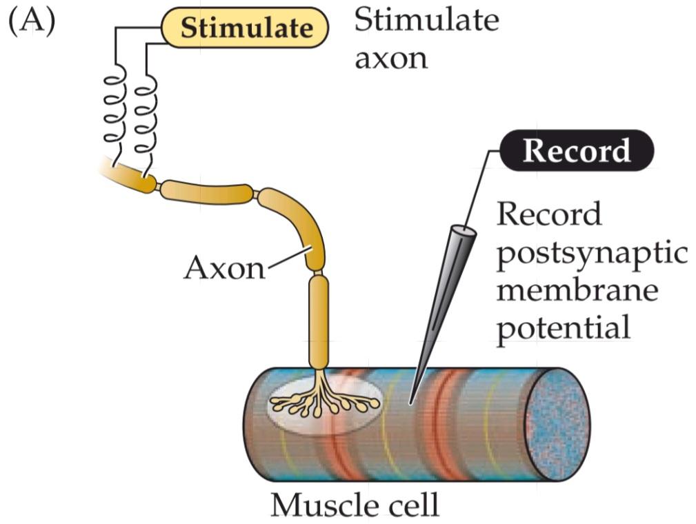


FIGURE 5.5 Synaptic transmission at the neuromuscular junction. (A) Experimental arrangement: The axon of the motor neuron innervating the muscle fiber is stimulated with an extracellular electrode, while an intracellular microelectrode is inserted into the postsynaptic muscle cell to record its electrical responses. (B) End plate potentials (shaded area) evoked by stimulation of a motor neuron are normally above threshold and therefore produce an action potential in the postsynaptic muscle cell. (C) Spontaneous miniature EPPs (MEPPs) occur in the absence of presynaptic stimulation. (D) When the neuromuscular junction is bathed in a solution that has a low concentration of Ca2+, stimulating the motor neuron evokes EPPs whose amplitudes are reduced to about the size of MEPPs. (After Fatt and Katz, 1952.)
شکل ۵.۵ انتقال سیناپسی در محل اتصال عصبی-عضلانی. (الف) چیدمان آزمایش: آکسون نورون حرکتی که فیبر عضلانی را عصبدهی میکند با یک الکترود خارج سلولی تحریک میشود، در حالی که یک میکروالکترود درون سلولی برای ثبت پاسخهای الکتریکی آن به سلول عضلانی پس سیناپسی وارد میشود. (ب) پتانسیلهای صفحه انتهایی (ناحیه سایهدار) که توسط تحریک یک نورون حرکتی برانگیخته میشوند، معمولاً بالاتر از آستانه هستند و بنابراین یک پتانسیل عمل در سلول عضلانی پس سیناپسی ایجاد میکنند. (ج) EPP های مینیاتوری خودبهخودی (MEPP) در غیاب تحریک پیش سیناپسی رخ میدهند. (د) هنگامی که محل اتصال عصبی-عضلانی در محلولی با غلظت کم +Ca2 غوطهور میشود، تحریک نورون حرکتی EPP هایی را برانگیخته میکند که دامنه آنها به اندازه MEPP ها کاهش مییابد. (بعد از Fatt and Katz، 1952.)
The idea that EPPs represent the simultaneous release of many MEPP-like units can be tested statistically. A method of statistical analysis based on the independent occurrence of unitary events (called Poisson statistics) predicts what the distribution of EPP amplitudes would look like during a large number of trials of motor neuron stimulation, under the assumption that EPPs are built up from unitary events represented by MEPPs (see Figure 5.6B). The distribution of EPP amplitudes determined experimentally was found to be just that expected if transmitter release from the motor neuron is indeed quantal (the red curve in Figure 5.6A). Such analyses confirmed the idea that release of acetylcholine does indeed occur in discrete packets, each equivalent to a MEPP. In short, a presynaptic action potential causes a postsynaptic EPP because it synchronizes the release of many transmitter quanta.
این ایده که EPPها نشاندهندهی آزادسازی همزمان بسیاری از واحدهای شبیه MEPP هستند، میتواند از نظر آماری مورد آزمایش قرار گیرد. یک روش تحلیل آماری مبتنی بر وقوع مستقل رویدادهای واحد (به نام آمار پواسون) پیشبینی میکند که توزیع دامنههای EPP در طول تعداد زیادی از آزمایشهای تحریک نورون حرکتی، با این فرض که EPPها از رویدادهای واحدی که توسط MEPPها نشان داده میشوند، ساخته شدهاند (شکل 5.6B را ببینید)، چگونه خواهد بود. توزیع دامنههای EPP که به صورت تجربی تعیین شدهاند، در صورتی که آزادسازی فرستنده از نورون حرکتی واقعاً کوانتالی باشد (منحنی قرمز در شکل 5.6A)، دقیقاً همانطور که انتظار میرفت، مشخص شد. چنین تحلیلهایی این ایده را تأیید کردند که آزادسازی استیل کولین در واقع در بستههای گسسته رخ میدهد که هر کدام معادل یک MEPP هستند. به طور خلاصه، یک پتانسیل عمل پیش سیناپسی باعث یک EPP پس سیناپسی میشود زیرا آزادسازی بسیاری از کوانتهای فرستنده را همزمان میکند.

FIGURE 5.6 Quantized distribution of EPP amplitudes evoked in a low-Ca2+ solution. Peaks of EPP amplitudes (A) tend to occur in integer multiples of the mean amplitude of MEPPs, whose amplitude distribution is shown in (B). The leftmost bar in the EPP amplitude distribution shows trials in which presynaptic stimulation failed to elicit an EPP in the muscle cell. The red curve indicates the prediction of a statistical model based on the assumption that the EPPs result from the independent release of multiple MEPP-like quanta. The observed match, including the predicted number of failures, supports this interpretation. (After Boyd and Martin, 1955.)
شکل ۵.۶ توزیع کوانتیزه دامنههای EPP ایجاد شده در محلول کم +Ca2. قلههای دامنههای EPP (A) معمولاً در مضربهای صحیحی از میانگین دامنه MEPPها رخ میدهند که توزیع دامنه آنها در (B) نشان داده شده است. سمت چپترین نوار در توزیع دامنه EPP، آزمایشهایی را نشان میدهد که در آنها تحریک پیشسیناپسی نتوانسته EPP را در سلول عضلانی ایجاد کند. منحنی قرمز نشاندهنده پیشبینی یک مدل آماری بر اساس این فرض است که EPPها ناشی از آزادسازی مستقل چندین کوانتوم شبیه MEPP هستند. تطابق مشاهده شده، از جمله تعداد پیشبینیشده شکستها، از این تفسیر پشتیبانی میکند. (به نقل از بوید و مارتین، 1955.)
Release of Transmitters from Synaptic Vesicles
🚀 با ما همراه شوید!
تازهترین مطالب و آموزشهای مغز و اعصاب را از دست ندهید. با فالو کردن کانال تلگرام آیندهنگاران مغز، از ما حمایت کنید!