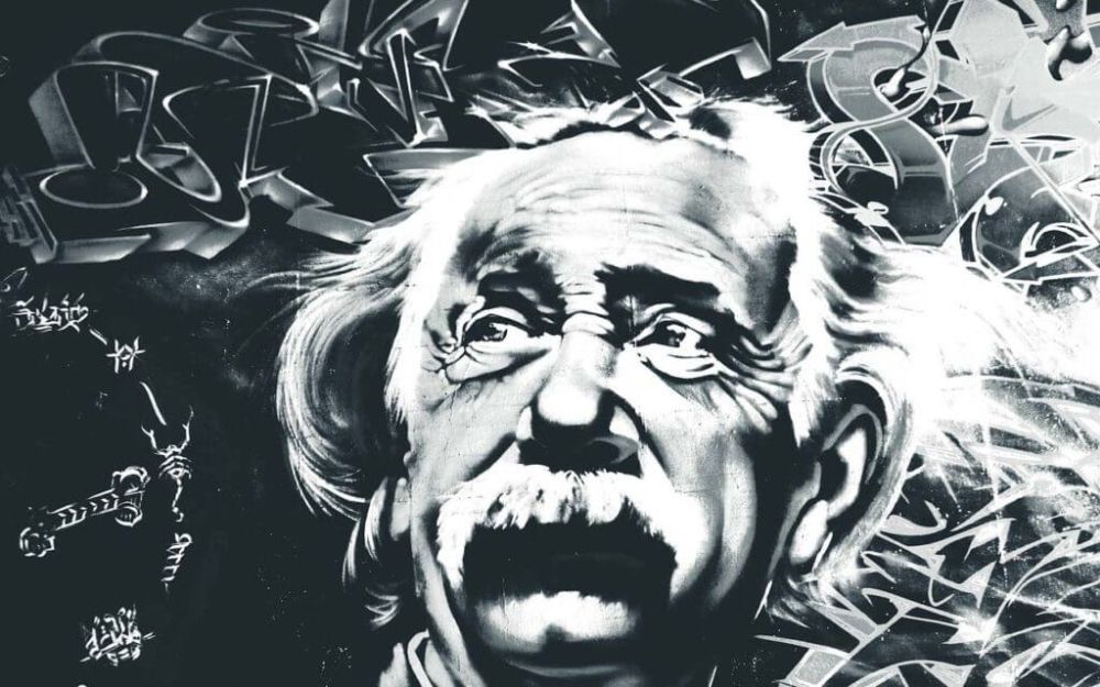
مغز اینشتین برخلاف سایر مغزها

مغز اینشتین برخلاف سایر مغزها، از سلولهای مسئول ساخت اطلاعات سرشار بود.
 برداشتن مغز آلبرت اینشتین، غیرقانونی انجام شد اما نتایج علمی این کار نشان میدهد که ویژگیهای مغز او ارزش انجام این کار غیرقانونی را داشت.(؟؟؟؟!!)تحقیقات روی مغز اینشتین اطلاعات ویژهای در مورد ویژگیهای مغز و نبوغ به محققان داده است.
سال ۱۹۵۵ بعد از مرگ اینشتین، «توماس هاروی» آسیبشناسی که کالبدشکافی اینشتین را انجام داد، طی یکی از مراحل استاندارد کالبدشکافی بخشی از مغز اینشتین را از جمجمهاش خارج کرد اما نتوانست آن را به جای خود بازگرداند. وی بعدها اظهار کرد که پسر اینشتین به وی اجازه داده این بخش از مغز را بردارد؛ اما خانواده اینشتین این ادعا را رد کردند.
هاروی در افتضاحی که در پی این رویداد به بار آمد، کار خود را از دست داد اما مغز اینشتین را نگه داشت. طی سالها او بخشهایی از مغز را برای عصبشناسانی که تلاش در درک ساختار مغزی داشتند که اینشتین را به یک نابغه تبدیل کرده بود، ارسال میکرد.
این برشها از مغز اینشتین از سال ۱۹۵۵ که وی در هفتاد و شش سالگی دارفانی را وداع گفت، سفر طولانی و عجیبی داشتهاند.
هاروی برای آموزش تکنیک برش به فیلادلفیا سفر کرد و در آنجا بود که قطعه به جامانده از مغز اینشتین در لابراتوار آسیبشناسی به نام «ویلیام اریک» برش خورد و به قطعات مختلفی تقسیم شد. هاروی به عنوان تشکر از اریک که این فن را به او آموخته و نمونه مغز را در لابراتوارش برش داده بود، ۴۶ برش از آن را به وی هدیه داد. ضخامت هر یک از این برشها ۲۰ تا ۵۰ میکرون است.
زمانی که اریک در سال ۱۹۶۷ درگذشت، همسرش قطعات را به پزشکی دیگر به نام «الن استینبرگ» اهدا کرد و در نهایت وی نیز برشها را به «لوسی رورک آدام» عصب ـ آسیبشناس بیمارستان کودکان فیلادلفیا سپرد.
مغز جوان در ۷۶ سالگی
محققانی که روی مغز این نابغه مطالعه کردهاند تعیین عاملی ویژه در مغز اینشتین را که به بروز نبوغ باورنکردنیاش منجر شده بسیار دشوار میدانند. با این همه شاید ساختار میکروسکوپی مغز اینشتین که پس از مرگش به صورت غیرطبیعی جوان به نظر میرسید، یکی از این عوامل باشد.
براساس گزارش لایو ساینس، محققان بر این باورند که مغز او توان ساخت «لیپوفوسین»، پسماند سلولی را که با سالخوردگی در ارتباط است نداشته و عروق خونی در مغز او نیز از ساختار و شکل بسیار خوبی برخوردار بوده است، به طوری که محققان میگویند مغز او در زمان مرگ، ساختار مغز فردی جوانتر از ۷۶ سال را داشته است، عواملی که شاید در نبوغ او تاثیرگذار بوده است.
مطالعه روی مغز اینشتین نشان داد که مغز او در مقایسه با میانگین متوسط انسانها، دارای مقدار زیادی سلولهای «گلیال» بوده که مسئول ساخت اطلاعات است
همچنین مطالعه روی مغز اینشتین نشان داد که مغز او در مقایسه با میانگین متوسط انسانها، دارای مقدار زیادی سلولهای «گلیال» بوده که مسئول ساخت اطلاعات است.
همچنین مغز اینشتین مقدار کمی چینخوردگی حقیقی موسوم به شیار «سیلویوس» داشته، که این مساله امکان ارتباط آسانتر سلولهای عصبی را با یکدیگر فراهم میسازد.
این تحقیقات روی لوب آهیانه انجام و مشخص شد که این منطقه از مغز اینشتین از دیگران ۱۵ درصد بزرگتر بوده است. لوب آهیانه برای درک ریاضیات، زبان و روابط سه بعدی و فضایی نقش مهمی ایفا میکند.
اگر میخواهید مغز یک نابغه را از نزدیک ببینید، باید به موزه «موتر» شهر فیلادلفیا سفر کنید تا برای اولینبار ۴۶ قطعه از مغز آلبرت اینشتین، فیزیکدان نظری که نظریه نسبیت عمومی را مطرح کرد، ببینید.
مغز اینشتین در برنامه اجرایی آیپد
سال ۱۹۵۵ یک موزه پزشکی در شیکاگو ۳۵۰ اسلاید ظریف و با ارزش از مغز اینشتین تهیه کرد. این برنامه اجرایی که با استفاده از اسلایدهای مغز اینشتین ساخته شده، به محققان این امکان را میدهد که مغز برنده جایزه نوبل را به گونهای بررسی کنند که گویی آن را زیر میکروسکوپ قرار دادهاند.
یک برنامه اجرایی از مغزی که فیزیک را متحول کرد، برای آیپد رونمایی شده تا دانشمندان بتوانند به تصاویری از جزئیات مغز اینشتین دسترسی پیدا کنند.
دکتر فیلیپ اپستین، عصبشناس و مشاور این موزه گفت: این برنامه جدید آیپد به محققان این امکان را میدهد که با نگاه کردن عمیقتر به مناطق مغز اینشتین که تجمع سلولهای عصبی در آن بیشتر از حد عادی است، علت نبوغ وی را کشف کنند.
اما از آنجا که این اسلایدها از این بافت پیش از ظهور فناوریهای مدرن تصویربرداری ارائه شده، ممکن است برای دانشمندان درک این مساله که دقیقا این اسلاید متعلق به کدام بخش از مغز اینشتین است دشوار باشد، اما این برنامه اجرایی اسلایدها را در منطقه خاص خود قرار داده است.
براساس اظهارات جاکوپو انسی از موسسه تحقیقات مغز دانشگاه کالیفرنیا در ساندیهگو، تصاویر سهبعدی از مغز اینشتین وجود ندارد، آنچه موجود است اسلایدهای یک در سه اینچی است که فقط یک بخش از مغز را تشکیل میدهد.
برنامه اجرایی مغز اینشتین برای غیر دانشمندان با قیمت ۹۹/۹ دلار قابل دانلود است.
Einstein’s brain was different from other people’s

Falk’s team used photographs to show that Einstein’s brain has a complex pattern of convolutions in the part of the brain that deals with abstract thought.
A new study led by Florida State University evolutionary anthropologist Dean Falk has revealed that portions of the brain of Albert Einstein are unlike those of most people. The differences could relate to Einstein’s unique discoveries about the nature of space and time. Falk’s team used photographs of Einstein’s brain, taken shortly after his death, but not previously analyzed in detail. The photographs showed that Einstein’s brain had an unusually complex pattern of convolutions in the prefrontal cortex, which is important for abstract thinking.
In other words, Einsteins’ brain actually looks different from yours or mine. Falk and her team published their work on November 16, 2012 in the journal Brain.
This is an actual photo of Einstein’s brain, which was preserved in formalin by pathologist Thomas Harvey after Einstein’s death in 1955. A new study of this photo and others of Einstein’s brain reveal an unusually complex pattern of convolutions in the prefrontal cortex, which is important for abstract thinking. Photo via the National Museum of Health and Medicine in Silver Spring, Maryland.
A 1920 photo of Einstein in his office at University of Berlin, released in the U.S. in 1920. Photo via Wikimedia Commons.
Falk and her colleagues obtained 12 original photographs of Einstein’s brain from the National Museum of Health and Medicine in Silver Spring, Maryland. They analyzed the photos and compared the patterns of convoluted ridges and furrows in Einstein’s prefrontal cortex with those of 85 brains described in other studies. According to an article in Nature, many of the photographs were taken from unusual angles. They apparently show brain structures that weren’t visible in previously analyzed photos.
How did Einstein’s brain come to undergo so much scrutiny? Pathologist Thomas Harvey performed an autopsy on Einstein shortly after his death in 1955. At that time, he removed Einstein’s brain and preserved it in formalin. He took dozens of black-and-white photos of the brain. Later, he cut Einstein’s brain up into 240 blocks, took tissue samples from each block, mounted them onto microscope slides and distributed the slides to some of the world’s best neuropathologists.
So studies of Einstein’s brain began, although the first detailed one didn’t appear for 30 more years. In 1985, a study revealed that two parts of Einstein’s brain contained an unusually large number of non-neuronal cells – called glia – for every neuron, or nerve-transmitting cell in the brain. Ten years after that, Einstein’s brain was found to lack a furrow normally seen in the parietal lobe. Scientists at that time said the missing furrow might have been related to Einstein’s enhanced ability to think in three dimensions, as well as to his mathematical skills.
Now the most recent study, by Falk et. al., suggests that the pattern of convolutions in Einstein’s prefrontal cortex looks different from most people’s. And if all this talk of removing Einstein’s brain, and photographing it, seems a bit ghoulish, well, the science journal Nature explains it this way:
Albert Einstein is considered to be one of the most intelligent people that ever lived, so researchers are naturally curious about what made his brain tick.
Einstein in 1947, at the age of 68. His special and general theories of relativity altered the way physicists, and the rest of us, think about space and time.
There’s no doubt that Einstein is the most famous abstract thinker known to most of us. His general and special theories of relativity changed the way the rest of us think about space and time, in ways you might take for granted. For example, Einstein said time is relative. It doesn’t click along steadily for everyone at the same rate. It’s Einstein who imagined such a thing, and can you picture yourself making that leap of thought, much less proving it to the world, using the tools of mathematics and physics?
Another example: Einstein altered scientists’ pre-existing understanding of gravity, and, in so doing, changed the way we think about the structure of space. In simple terms, Einstein said that matter causes space to curve. That’s what Einstein’s brain suggested to him and, ultimately, it’s what caused the 20th century revolution in physics.
Einstein’s ability to think abstractly – to think about fundamental properties of the universe in ways no one ever had – is why he’s considered the father of modern physics and the most influential physicist of the 20th century.
Bottom line: Florida State University evolutionary anthropologist Dean Falk led a study showing that portions of the brain of Albert Einstein are different from those of most people. Falk’s team used photographs of Einstein’s brain, taken shortly after his death, and showed that Einstein’s brain has an unusually complex pattern of convolutions in the prefrontal cortex. This part of the brain is important for abstract thought. Falk and her team published their work on November 16, 2012 in the journal Brain.


واقعا جالب و خواندنی بود دست سپاس از شما
انیشتین چنددرصد از مغز خود را استفاده کرده بود؟
خیلی جالب بود
😍
خیلی جالب بود 😍😍
واقعا جالب بود خسته نباشید استاد
عالی بود🌺
سلام
مقاله خوبی هست
میگن جسم پینه ای مغز انیشتین بزرگتر از بقیه بوده
عالی بسیار مفید سودمند
تشکر از شما